|
Pharyngeal Branch Of Maxillary Artery
Pharyngeal may refer to: Anatomy * Pharynx, for pharyngeal anatomy * Pharyngeal muscles **Superior pharyngeal constrictor muscle ** Middle pharyngeal constrictor muscle ** Inferior pharyngeal constrictor muscle * Pharyngeal artery * Pharyngeal slit * Pharyngeal tonsil, a mass of lymphoid tissue in the pharynx Other * Pharyngeal consonant A pharyngeal consonant is a consonant that is articulated primarily in the pharynx. Some phoneticians distinguish upper pharyngeal consonants, or "high" pharyngeals, pronounced by retracting the root of the tongue in the mid to upper pharynx ..., for pharyngeal sounds in phonetics See also * * {{disambiguation ... [...More Info...] [...Related Items...] OR: [Wikipedia] [Google] [Baidu] |
Pharynx
The pharynx (: pharynges) is the part of the throat behind the human mouth, mouth and nasal cavity, and above the esophagus and trachea (the tubes going down to the stomach and the lungs respectively). It is found in vertebrates and invertebrates, though its structure varies across species. The pharynx carries food to the esophagus and air to the larynx. The flap of cartilage called the epiglottis stops food from entering the larynx. In humans, the pharynx is part of the Digestion, digestive system and the conducting zone of the respiratory system. (The conducting zone—which also includes the nostrils of the Human nose, nose, the larynx, trachea, bronchus, bronchi, and bronchioles—filters, warms, and moistens air and conducts it into the lungs). The human pharynx is conventionally divided into three sections: the nasopharynx, oropharynx, and laryngopharynx (hypopharynx). In humans, two sets of pharyngeal muscles form the pharynx and determine the shape of its lumen (anatomy), ... [...More Info...] [...Related Items...] OR: [Wikipedia] [Google] [Baidu] |
Pharyngeal Muscles
The pharyngeal muscles are a group of muscles that form the pharynx, which is posterior to the oral cavity, determining the shape of its lumen, and affecting its sound properties as the primary resonating cavity. The pharyngeal muscles (involuntary skeletal) push food into the esophagus. There are two muscular layers of the pharynx: the outer circular layer and the inner longitudinal layer. The outer circular layer includes: * Superior constrictor muscle * Middle constrictor muscle * Inferior constrictor muscle During swallowing, these muscles constrict to propel a bolus downwards (an involuntary process). The inner longitudinal layer includes: * Stylopharyngeus muscle * Salpingopharyngeus muscle * Palatopharyngeus muscle During swallowing, these muscles act to shorten and widen the pharynx. They are innervated by the pharyngeal branch of the vagus nerve (CN X) with the exception of the stylopharyngeus muscle which is innervated by the glossopharyngeal nerve The gl ... [...More Info...] [...Related Items...] OR: [Wikipedia] [Google] [Baidu] |
Superior Pharyngeal Constrictor Muscle
The superior pharyngeal constrictor muscle is a quadrilateral muscle of the pharynx. It is the uppermost and thinnest of the three pharyngeal constrictors. The muscle is divided into four parts according to its four distincts origins: a pterygopharyngeal, buccopharyngeal, mylopharyngeal, and a glossopharyngeal part. The muscle inserts onto the pharyngeal raphe, and pharyngeal spine. It is innervated by pharyngeal branch of the vagus nerve via the pharyngeal plexus. It acts to convey a bolus down towards the esophagus, facilitating swallowing. Anatomy The superior constrictor muscle is a quadrilateral, sheet-like muscle. It is thinner than the middle and inferior constrictor muscles. Origin The sites of origin of the muscles collectively are the pterygoid hamulus (and occasionally the adjoining posterior margin of the medial pterygoid plate) anteriorly, (the posterior margin of) the pterygomandibular raphe, the posterior extremity of the mylohyoid line of mandible, a ... [...More Info...] [...Related Items...] OR: [Wikipedia] [Google] [Baidu] |
Middle Pharyngeal Constrictor Muscle
The middle pharyngeal constrictor is a fan-shaped muscle located in the neck. It is one of three pharyngeal constrictor muscles. It is smaller than the inferior pharyngeal constrictor muscle. The middle pharyngeal constrictor originates from the greater cornu and lesser cornu of the hyoid bone, and the stylohyoid ligament. It inserts onto the pharyngeal raphe. It is innervated by a branch of the vagus nerve through the pharyngeal plexus. It acts to propel a bolus downwards along the pharynx towards the esophagus, facilitating swallowing. Structure The middle pharyngeal constrictor is a sheet-like, fan-shaped muscle. The muscle's fibers diverge from their origin: the more inferior fibres descend deep to the inferior pharyngeal constrictor muscle; the middle portion of fibres pass transversely; the more superior fibers ascend and overlap the superior pharyngeal constrictor muscle. Origin Two parts of the middle pharyngeal constrictor muscle are distinguished according to i ... [...More Info...] [...Related Items...] OR: [Wikipedia] [Google] [Baidu] |
Inferior Pharyngeal Constrictor Muscle
The inferior pharyngeal constrictor muscle is a skeletal muscle of the neck. It is the thickest of the three outer pharyngeal muscles. It arises from the sides of the cricoid cartilage and the thyroid cartilage. It is supplied by the vagus nerve (CN X). It is active during swallowing, and partially during breathing and speech. It may be affected by Zenker's diverticulum. Structure The inferior pharyngeal constrictor muscle is composed of two parts. The first part (and more superior) arises from the thyroid cartilage (thyropharyngeal part), and the second part arises from the cricoid cartilage (cricopharyngeal part). * On the ''thyroid cartilage'', it arises from the oblique line on the side of the lamina, from the surface behind this nearly as far as the posterior border and from the inferior horn of the thyroid cartilage. * From the ''cricoid cartilage'', it arises in the interval between the cricothyroid muscle in front, and the articular facet for the inferior horn of ... [...More Info...] [...Related Items...] OR: [Wikipedia] [Google] [Baidu] |
Pharyngeal Artery
The pharyngeal artery is a branch of the ascending pharyngeal artery. The pharyngeal artery passes inferior-ward in between the superior margin of the superior pharyngeal constrictor muscle, and the levator veli palatini muscle. It issues branches to the constrictor muscles of the pharynx, the stylopharyngeus muscle, the pharyngotympanic tube, and palatine tonsil Palatine tonsils, commonly called the tonsils and occasionally called the faucial tonsils, are tonsils located on the left and right sides at the back of the throat in humans and other mammals, which can often be seen as flesh-colored, pinkish ...; a palatine branch may sometimes be present, replacing the ascending palatine branch of facial artery. References __NOTOC__ Physiology Anatomy Arteries {{circulatory-stub ... [...More Info...] [...Related Items...] OR: [Wikipedia] [Google] [Baidu] |
Pharyngeal Slit
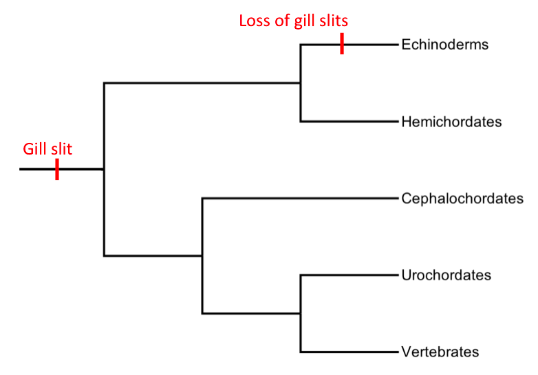
Pharyngeal slits are Filter feeder, filter-feeding organs found among deuterostomes. Pharyngeal slits are repeated openings that appear along the pharynx caudal to the mouth. With this position, they allow for the movement of water in the mouth and out the pharyngeal slits. It is postulated that this is how pharyngeal slits first assisted in filter-feeding, and later, with the addition of gills along their walls, aided in respiration of aquatic chordates. These repeated segments are controlled by similar developmental mechanisms. Some hemichordate species can have as many as 200 gill slits. Pharynx, Pharyngeal clefts resembling gill slits are transiently present during the embryonic stages of tetrapod development. The presence of pharyngeal arches and clefts in the neck of the developing human embryo famously led Ernst Haeckel to postulate that "ontogeny recapitulates phylogeny"; this hypothesis, while false, contains elements of truth, as explored by Stephen Jay Gould in ''Onto ... [...More Info...] [...Related Items...] OR: [Wikipedia] [Google] [Baidu] |
Pharyngeal Tonsil
In anatomy, the pharyngeal tonsil, also known as the nasopharyngeal tonsil or adenoid, is the superior-most of the tonsils. It is a mass of lymphoid tissue located behind the nasal cavity, in the roof and the posterior wall of the nasopharynx, where the nose blends into the throat. In children, it normally forms a soft mound in the roof and back wall of the nasopharynx, just above and behind the uvula. The term ''adenoid'' is also used to represent adenoid hypertrophy, the abnormal growth of the pharyngeal tonsils. Structure The adenoid is a mass of lymphoid tissue located behind the nasal cavity, in the roof and the posterior wall of the nasopharynx, where the nose blends into the throat. The adenoid, unlike the palatine tonsils, has pseudostratified epithelium. The adenoids are part of the so-called Waldeyer ring of lymphoid tissue which also includes the palatine tonsils, the lingual tonsils and the tubal tonsils. Development Adenoids develop from a subepithelial infi ... [...More Info...] [...Related Items...] OR: [Wikipedia] [Google] [Baidu] |


