|
Pellucid Marginal Degeneration
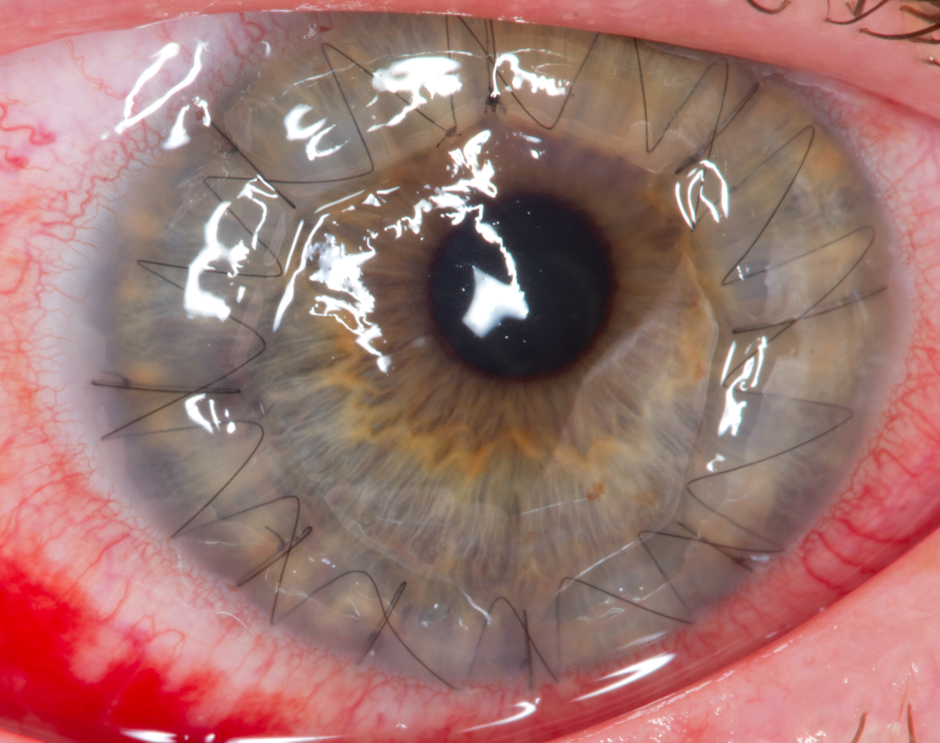
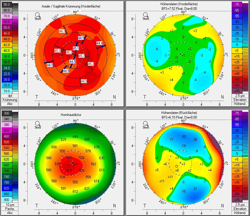
Pellucid marginal degeneration (PMD) is a degenerative corneal condition, often confused with keratoconus. It typically presents with painless vision loss affecting both eyes. Rarely, it may cause acute vision loss with severe pain due to perforation of the cornea. It is typically characterized by a clear, bilateral thinning ( ectasia) in the inferior and peripheral region of the cornea, although some cases affect only one eye. The cause of the disease remains unclear. Pellucid marginal degeneration is diagnosed by corneal topography. Corneal pachymetry may be useful in confirming the diagnosis. Treatment usually consists of vision correction with eyeglasses or contact lenses. Intacs implants, corneal collagen cross-linking, and corneal transplant surgery are additional options. Surgery is reserved for individuals who do not tolerate contact lenses. The term "pellucid marginal degeneration" was first coined in 1957 by the ophthalmologist Schalaeppi. The word "pellucid" means c ... [...More Info...] [...Related Items...] OR: [Wikipedia] [Google] [Baidu] |
Corneal Hydrops
Corneal hydrops is an uncommon complication seen in people with advanced keratoconus or other corneal ectatic disorders, and is characterized by stromal edema due to leakage of aqueous humor through a tear in Descemet's membrane. Although a hydrops usually causes increased scarring of the cornea, occasionally it will benefit a patient by creating a flatter cone, aiding the fitting of contact lenses. Corneal transplantation is not usually indicated during corneal hydrops. Signs and symptoms The person experiences pain and a sudden severe clouding of vision, with the cornea taking on a translucent milky-white appearance known as a corneal hydrops. Diagnosis Patients are recommended to take a sodium chloride eye drop solution as well as a dexamethasone solution for a period of 4-6 weeks, timeframes may vary depending on the severity of a patient's condition. Once the medication cycle is complete and the cloud clears, scarring will be left on the cornea. Management The effect is ... [...More Info...] [...Related Items...] OR: [Wikipedia] [Google] [Baidu] |
Corneal Stroma
The stroma of the cornea (or substantia propria) is a fibrous, tough, unyielding, perfectly transparent and the thickest layer of the cornea of the eye. It is between Bowman's layer anteriorly, and Descemet's membrane posteriorly. At its centre, a human corneal stroma is composed of about 200 flattened ''lamellae'' (layers of collagen fibrils), superimposed one on another. They are each about 1.5-2.5 μm in thickness. The anterior lamellae interweave more than posterior lamellae. The fibrils of each lamella are parallel with one another, but at different angles to those of adjacent lamellae. The lamellae are produced by keratocytes (corneal connective tissue cells), which occupy about 10% of the substantia propria. Apart from the cells, the major non-aqueous constituents of the stroma are collagen fibrils and proteoglycans. The collagen fibrils are made of a mixture of type I and type V collagens. These molecules are tilted by about 15 degrees to the fibril axis, and because of ... [...More Info...] [...Related Items...] OR: [Wikipedia] [Google] [Baidu] |
Corneal Transplantation
Corneal transplantation, also known as corneal grafting, is a surgical procedure where a damaged or diseased cornea is replaced by donated corneal tissue (the graft). When the entire cornea is replaced it is known as penetrating keratoplasty and when only part of the cornea is replaced it is known as lamellar keratoplasty. Keratoplasty simply means surgery to the cornea. The graft is taken from a recently deceased individual with no known diseases or other factors that may affect the chance of survival of the donated tissue or the health of the recipient. The cornea is the transparent front part of the eye that covers the iris, pupil and anterior chamber. The surgical procedure is performed by ophthalmologists, physicians who specialize in eyes, and is often done on an outpatient basis. Donors can be of any age, as is shown in the case of Janis Babson, who donated her eyes after dying at the age of 10. Corneal transplantation is performed when medicines, keratoconus conser ... [...More Info...] [...Related Items...] OR: [Wikipedia] [Google] [Baidu] |
Ablation
Ablation ( – removal) is the removal or destruction of something from an object by vaporization, chipping, erosion, erosive processes, or by other means. Examples of ablative materials are described below, including spacecraft material for ascent and atmospheric reentry, ice and snow in glaciology, biological tissues in medicine and passive fire protection materials. Artificial intelligence In artificial intelligence (AI), especially machine learning, Ablation (artificial intelligence), ablation is the removal of a component of an AI system. The term is by analogy with biology: removal of components of an organism. Biology Biological ablation is the removal of a biological structure or functionality. Genetic ablation is another term for gene silencing, in which gene expression is abolished through the alteration or deletion of genetic sequence information. In cell ablation, individual cells in a population or culture are destroyed or removed. Both can be used as experimen ... [...More Info...] [...Related Items...] OR: [Wikipedia] [Google] [Baidu] |
Photorefractive Keratectomy
Photorefractive keratectomy (PRK) and laser-assisted sub-epithelial keratectomy (or laser epithelial keratomileusis) (LASEK) are laser eye surgery procedures intended to correct a person's vision, reducing dependency on glasses or contact lenses. LASEK and PRK permanently change the shape of the anterior central cornea using an excimer laser to ablate (remove by vaporization) a small amount of tissue from the corneal stroma at the front of the eye, just under the corneal epithelium. The outer layer of the cornea is removed prior to the ablation. A computer system tracks the patient's eye position 60 to 4,000 times per second, depending on the specifications of the laser that is used. The computer system redirects laser pulses for precise laser placement. Most modern lasers will automatically center on the patient's visual axis and will pause if the eye moves out of range and then resume ablating at that point after the patient's eye is re-centered. The outer layer of the corne ... [...More Info...] [...Related Items...] OR: [Wikipedia] [Google] [Baidu] |
Intacs
An intrastromal corneal ring segment (ICRS) (also known as intrastromal corneal ring, corneal implant or corneal insert) is a small device surgically implanted in the cornea of the eye to correct vision. Two crescent or semi-circular shaped ring segments are inserted between the layers of the corneal stroma, one on each side of the pupil, This is intended to flatten the cornea and change the refraction of light passing through the cornea on its way into the eye. Design Intrastromal corneal ring segments have many different types and designs. Manufacturers include Intacs (US), Cornealring (Brazil), Mediphacos Keraring (Brazil), Ferrara ring (Brazil), Myoring (Austria) and Intraseg (UK). Medical uses Intrastromal corneal rings were originally used to treat mild myopia. For this purpose, they have largely been superseded by excimer lasers, which have better accuracy. They are now mostly used to treat mild to moderate keratoconus. Intrastromal corneal rings were approved in 2004 by ... [...More Info...] [...Related Items...] OR: [Wikipedia] [Google] [Baidu] |
Scleral Lens
A scleral lens, also known as a scleral contact lens, is a large contact lens that rests on the sclera and creates a tear-filled vault over the cornea. Scleral lenses are designed to treat a variety of eye conditions, many of which do not respond to other forms of treatment. Uses Medical uses Scleral lenses may be used to improve vision and reduce pain and light sensitivity for people with a growing number of disorders or injuries to the eye, such as severe dry eye syndrome, microphthalmia, keratoconus, corneal ectasia, Stevens–Johnson syndrome, Sjögren's syndrome, aniridia, neurotrophic keratitis (anesthetic corneas), complications post-LASIK, higher-order aberrations of the eye, complications post-corneal transplant and pellucid degeneration. Injuries to the eye such as surgical complications, distorted corneal implants, as well as chemical and burn injuries also may be treated by the use of scleral lenses. Sclerals may also be used in people with eyes that are t ... [...More Info...] [...Related Items...] OR: [Wikipedia] [Google] [Baidu] |
Refraction
In physics, refraction is the redirection of a wave as it passes from one transmission medium, medium to another. The redirection can be caused by the wave's change in speed or by a change in the medium. Refraction of light is the most commonly observed phenomenon, but other waves such as sound waves and Wind wave, water waves also experience refraction. How much a wave is refracted is determined by the change in wave speed and the initial direction of wave propagation relative to the direction of change in speed. Optical Prism (optics), prisms and Lens (optics), lenses use refraction to redirect light, as does the human eye. The refractive index of materials varies with the wavelength of light,R. Paschotta, article ochromatic dispersion in th, accessed on 2014-09-08 and thus the angle of the refraction also varies correspondingly. This is called dispersion (optics), dispersion and causes prism (optics), prisms and rainbows to divide white light into its constituent spectral ... [...More Info...] [...Related Items...] OR: [Wikipedia] [Google] [Baidu] |
Corneal Pachymetry
Corneal pachymetry is the process of measuring the thickness of the cornea. A pachymeter is a medical device used to measure the thickness of the eye's cornea. It is used to perform corneal pachymetry prior to refractive surgery, for Keratoconus screening, LRI surgery and is useful in screening for patients suspected of developing glaucoma among other uses. Process It can be done using either ultrasonic or optical methods . The contact methods, such as ultrasound and optical such as confocal microscopy (CONFOSCAN), or noncontact methods such as optical biometry with a single Scheimpflug camera (such as SIRIUS or PENTACAM), or a Dual Scheimpflug camera (such as GALILEI), or Optical Coherence Tomography (OCT, such as Visante) and online Optical Coherence Pachymetry (OCP, such as ORBSCAN). Corneal Pachymetry is essential prior to a refractive surgery procedure for ensuring sufficient corneal thickness to prevent abnormal bulging of the cornea, a side effect known as ectasia. Pa ... [...More Info...] [...Related Items...] OR: [Wikipedia] [Google] [Baidu] |
Specificity (tests)
In medicine and statistics, sensitivity and specificity mathematically describe the accuracy of a test that reports the presence or absence of a medical condition. If individuals who have the condition are considered "positive" and those who do not are considered "negative", then sensitivity is a measure of how well a test can identify true positives and specificity is a measure of how well a test can identify true negatives: * Sensitivity (true positive rate) is the probability of a positive test result, conditioned on the individual truly being positive. * Specificity (true negative rate) is the probability of a negative test result, conditioned on the individual truly being negative. If the true status of the condition cannot be known, sensitivity and specificity can be defined relative to a " gold standard test" which is assumed correct. For all testing, both diagnoses and screening, there is usually a trade-off between sensitivity and specificity, such that higher sensiti ... [...More Info...] [...Related Items...] OR: [Wikipedia] [Google] [Baidu] |
Gold Standard (test)
In medicine and medical statistics, the gold standard, criterion standard, or reference standard is the diagnostic test or benchmark that is the best available under ''reasonable'' conditions. It is the test against which new tests are compared to gauge their validity, and it is used to evaluate the efficacy of treatments. The meaning of "gold standard" may differ between practical medicine and the statistical ideal. With some medical conditions, only an autopsy can guarantee diagnostic certainty. In these cases, the gold standard test is the best test that keeps the patient alive, and even gold standard tests can require follow-up to confirm or refute the diagnosis. History The term 'gold standard' in its current sense in medical research was coined by Rudd in 1979, in reference to the monetary gold standard. In medicine "Gold standard" can refer to popular clinical endpoints by which scientific evidence is evaluated. For example, in resuscitation research, the "gold standar ... [...More Info...] [...Related Items...] OR: [Wikipedia] [Google] [Baidu] |
Munson's Sign
Munson's sign is a V-shaped indentation observed in the lower eyelid when the patient's gaze is directed downwards. The medical sign is characteristic of advanced cases of keratoconus and is caused by the cone-shaped cornea The cornea is the transparency (optics), transparent front part of the eyeball which covers the Iris (anatomy), iris, pupil, and Anterior chamber of eyeball, anterior chamber. Along with the anterior chamber and Lens (anatomy), lens, the cornea ... pressing down into the eyelid. It is named after American ophthalmologist Edwin Sterling Munson (born May 8, 1870 – died Feb. 2, 1958). References Medical signs Eye diseases {{eye-stub ... [...More Info...] [...Related Items...] OR: [Wikipedia] [Google] [Baidu] |


