|
Passavant's Ridge
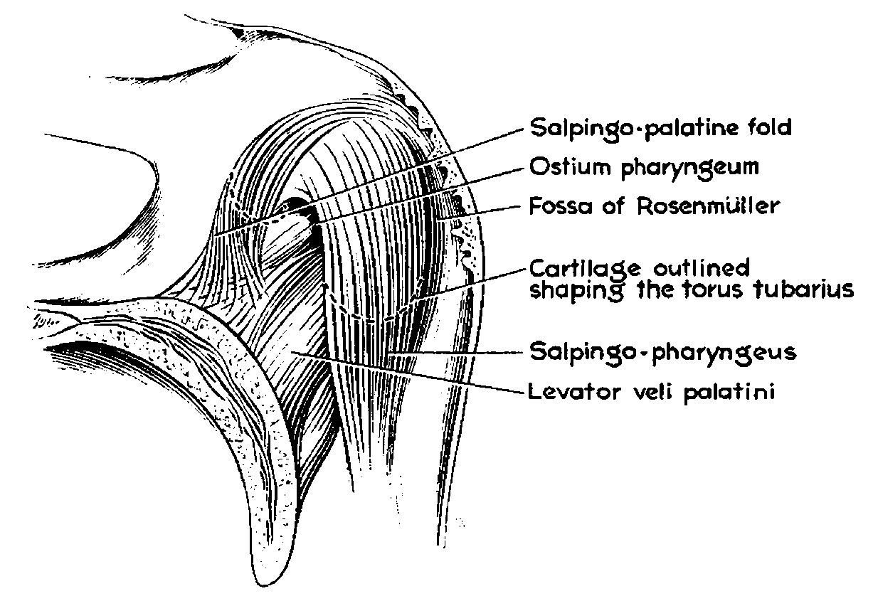
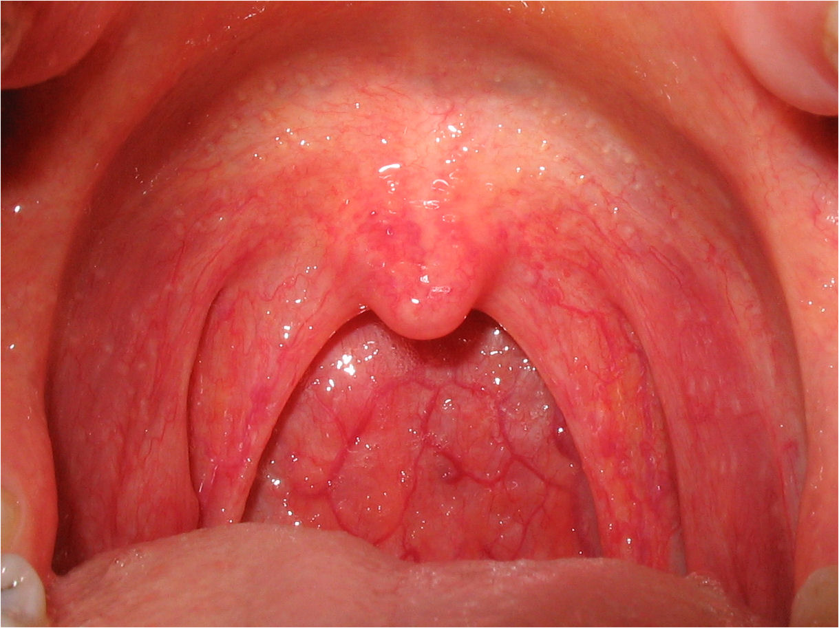
Passavant's ridge is a mucous elevation situated behind the floor of the naso-pharynx. Anatomy It is also known as Passavant's pad or palatopharyngeal ridge. The prominence of mucous tissue is formed by the contraction of superior constrictor during swallowing. Palatopharyngeus muscle originates from the upper surface of the palatal aponeurosis by anterior and posterior fascicle, which are separated by the insertion of levator veli palatini. Both fasciculi join laterally to form a single muscle that passes downward and backward under cover of the palatopharyngeal arch. In the pharynx, it joins with the salpingopharyngeus muscles and is inserted. A few fibers of palatopharyngeus muscle sweep backward under cover of the Passavant's ridge and form a U-shaped sling of palatopharyngeal sphincter. When the soft palate is elevated it comes in contact with ridge, the two together closing pharyngeal isthmus between nasopharynx and oropharynx The pharynx (plural: pharynges) is the part ... [...More Info...] [...Related Items...] OR: [Wikipedia] [Google] [Baidu] |
Mucous
Mucus ( ) is a slippery aqueous secretion produced by, and covering, mucous membranes. It is typically produced from cells found in mucous glands, although it may also originate from mixed glands, which contain both serous and mucous cells. It is a viscous colloid containing inorganic salts, antimicrobial enzymes (such as lysozymes), immunoglobulins (especially IgA), and glycoproteins such as lactoferrin and mucins, which are produced by goblet cells in the mucous membranes and submucosal glands. Mucus serves to protect epithelial cells in the linings of the respiratory, digestive, and urogenital systems, and structures in the visual and auditory systems from pathogenic fungi, bacteria and viruses. Most of the mucus in the body is produced in the gastrointestinal tract. Amphibians, fish, snails, slugs, and some other invertebrates also produce external mucus from their epidermis as protection against pathogens, and to help in movement and is also produced in ... [...More Info...] [...Related Items...] OR: [Wikipedia] [Google] [Baidu] |
Naso-pharynx
The pharynx (plural: pharynges) is the part of the throat behind the mouth and nasal cavity, and above the oesophagus and trachea (the tubes going down to the stomach and the lungs). It is found in vertebrates and invertebrates, though its structure varies across species. The pharynx carries food and air to the esophagus and larynx respectively. The flap of cartilage called the epiglottis stops food from entering the larynx. In humans, the pharynx is part of the digestive system and the conducting zone of the respiratory system. (The conducting zone—which also includes the nostrils of the nose, the larynx, trachea, bronchi, and bronchioles—filters, warms and moistens air and conducts it into the lungs). The human pharynx is conventionally divided into three sections: the nasopharynx, oropharynx, and laryngopharynx. It is also important in vocalization. In humans, two sets of pharyngeal muscles form the pharynx and determine the shape of its lumen. They are arranged as ... [...More Info...] [...Related Items...] OR: [Wikipedia] [Google] [Baidu] |
Palatopharyngeus Muscle
The palatopharyngeus (palatopharyngeal or pharyngopalatinus) muscle is a small muscle in the roof of the mouth. It is a long, fleshy fasciculus, narrower in the middle than at either end, forming, with the mucous membrane covering its surface, the palatopharyngeal arch. Structure It is separated from the palatoglossus muscle by an angular interval, in which the palatine tonsil is lodged. It arises from the soft palate, where it is divided into two fasciculi by the levator veli palatini and musculus uvulae. * The ''posterior fasciculus'' lies in contact with the mucous membrane, and joins with that of the opposite muscle in the middle line. * The ''anterior fasciculus'', the thicker, lies in the soft palate between the levator and tensor veli palatini muscles, and joins in the middle line the corresponding part of the opposite muscle. Passing laterally and downward behind the palatine tonsil, the palatopharyngeus joins the stylopharyngeus and is inserted with that muscle into t ... [...More Info...] [...Related Items...] OR: [Wikipedia] [Google] [Baidu] |
Fascicle (botany)
In botany, a fascicle is a bundle of leaves or flowers growing crowded together; alternatively the term might refer to the vascular tissues that supply such an organ with nutrients.Shashtri, Varun. Dictionary of Botany. Publisher: Isha Books 2005. However, vascular tissues may occur in fascicles even when the organs they supply are not fascicled. Etymology of fascicle and related terms The term ''fascicle'' and its derived terms such as ''fasciculation'' are from the Latin ''fasciculus'', the diminutive of ''fascis'', a bundle. Accordingly, such words occur in many forms and contexts wherever they are convenient for descriptive purposes. A fascicle may be leaves or flowers on a short shoot where the nodes of a shoot are crowded without clear internodes, such as in species of ''Pinus'' or ''Rhigozum''. However, bundled fibres, nerves or bristles as in tissues or the glochid fascicles of ''Opuntia'' may have little or nothing to do with branch morphology. In pines Leaf fascic ... [...More Info...] [...Related Items...] OR: [Wikipedia] [Google] [Baidu] |
Levator Veli Palatini
The levator veli palatini () is the elevator muscle of the soft palate in the human body. It is supplied via the pharyngeal plexus. During swallowing, it contracts, elevating the soft palate to help prevent food from entering the nasopharynx. Structure The levator veli palatini muscle is found in the soft palate of the mouth. It arises from the under surface of the apex of the petrous part of the temporal bone, and from the surface inferolateral to the medial lamina of the cartilage of the Eustachian tube. It does not connect with the medial lamina. It passes above the upper concave margin of the superior pharyngeal constrictor muscle. It spreads out in the palatine velum, its fibers extending obliquely downward and medially to the middle line, where they blend with those of the opposite side. It lies lateral to the choana. Nerve supply The levator veli palatini muscle is supplied by the pharyngeal plexus, which is supplied by the vagus nerve (CN X). Function The ... [...More Info...] [...Related Items...] OR: [Wikipedia] [Google] [Baidu] |
Pharynx
The pharynx (plural: pharynges) is the part of the throat behind the mouth and nasal cavity, and above the oesophagus and trachea (the tubes going down to the stomach and the lungs). It is found in vertebrates and invertebrates, though its structure varies across species. The pharynx carries food and air to the esophagus and larynx respectively. The flap of cartilage called the epiglottis stops food from entering the larynx. In humans, the pharynx is part of the digestive system and the conducting zone of the respiratory system. (The conducting zone—which also includes the nostrils of the nose, the larynx, trachea, bronchi, and bronchioles—filters, warms and moistens air and conducts it into the lungs). The human pharynx is conventionally divided into three sections: the nasopharynx, oropharynx, and laryngopharynx. It is also important in vocalization. In humans, two sets of pharyngeal muscles form the pharynx and determine the shape of its lumen. They are ... [...More Info...] [...Related Items...] OR: [Wikipedia] [Google] [Baidu] |
Salpingopharyngeus
The salpingopharyngeus muscle is a muscle of the pharynx. It arises from cartilage around the Eustachian tube, and inserts into the palatopharyngeus muscle by blending with its posterior fasciculus. It raises the pharynx and larynx during deglutition (swallowing) and laterally draws the pharyngeal walls up. It opens the pharyngeal orifice of the Eustachian tube during swallowing allowing for the equalization of pressure between it and the pharynx. Structure The salpingopharyngeus muscle arises from the superior border of the medial cartilage of the Eustachian tube, in the nasal cavity. This makes the posterior welt of the torus tubarius. It passes downward and blends with the posterior fasciculus of the palatopharyngeus muscle. Nerve supply The salpingopharyngeus is supplied by the vagus nerve (CN X) via the pharyngeal plexus. Blood supply The salpingopharyngeus muscle is supplied by the ascending pharyngeal artery. Function The salpingopharyngeus muscle raises th ... [...More Info...] [...Related Items...] OR: [Wikipedia] [Google] [Baidu] |
Nasopharynx
The pharynx (plural: pharynges) is the part of the throat behind the mouth and nasal cavity, and above the oesophagus and trachea (the tubes going down to the stomach and the lungs). It is found in vertebrates and invertebrates, though its structure varies across species. The pharynx carries food and air to the esophagus and larynx respectively. The flap of cartilage called the epiglottis stops food from entering the larynx. In humans, the pharynx is part of the digestive system and the conducting zone of the respiratory system. (The conducting zone—which also includes the nostrils of the nose, the larynx, trachea, bronchi, and bronchioles—filters, warms and moistens air and conducts it into the lungs). The human pharynx is conventionally divided into three sections: the nasopharynx, oropharynx, and laryngopharynx. It is also important in vocalization. In humans, two sets of pharyngeal muscles form the pharynx and determine the shape of its lumen. They are arranged a ... [...More Info...] [...Related Items...] OR: [Wikipedia] [Google] [Baidu] |


_Taub._(Flame_of_the_forest)_-_Flickr_-_lalithamba.jpg)

