|
Paraventricular Thalamus
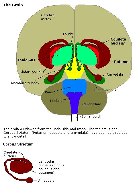
The paraventricular thalamus (PVT) is a midline thalamic nucleus with broad connectivity with other brain structures such as the hypothalamus, striatum, and amygdala. Rodent studies suggest that the PVT plays a role in modulating reward-seeking behavior, threat avoidance, and wakefulness via the hypothalamic-thalamic-striatal circuit, while contributing to the retrieval of fear and the regulation of stress through other circuits. Anatomy The PVT is a nucleus in the midline nuclear group of the thalamus. It has an elongated ovoid shape and an approximate volume of 7 mm³. At the cellular level, the PVT is composed predominantly of glutamatergic neurons, neurons that release glutamate as the primary neurotransmitter. Connectivity The PVT is connected to several structures, including the brainstem, midbrain, striatum, and medial temporal lobe. For the brainstem and midbrain, the PVT receives inputs in the form of orexin from the hypothalamus, which are essential for regulating ... [...More Info...] [...Related Items...] OR: [Wikipedia] [Google] [Baidu] |
Nucleus (neuroanatomy)
In neuroanatomy, a nucleus (: nuclei) is a cluster of neurons in the central nervous system, located deep within the cerebral hemispheres and brainstem. The neurons in one nucleus usually have roughly similar connections and functions. Nuclei are connected to other nuclei by tracts, the bundles (fascicles) of axons (nerve fibers) extending from the cell bodies. A nucleus is one of the two most common forms of nerve cell organization, the other being layered structures such as the cerebral cortex or cerebellar cortex. In anatomical sections, a nucleus shows up as a region of gray matter, often bordered by white matter. The vertebrate brain contains hundreds of distinguishable nuclei, varying widely in shape and size. A nucleus may itself have a complex internal structure, with multiple types of neurons arranged in clumps (subnuclei) or layers. The term "nucleus" is in some cases used rather loosely, to mean simply an identifiably distinct group of neurons, even if they are sprea ... [...More Info...] [...Related Items...] OR: [Wikipedia] [Google] [Baidu] |
Amygdala
The amygdala (; : amygdalae or amygdalas; also '; Latin from Greek language, Greek, , ', 'almond', 'tonsil') is a paired nucleus (neuroanatomy), nuclear complex present in the Cerebral hemisphere, cerebral hemispheres of vertebrates. It is considered part of the limbic system. In Primate, primates, it is located lateral and medial, medially within the temporal lobes. It consists of many nuclei, each made up of further subnuclei. The subdivision most commonly made is into the Basolateral amygdala, basolateral, Central nucleus of the amygdala, central, cortical, and medial nuclei together with the intercalated cells of the amygdala, intercalated cell clusters. The amygdala has a primary role in the processing of memory, decision making, decision-making, and emotions, emotional responses (including fear, anxiety, and aggression). The amygdala was first identified and named by Karl Friedrich Burdach in 1822. Structure Thirteen Nucleus (neuroanatomy), nuclei have been identif ... [...More Info...] [...Related Items...] OR: [Wikipedia] [Google] [Baidu] |
Protein C-Fos
Protein c-Fos is a proto-oncogene that is the human homolog of the retroviral oncogene v-fos. It is encoded in humans by the ''FOS'' gene. It was first discovered in rat fibroblasts as the transforming gene of the FBJ MSV (Finkel–Biskis–Jinkins murine osteogenic sarcoma virus) (Curran and Tech, 1982). It is a part of a bigger Fos family of transcription factors which includes c-Fos, FosB, Fra-1 and Fra-2. It has been mapped to chromosome region 14q21→q31. c-Fos encodes a 62 kDa protein, which forms heterodimer with c-jun (part of Jun family of transcription factors), resulting in the formation of AP-1 (Activator Protein-1) complex which binds DNA at AP-1 specific sites at the promoter and enhancer regions of target genes and converts extracellular signals into changes of gene expression. It plays an important role in many cellular functions and has been found to be overexpressed in a variety of cancers. Structure and function c-Fos is a 380 amino acid protein with a b ... [...More Info...] [...Related Items...] OR: [Wikipedia] [Google] [Baidu] |
Fear Conditioning
Pavlovian fear conditioning is a behavioral paradigm in which organisms learn to predict aversive events. It is a form of learning in which an aversive stimulus (e.g. an electrical shock) is associated with a particular neutral context (e.g., a room) or neutral stimulus (e.g., a tone), resulting in the expression of fear responses to the originally neutral stimulus or context. This can be done by pairing the neutral stimulus with an aversive stimulus (e.g., an electric shock, loud noise, or unpleasant odor). Eventually, the neutral stimulus alone can elicit the state of fear. In the vocabulary of classical conditioning, the neutral stimulus or context is the "conditional stimulus" (CS), the aversive stimulus is the "unconditional stimulus" (US), and the fear is the "conditional response" (CR). Fear conditioning has been studied in numerous species, from snails to humans. In humans, conditioned fear is often measured with verbal report and galvanic skin response. In other animals ... [...More Info...] [...Related Items...] OR: [Wikipedia] [Google] [Baidu] |
Dopamine Receptor D1
Dopamine receptor D1, also known as DRD1. It is one of the two types of D1-like receptor family receptors D1 and D5. It is a protein that in humans is encoded by the DRD1 gene. Tissue distribution D1 receptors are the most abundant kind of dopamine receptor in the central nervous system. Northern blot and in situ hybridization show that the gene expression, mRNA expression of DRD1 is highest in the dorsal striatum (caudate nucleus, caudate and putamen) and ventral striatum (nucleus accumbens and olfactory tubercle). Lower levels occur in the basolateral amygdala, cerebral cortex, septum, thalamus, and hypothalamus. The DRD1 gene expresses primarily in the caudate putamen in humans, and in the caudate putamen, the nucleus accumbens and the olfactory tubercle in mouse. Structure The dopamine receptor D1 (D1R) is a Gs-coupled GPCR characterized by a canonical seven-transmembrane (TM) helical domain, with a ligand-binding pocket located Extracellular space, extracellularl ... [...More Info...] [...Related Items...] OR: [Wikipedia] [Google] [Baidu] |
Optogenetics
Optogenetics is a biological technique to control the activity of neurons or other cell types with light. This is achieved by Gene expression, expression of Channelrhodopsin, light-sensitive ion channels, Halorhodopsin, pumps or Photoactivated adenylyl cyclase, enzymes specifically in the target cells. On the level of individual Cell (biology), cells, Photoactivated adenylyl cyclase, light-activated enzymes and transcription factors allow precise control of biochemical signaling pathways. In Neuroscience, systems neuroscience, the ability to control the activity of a genetically defined set of neurons has been used to understand their contribution to decision making, learning, fear memory, mating, addiction, feeding, and locomotion. In a first medical application of optogenetic technology, vision was partially restored in a blind patient with Retinitis pigmentosa. Optogenetic techniques have also been introduced to map the Brain connectivity estimators, functional connectivity of t ... [...More Info...] [...Related Items...] OR: [Wikipedia] [Google] [Baidu] |
Chemogenetics
Chemogenetics is the process by which macromolecules can be engineered to interact with previously unrecognized small molecules. Chemogenetics as a term was originally coined to describe the observed effects of mutations on chalcone isomerase activity on substrate specificities in the flowers of ''Dianthus caryophyllus''. This method is very similar to optogenetics; however, it uses chemically engineered molecules and ligands instead of light and light-sensitive channels known as opsins. In recent research projects, chemogenetics has been widely used to understand the relationship between brain activity and behavior. Prior to chemogenetics, researchers used methods such as transcranial magnetic stimulation and deep brain stimulation to study the relationship between neuronal activity and behavior. Comparison to optogenetics Optogenetics and chemogenetics are the more recent and popular methods used to study this relationship. Both of these methods target specific brain circuits ... [...More Info...] [...Related Items...] OR: [Wikipedia] [Google] [Baidu] |
Lateral Hypothalamus
The lateral hypothalamus (LH), also called the lateral hypothalamic area (LHA), contains the primary orexinergic nucleus within the hypothalamus that widely projects throughout the nervous system; this system of neurons mediates an array of cognitive and physical processes, such as promoting feeding behavior and arousal, reducing pain perception, and regulating body temperature, digestive functions, and blood pressure, among many others. Clinically significant disorders that involve dysfunctions of the orexinergic projection system include narcolepsy, motility disorders or functional gastrointestinal disorders involving visceral hypersensitivity (e.g., irritable bowel syndrome), and eating disorders. The neurotransmitter glutamate and the endocannabinoids (e.g., anandamide) and the orexin neuropeptides orexin-A and orexin-B are the primary signaling neurochemicals in orexin neurons; pathway-specific neurochemicals include GABA, melanin-concentrating hormone, nociceptin, gluc ... [...More Info...] [...Related Items...] OR: [Wikipedia] [Google] [Baidu] |
Hypocretin
Orexin (), also known as hypocretin, is a neuropeptide that regulates arousal, wakefulness, and appetite. It exists in the forms of orexin-A and orexin-B. The most common form of narcolepsy, type 1, in which the individual experiences brief losses of muscle tone ("drop attacks" or cataplexy), is caused by a lack of orexin in the brain due to destruction of the cells that produce it.Stanford Center for NarcolepsFAQ(retrieved 27-Mar-2012) There are 50,000–80,000 orexin-producing neurons in the human brain, located predominantly in the perifornical area and lateral hypothalamus. They project widely throughout the central nervous system, regulating wakefulness, feeding, and other behaviours. There are two types of orexin peptide and two types of orexin receptor. Orexin was discovered in 1998 almost simultaneously by two independent groups of researchers working on the rat brain. One group named it orexin, from ''orexis,'' meaning "appetite" in Greek; the other group named it hypocr ... [...More Info...] [...Related Items...] OR: [Wikipedia] [Google] [Baidu] |
Acetylcholine
Acetylcholine (ACh) is an organic compound that functions in the brain and body of many types of animals (including humans) as a neurotransmitter. Its name is derived from its chemical structure: it is an ester of acetic acid and choline. Parts in the body that use or are affected by acetylcholine are referred to as cholinergic. Acetylcholine is the neurotransmitter used at the neuromuscular junction. In other words, it is the chemical that motor neurons of the nervous system release in order to activate muscles. This property means that drugs that affect cholinergic systems can have very dangerous effects ranging from paralysis to convulsions. Acetylcholine is also a neurotransmitter in the autonomic nervous system, both as an internal transmitter for both the sympathetic nervous system, sympathetic and the parasympathetic nervous system, and as the final product released by the parasympathetic nervous system. Acetylcholine is the primary neurotransmitter of the parasympathet ... [...More Info...] [...Related Items...] OR: [Wikipedia] [Google] [Baidu] |
Medium Spiny Neuron
Medium spiny neurons (MSNs), also known as spiny projection neurons (SPNs), are a special type of inhibitory GABAergic neuron representing approximately 90% of neurons within the human striatum, a basal ganglia structure. Medium spiny neurons have two primary phenotypes (characteristic types): D1-type MSNs of the direct pathway and D2-type MSNs of the indirect pathway. Most striatal MSNs contain only D1-type or D2-type dopamine receptors, but a subpopulation of MSNs exhibit both phenotypes. Direct pathway MSNs excite their ultimate basal ganglia output structure (such as the thalamus) and promote associated behaviors; these neurons express D1-type dopamine receptors, adenosine A1 receptors, dynorphin peptides, and substance P peptides. Indirect pathway MSNs inhibit their output structure and in turn inhibit associated behaviors; these neurons express D2-type dopamine receptors, adenosine A2A receptors (A2A), heterotetramers, and enkephalin. Both types express glut ... [...More Info...] [...Related Items...] OR: [Wikipedia] [Google] [Baidu] |


