|
Paraphaeosphaeria Pilleata
''Paraphaeosphaeria pilleata'' is a species of fungus in the Didymosphaeriaceae family. The species fruits exclusively in the lower parts of the culms of the black needlerush (''Juncus roemerianus''). It is found on the Atlantic Coast of North Carolina. Taxonomy and naming The species was first described by mycologists Jan Kohlmeyer, Brigitte Volkmann-Kohlmeyer, and Ove Eriksson in a 1996 '' Mycological Research'' publication. The specific epithet is derived from Latin ''pilleatus'' ("capped"), and refers to the small cap on the top of the ascus. Description The roughly spherical to ellipsoidal leathery fruit bodies (ascomata) are 120–300 μm high by 150–350 μm wide, with a short neck, and an ostiole (opening). The neck is 15–50 μm high, 42–70 μm in diameter, cylindrical to conical, and dark brown. The ascomata are immersed in the hard cortex of the host plant, with bases embedded in the pith. The ascomata are light brown in color, but darker around the ostiole. The ... [...More Info...] [...Related Items...] OR: [Wikipedia] [Google] [Baidu] |
East Coast Of The United States
The East Coast of the United States, also known as the Eastern Seaboard, the Atlantic Coast, and the Atlantic Seaboard, is the region encompassing the coast, coastline where the Eastern United States meets the Atlantic Ocean; it has always played a major socioeconomic role in the development of the United States. The region is generally understood to include the U.S. states that border the Atlantic Ocean: Connecticut, Delaware, Florida, Georgia (U.S. state), Georgia, Maine, Maryland, Massachusetts, New Hampshire, New Jersey, New York (state), New York, North Carolina, Rhode Island, South Carolina, and Virginia, as well as some landlocked territories (Pennsylvania, Vermont, West Virginia and Washington, D.C.). Toponymy and composition The Toponymy, toponym derives from the concept that the contiguous 48 states are defined by two major coastlines, one at the West Coast of the United States, western edge and one on the eastern edge. Other terms for referring to this area include ... [...More Info...] [...Related Items...] OR: [Wikipedia] [Google] [Baidu] |
Peridium
The peridium is the protective layer that encloses a mass of spores in fungi. This outer covering is a distinctive feature of gasteroid fungi. Description Depending on the species, the peridium may vary from being paper-thin to thick and rubbery or even hard. Typically, peridia consist of one to three layers. If there is only a single layer, it is called a peridium. If two layers are present, the outer layer is called the exoperidium and the inner layer the endoperidium. If three layers are present, they are the exoperidium, the mesoperidium and the endoperidium. In the simplest subterranean forms, the peridium remains closed until the spores are mature, and even then shows no special arrangement for dehiscence or opening, but has to decay before the spores are liberated. Puffballs For most fungi, the peridium is ornamented with scales or spines. In species that become raised above ground during their development, generally known as the "puffballs", the peridium is usually ... [...More Info...] [...Related Items...] OR: [Wikipedia] [Google] [Baidu] |
Conidia
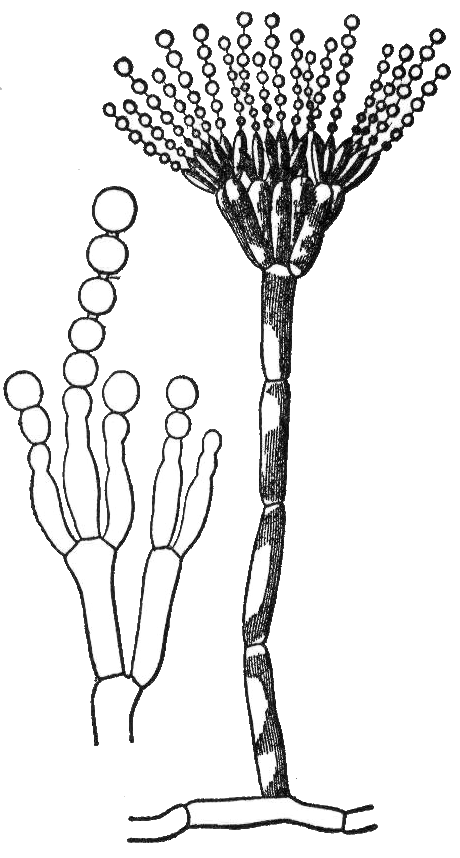
A conidium ( ; : conidia), sometimes termed an asexual chlamydospore or chlamydoconidium (: chlamydoconidia), is an asexual, non- motile spore of a fungus. The word ''conidium'' comes from the Ancient Greek word for dust, ('). They are also called mitospores due to the way they are generated through the cellular process of mitosis. They are produced exogenously. The two new haploid cells are genetically identical to the haploid parent, and can develop into new organisms if conditions are favorable, and serve in biological dispersal. Asexual reproduction in ascomycetes (the phylum Ascomycota) is by the formation of conidia, which are borne on specialized stalks called conidiophores. The morphology of these specialized conidiophores is often distinctive between species and, before the development of molecular techniques at the end of the 20th century, was widely used for identification of (''e.g.'' '' Metarhizium'') species. The terms microconidia and macroconidia are some ... [...More Info...] [...Related Items...] OR: [Wikipedia] [Google] [Baidu] |
Conidiomata
Conidiomata (singular: Conidioma) are blister-like fruiting structures produced by a specific type of fungus called a coelomycete. They are formed as a means of dispersing asexual spores call conidia, which they accomplish by creating the blister-like formations which then rupture to release the contained spores. Structure Conidiomata mainly consist of a mass of densely packed hypha which develops below the surface of the host cuticle, and the fungus may or may not use some of the host’s own tissue to construct the structure. Development of these structures can either occur just below the cuticle or below the epidermal layer of the tissue. Formation on the differing levels is dependent upon the type of conidiomata being formed. Five types of conidioma have been found and are classified as acervuli, pycnidia, sporodochia, synnemata and corenima. Types Acervuli Acervuli is one of the two major groups of conidiomata (the other being pycnidia). Conidiomata of this type for ... [...More Info...] [...Related Items...] OR: [Wikipedia] [Google] [Baidu] |
Pure Culture
A microbiological culture, or microbial culture, is a method of multiplying microbial organisms by letting them reproduce in predetermined culture medium under controlled laboratory conditions. Microbial cultures are foundational and basic diagnostic methods used as research tools in molecular biology. The term ''culture'' can also refer to the microorganisms being grown. Microbial cultures are used to determine the type of organism, its abundance in the sample being tested, or both. It is one of the primary diagnostic methods of microbiology and used as a tool to determine the cause of infectious disease by letting the agent multiply in a predetermined medium. For example, a throat culture is taken by scraping the lining of tissue in the back of the throat and blotting the sample into a medium to be able to screen for harmful microorganisms, such as ''Streptococcus pyogenes'', the causative agent of strep throat. Furthermore, the term culture is more generally used informally ... [...More Info...] [...Related Items...] OR: [Wikipedia] [Google] [Baidu] |
Ascospore
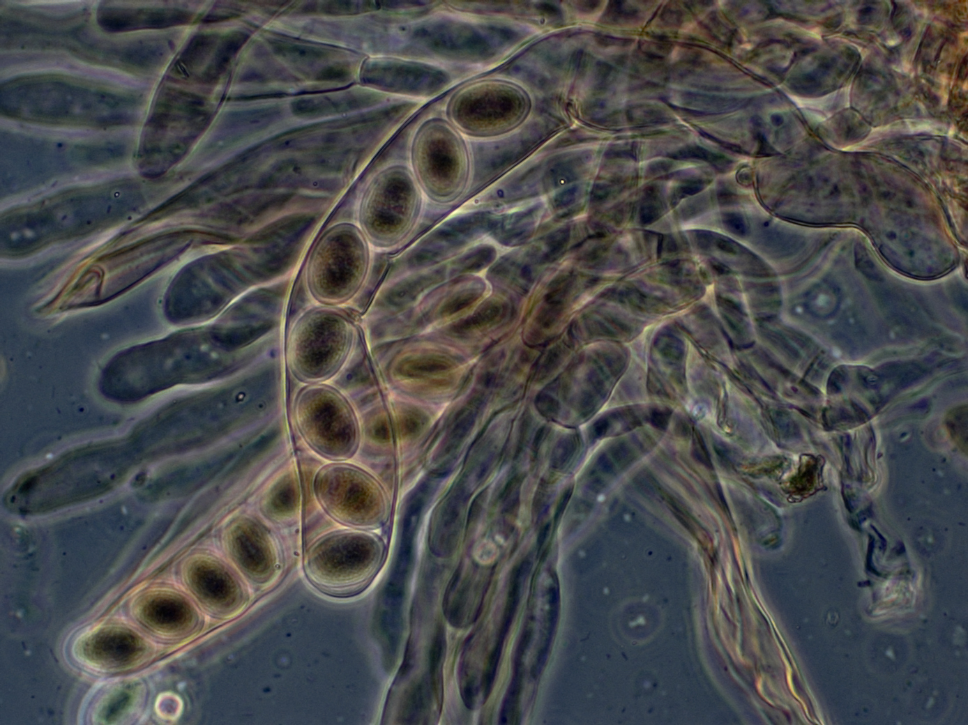
In fungi, an ascospore is the sexual spore formed inside an ascus—the sac-like cell that defines the division Ascomycota, the largest and most diverse Division (botany), division of fungi. After two parental cell nucleus, nuclei fuse, the ascus undergoes meiosis (halving of genetic material) followed by a mitosis (cell division), ordinarily producing eight genetically distinct haploid spores; most yeasts stop at four ascospores, whereas some moulds carry out extra post-meiotic divisions to yield dozens. Many asci build turgor, internal pressure and shoot their spores clear of the calm boundary layer, thin layer of still air enveloping the fruit body, whereas subterranean truffles depend on animals for biological dispersal, dispersal. Ontogeny, Development shapes both form and endurance of ascospores. A hook-shaped crozier aligns the paired nuclei; a double-biological membrane, membrane system then parcels each daughter nucleus, and successive wall layers of β-glucan, chitosan ... [...More Info...] [...Related Items...] OR: [Wikipedia] [Google] [Baidu] |
Dehiscence (botany)
Dehiscence is the splitting of a mature plant structure along a built-in line of weakness to release its contents. This is common among fruits, anthers and sporangia. Sometimes this involves the complete detachment of a part. Structures that open in this way are said to be dehiscent. Structures that do not open in this way are called indehiscent, and rely on other mechanisms such as decay, digestion by herbivores, or predation to release the contents. A similar process to dehiscence occurs in some flower buds (e.g., '' Platycodon'', '' Fuchsia''), but this is rarely referred to as dehiscence unless circumscissile dehiscence is involved; anthesis is the usual term for the opening of flowers. Dehiscence may or may not involve the loss of a structure through the process of abscission. The lost structures are said to be caducous. Association with crop breeding Manipulation of dehiscence can improve crop yield since a trait that causes seed dispersal is a disadvantage for fa ... [...More Info...] [...Related Items...] OR: [Wikipedia] [Google] [Baidu] |
Fissitunicate
{{Short pages monitor ... [...More Info...] [...Related Items...] OR: [Wikipedia] [Google] [Baidu] |
Septum
In biology, a septum (Latin language, Latin for ''something that encloses''; septa) is a wall, dividing a Body cavity, cavity or structure into smaller ones. A cavity or structure divided in this way may be referred to as septate. Examples Human anatomy * Interatrial septum, the wall of tissue that is a sectional part of the left and right atria of the heart * Interventricular septum, the wall separating the left and right ventricles of the heart * Lingual septum, a vertical layer of fibrous tissue that separates the halves of the tongue *Nasal septum: the cartilage wall separating the nostrils of the nose * Alveolar septum: the thin wall which separates the Pulmonary alveolus, alveoli from each other in the lungs * Orbital septum, a palpebral ligament in the upper and lower eyelids * Septum pellucidum or septum lucidum, a thin structure separating two fluid pockets in the brain * Uterine septum, a malformation of the uterus * Septum of the penis, Penile septum, a fibrous w ... [...More Info...] [...Related Items...] OR: [Wikipedia] [Google] [Baidu] |
Pseudoparaphyses
{{Short pages monitor ... [...More Info...] [...Related Items...] OR: [Wikipedia] [Google] [Baidu] |
Hymenium
The hymenium is the tissue layer on the hymenophore of a fungal fruiting body where the cells develop into basidia or asci, which produce spores. In some species all of the cells of the hymenium develop into basidia or asci, while in others some cells develop into sterile cells called cystidia ( basidiomycetes) or paraphyses ( ascomycetes). Cystidia are often important for microscopic identification. The subhymenium consists of the supportive hyphae from which the cells of the hymenium grow, beneath which is the hymenophoral trama, the hyphae that make up the mass of the hymenophore. The position of the hymenium is traditionally the first characteristic used in the classification and identification of mushrooms. Below are some examples of the diverse types which exist among the macroscopic Basidiomycota and Ascomycota. * In agarics, the hymenium is on the vertical faces of the gills. * In boletes and polypores, it is in a spongy mass of downward-pointing tubes ... [...More Info...] [...Related Items...] OR: [Wikipedia] [Google] [Baidu] |
Hypha
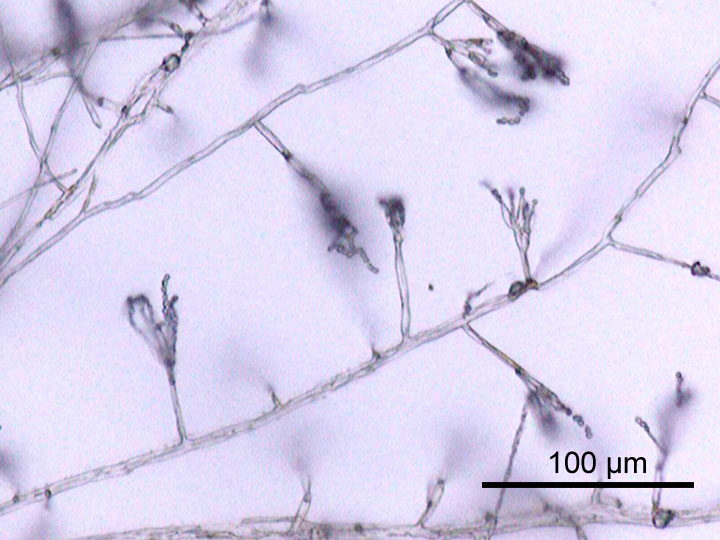
A hypha (; ) is a long, branching, filamentous structure of a fungus, oomycete, or actinobacterium. In most fungi, hyphae are the main mode of vegetative growth, and are collectively called a mycelium. Structure A hypha consists of one or more cells surrounded by a tubular cell wall. In most fungi, hyphae are divided into cells by internal cross-walls called "septa" (singular septum). Septa are usually perforated by pores large enough for ribosomes, mitochondria, and sometimes nuclei to flow between cells. The major structural polymer in fungal cell walls is typically chitin, in contrast to plants and oomycetes that have cellulosic cell walls. Some fungi have aseptate hyphae, meaning their hyphae are not partitioned by septa. Hyphae have an average diameter of 4–6 μm. Growth Hyphae grow at their tips. During tip growth, cell walls are extended by the external assembly and polymerization of cell wall components, and the internal production of new cell membrane. ... [...More Info...] [...Related Items...] OR: [Wikipedia] [Google] [Baidu] |


