|
Pamelaria Scale Diagram
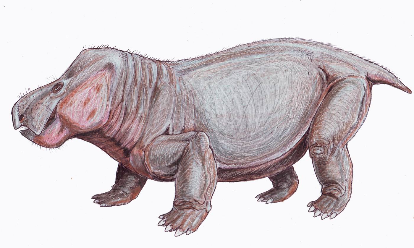
''Pamelaria'' is an extinct genus of allokotosaurian archosauromorph reptile known from a single species, ''Pamelaria dolichotrachela'', from the Middle Triassic of India. ''Pamelaria'' has sprawling legs, a long neck, and a pointed skull with nostrils positioned at the very tip of the snout. Among early archosauromorphs, ''Pamelaria'' is most similar to '' Prolacerta'' from the Early Triassic of South Africa and Antarctica. Both have been placed in the family Prolacertidae. ''Pamelaria'', ''Prolacerta'', and various other Permo-Triassic reptiles such as '' Protorosaurus'' and '' Tanystropheus'' have often been placed in a group of archosauromorphs called Protorosauria (alternatively called Prolacertiformes), which was regarded as one of the most basal group of archosauromorphs. However, more recent phylogenetic analyses indicate that ''Pamelaria'' and ''Prolacerta'' are more closely related to Archosauriformes than are ''Protorosaurus'', ''Tanystropheus'', and other protorosa ... [...More Info...] [...Related Items...] OR: [Wikipedia] [Google] [Baidu] |
Middle Triassic
In the geologic timescale, the Middle Triassic is the second of three epochs of the Triassic period or the middle of three series in which the Triassic system is divided in chronostratigraphy. The Middle Triassic spans the time between Ma and Ma (million years ago). It is preceded by the Early Triassic Epoch and followed by the Late Triassic Epoch. The Middle Triassic is divided into the Anisian and Ladinian ages or stages. Formerly the middle series in the Triassic was also known as Muschelkalk. This name is now only used for a specific unit of rock strata with approximately Middle Triassic age, found in western Europe. Middle Triassic fauna Following the Permian–Triassic extinction event, the most devastating of all mass-extinctions, life recovered slowly. In the Middle Triassic, many groups of organisms reached higher diversity again, such as the marine reptiles (e.g. ichthyosaurs, sauropterygians, thallatosaurs), ray-finned fish and many invertebrate g ... [...More Info...] [...Related Items...] OR: [Wikipedia] [Google] [Baidu] |
Trilophosauridae
Trilophosaurs are lizard-like Triassic allokotosaur reptiles related to the archosaurs. The best known genus is '' Trilophosaurus'', a herbivore up to long. It had a short, unusually heavily built skull, equipped with massive, broad flattened cheek teeth with sharp shearing surfaces for cutting up tough plant material. Teeth are absent from the premaxilla and front of the lower jaw, which in life were probably equipped with a horny beak. The skull is also unusual in that the lower temporal opening is missing, giving the appearance of a euryapsid skull, and originally the Trilophosaurs were classified with placodonts and sauropterygia. Carroll (1988) suggests that the lower opening may have been lost to strengthen the skull. Trilophosaurs are so far known only from the Late Triassic of North America and Europe. Below is a cladogram showing the phylogenetic relationships of Trilophosauridae within Archosauromorpha Archosauromorpha ( Greek for "ruling lizard forms") is a c ... [...More Info...] [...Related Items...] OR: [Wikipedia] [Google] [Baidu] |
Yerrapalli Formation
The Yerrapalli Formation is a Triassic rock formation consisting primarily of mudstones that outcrops in the Pranhita–Godavari Basin in southeastern India. The Yerrapalli Formation preserves fossils of freshwater and terrestrial vertebrates as well as trace fossils of invertebrates.Yerrapalli Formation at .org The fauna includes amphibians, |
Coronoid Process Of The Mandible
In human anatomy, the mandible's coronoid process (from Greek ''korōnē'', denoting something hooked) is a thin, triangular eminence, which is flattened from side to side and varies in shape and size. Its anterior border is convex and is continuous below with the anterior border of the ramus. Its ''posterior border'' is concave and forms the anterior boundary of the mandibular notch. The ''lateral surface'' is smooth, and affords insertion to the temporalis and masseter muscles. Its ''medial surface'' gives insertion to the temporalis, and presents a ridge which begins near the apex of the process and runs downward and forward to the inner side of the last molar tooth. Between this ridge and the anterior border is a grooved triangular area, the upper part of which gives attachment to the temporalis, the lower part to some fibers of the buccinator. Clinical significance Fractures of the mandible are common. However, coronoid process fractures are very rare. Isolated fractures o ... [...More Info...] [...Related Items...] OR: [Wikipedia] [Google] [Baidu] |
Jugal Bone
The jugal is a skull bone found in most reptiles, amphibians and birds. In mammals, the jugal is often called the malar or zygomatic. It is connected to the quadratojugal and maxilla, as well as other bones, which may vary by species. Anatomy The jugal bone is located on either side of the skull in the circumorbital region. It is the origin of several masticatory muscles in the skull. The jugal and lacrimal bones are the only two remaining from the ancestral circumorbital series: the prefrontal, postfrontal, postorbital, jugal, and lacrimal bones. During development, the jugal bone originates from dermal bone. In dinosaurs This bone is considered key in the determination of general traits in cases in which the entire skull has not been found intact (for instance, as with dinosaurs in paleontology). In some dinosaur genera the jugal also forms part of the lower margin of either the antorbital fenestra or the infratemporal fenestra, or both. Most commonly, this bone art ... [...More Info...] [...Related Items...] OR: [Wikipedia] [Google] [Baidu] |
Quadrate Bone
The quadrate bone is a skull bone in most tetrapods, including amphibians, sauropsids (reptiles, birds), and early synapsids. In most tetrapods, the quadrate bone connects to the quadratojugal and squamosal bones in the skull, and forms upper part of the jaw joint. The lower jaw articulates at the articular bone, located at the rear end of the lower jaw. The quadrate bone forms the lower jaw articulation in all classes except mammals. Evolutionarily, it is derived from the hindmost part of the primitive cartilaginous upper jaw. Function in reptiles In certain extinct reptiles, the variation and stability of the morphology of the quadrate bone has helped paleontologists in the species-level taxonomy and identification of mosasaur squamates and spinosaurine dinosaurs. In some lizards and dinosaurs, the quadrate is articulated at both ends and movable. In snakes, the quadrate bone has become elongated and very mobile, and contributes greatly to their ability to swallow ve ... [...More Info...] [...Related Items...] OR: [Wikipedia] [Google] [Baidu] |
Temporal Fenestra
An infratemporal fenestra, also called the lateral temporal fenestra or simply temporal fenestra, is an opening in the skull behind the orbit in some animals. It is ventrally bordered by a zygomatic arch. An opening in front of the eye sockets, conversely, is called an antorbital fenestra. Both of these openings reduce the weight of the skull. Infratemporal fenestrae are commonly (although not universally) seen in the fossilized skulls of dinosaurs. Synapsids, including mammals Mammals () are a group of vertebrate animals constituting the class Mammalia (), characterized by the presence of mammary glands which in females produce milk for feeding (nursing) their young, a neocortex (a region of the brain), fu ..., have one temporal fenestra, while sauropsids, the birds and reptiles, have two. References {{ref list Dinosaur anatomy Foramina of the skull ... [...More Info...] [...Related Items...] OR: [Wikipedia] [Google] [Baidu] |
Orbit (anatomy)
In anatomy, the orbit is the cavity or socket of the skull in which the eye and its appendages are situated. "Orbit" can refer to the bony socket, or it can also be used to imply the contents. In the adult human, the volume of the orbit is , of which the eye occupies . The orbital contents comprise the eye, the orbital and retrobulbar fascia, extraocular muscles, cranial nerves II, III, IV, V, and VI, blood vessels, fat, the lacrimal gland with its sac and duct, the eyelids, medial and lateral palpebral ligaments, cheek ligaments, the suspensory ligament, septum, ciliary ganglion and short ciliary nerves. Structure The orbits are conical or four-sided pyramidal cavities, which open into the midline of the face and point back into the head. Each consists of a base, an apex and four walls."eye, human."Encyclopædia Britannica from Encyclopædia Britannica 2006 Ultimate Reference Suite DVD 2009 Openings There are two important foramina, or windows, two important f ... [...More Info...] [...Related Items...] OR: [Wikipedia] [Google] [Baidu] |
Naris
A nostril (or naris , plural ''nares'' ) is either of the two orifices of the nose. They enable the entry and exit of air and other gasses through the nasal cavities. In birds and mammals, they contain branched bones or cartilages called turbinates, whose function is to warm air on inhalation and remove moisture on exhalation. Fish do not breathe through noses, but they do have two (but cyclostomes have merged into one) small holes used for smelling, which can also be referred to as nostrils. In humans, the nasal cycle is the normal ultradian cycle of each nostril's blood vessels becoming engorged in swelling, then shrinking. The nostrils are separated by the septum. The septum can sometimes be deviated, causing one nostril to appear larger than the other. With extreme damage to the septum and columella, the two nostrils are no longer separated and form a single larger external opening. Like other tetrapods, humans have two external nostrils (anterior nares) and two additi ... [...More Info...] [...Related Items...] OR: [Wikipedia] [Google] [Baidu] |
Pectoral Girdle
The shoulder girdle or pectoral girdle is the set of bones in the appendicular skeleton which connects to the arm on each side. In humans it consists of the clavicle and scapula; in those species with three bones in the shoulder, it consists of the clavicle, scapula, and coracoid. Some mammalian species (such as the dog and the horse) have only the scapula. The pectoral girdles are to the upper limbs as the pelvic girdle is to the lower limbs; the girdles are the parts of the appendicular skeleton that anchor the appendages to the axial skeleton. In humans, the only true anatomical joints between the shoulder girdle and the axial skeleton are the sternoclavicular joints on each side. No anatomical joint exists between each scapula and the rib cage; instead the muscular connection or physiological joint between the two permits great mobility of the shoulder girdle compared to the compact pelvic girdle; because the upper limb is not usually involved in weight bearing, ... [...More Info...] [...Related Items...] OR: [Wikipedia] [Google] [Baidu] |
Cervical Vertebrae
In tetrapods, cervical vertebrae (singular: vertebra) are the vertebrae of the neck, immediately below the skull. Truncal vertebrae (divided into thoracic and lumbar vertebrae in mammals) lie caudal (toward the tail) of cervical vertebrae. In sauropsid species, the cervical vertebrae bear cervical ribs. In lizards and saurischian dinosaurs, the cervical ribs are large; in birds, they are small and completely fused to the vertebrae. The vertebral transverse processes of mammals are homologous to the cervical ribs of other amniotes. Most mammals have seven cervical vertebrae, with the only three known exceptions being the manatee with six, the two-toed sloth with five or six, and the three-toed sloth with nine. In humans, cervical vertebrae are the smallest of the true vertebrae and can be readily distinguished from those of the thoracic or lumbar regions by the presence of a foramen (hole) in each transverse process, through which the vertebral artery, vertebral veins, ... [...More Info...] [...Related Items...] OR: [Wikipedia] [Google] [Baidu] |
Pamelaria Scale Diagram
''Pamelaria'' is an extinct genus of allokotosaurian archosauromorph reptile known from a single species, ''Pamelaria dolichotrachela'', from the Middle Triassic of India. ''Pamelaria'' has sprawling legs, a long neck, and a pointed skull with nostrils positioned at the very tip of the snout. Among early archosauromorphs, ''Pamelaria'' is most similar to '' Prolacerta'' from the Early Triassic of South Africa and Antarctica. Both have been placed in the family Prolacertidae. ''Pamelaria'', ''Prolacerta'', and various other Permo-Triassic reptiles such as '' Protorosaurus'' and '' Tanystropheus'' have often been placed in a group of archosauromorphs called Protorosauria (alternatively called Prolacertiformes), which was regarded as one of the most basal group of archosauromorphs. However, more recent phylogenetic analyses indicate that ''Pamelaria'' and ''Prolacerta'' are more closely related to Archosauriformes than are ''Protorosaurus'', ''Tanystropheus'', and other protorosa ... [...More Info...] [...Related Items...] OR: [Wikipedia] [Google] [Baidu] |

.jpg)

.jpg)


