|
Palmaris Brevis Muscle
Palmaris brevis muscle is a thin, quadrilateral muscle, placed beneath the integument of the ulnar side of the hand. It acts to fold the skin of the hypothenar eminence transversally. Structure Origin and insertion Palmaris brevis muscle is located on the ulnar side of the hand. It arises from the tendinous fasciculi from the transverse carpal ligament and palmar aponeurosis. The muscle fibres are inserted into the skin on the ulnar border of the palm of the hand, and occasionally on the pisiform bone. Innervation Palmaris brevis muscle is the only muscle innervated by the superficial branch of the ulnar nerve (C8, T1). Blood supply Palmaris brevis muscle is supplied by the palmar metacarpal artery of the deep palmar arch. Discovery The first recorded observation of the muscle is by Italian anatomist Giambattista Canano sometime before 1543. The muscle was independently discovered a few years later by Realdo Colombo before being pushed to general acceptance in the ... [...More Info...] [...Related Items...] OR: [Wikipedia] [Google] [Baidu] |
Flexor Retinaculum Of The Hand
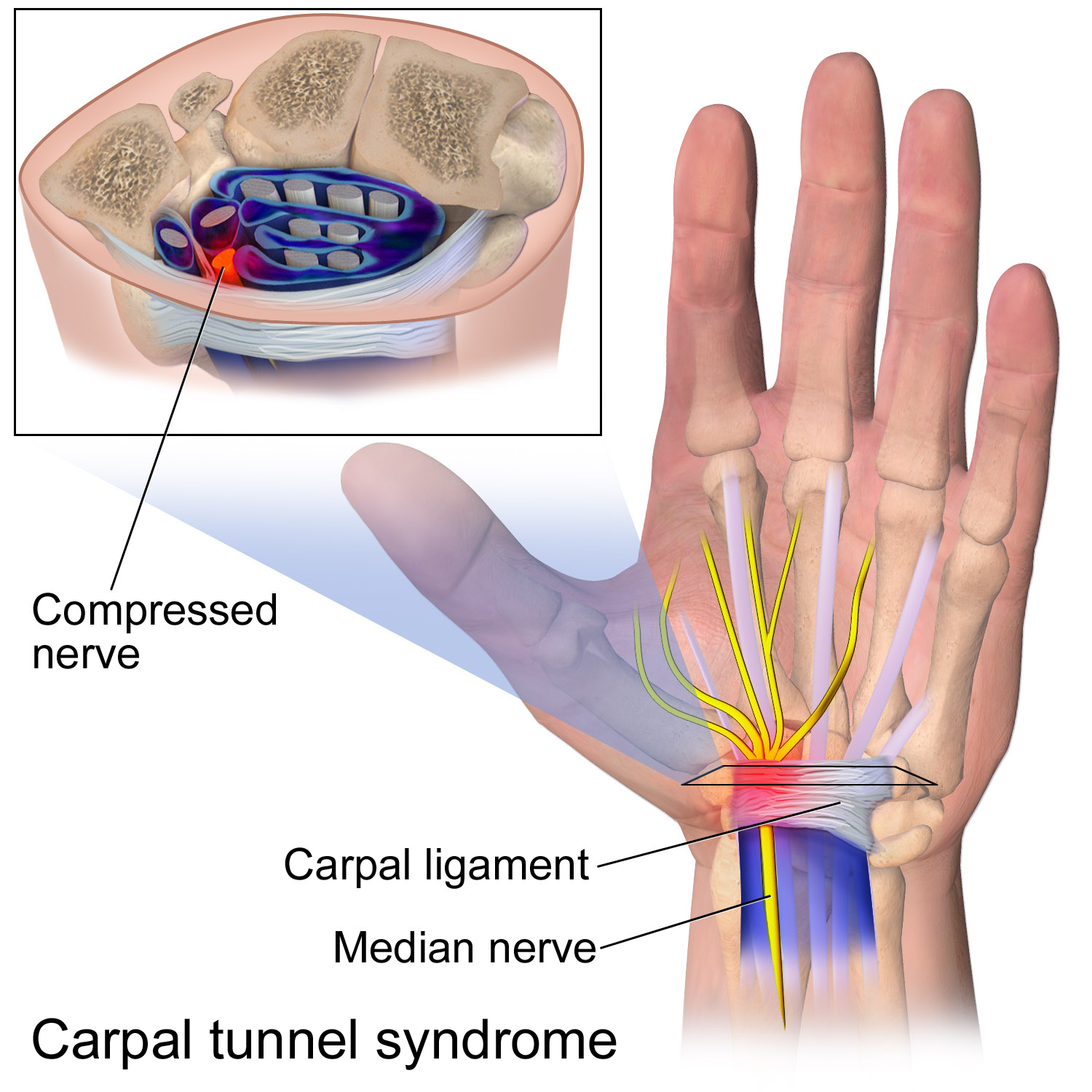
The flexor retinaculum (transverse carpal ligament or anterior annular ligament) is a fibrous band on the palmar side of the hand near the wrist. It arches over the carpal bones of the hands, covering them and forming the carpal tunnel. Structure The flexor retinaculum is a strong, fibrous band that covers the carpal bones on the palmar side of the hand near the wrist. It attaches to the bones near the radius and ulna. On the ulnar side, the flexor retinaculum attaches to the pisiform bone and the hook of the hamate bone. On the radial side, it attaches to the tubercle of the scaphoid bone, and to the medial part of the palmar surface and the ridge of the trapezium bone. The flexor retinaculum is continuous with the palmar carpal ligament, and deeper with the palmar aponeurosis. The ulnar artery and ulnar nerve, and the cutaneous branches of the median and ulnar nerves, pass on top of the flexor retinaculum. On the radial side of the retinaculum is the tendon of the flexor car ... [...More Info...] [...Related Items...] OR: [Wikipedia] [Google] [Baidu] |
Palmar Metacarpal Artery
The palmar metacarpal arteries (volar metacarpal arteries, palmar interosseous arteries) are three or four arteries that arise from the convexity of the deep palmar arch. Structure The palmar metacarpal arteries arise from the convexity of the deep palmar arch. They run distally upon the palmar interossei muscles. They anastomose at the clefts of the fingers with the common palmar digital arteries which arise from the superficial palmar arch The superficial palmar arch is formed predominantly by the ulnar artery, with a contribution from the superficial palmar branch of the radial artery. However, in some individuals the contribution from the radial artery might be absent, and instea .... References External links * ("Palm of the hand, deep dissection, anterior view") Arteries of the upper limb {{circulatory-stub ... [...More Info...] [...Related Items...] OR: [Wikipedia] [Google] [Baidu] |
Palmar Interossei Muscles
In human anatomy, the palmar or volar interossei (interossei volares in older literature) are four muscles, one on the thumb that is occasionally missing, and three small, unipennate, central muscles in the hand that lie between the Metacarpus, metacarpal bones and are attached to the Index finger, index, Ring finger, ring, and Little finger, little fingers. They are smaller than the dorsal interossei of the hand. Structure All palmar interossei originate along the shaft of the metacarpal bone of the digit on which they act. They are inserted into the base of the Phalanx bone, proximal phalanx and the extensor expansion of the Extensor digitorum muscle, extensor digitorum of the same digit. Pollical palmar interosseous The first palmar interosseous is located at the thumb's medial side. Passing between the first dorsal interosseous and the oblique head of Adductor pollicis muscle, adductor pollicis, it is inserted on the base of the thumb's proximal phalanx together with Adductor ... [...More Info...] [...Related Items...] OR: [Wikipedia] [Google] [Baidu] |
Thenar Eminence
The thenar eminence is the mound formed at the base of the thumb on the palm of the hand by the intrinsic group of muscles of the thumb. The skin overlying this region is the area stimulated when trying to elicit a palmomental reflex. The word thenar comes . Structure The following three muscles are considered part of the thenar eminence: * Abductor pollicis brevis abducts the thumb. This muscle is the most superficial of the thenar group. * Flexor pollicis brevis, which lies next to the abductor, will flex the thumb, curling it up in the palm. (The flexor pollicis longus, which is inserted into the distal phalanx of the thumb, is not considered part of the thenar eminence.) * Opponens pollicis lies deep to abductor pollicis brevis. As its name suggests it opposes the thumb, bringing it against the fingers. This is a very important movement, as most of human hand dexterity comes from this action. Another muscle that controls movement of the thumb is adductor pollicis ... [...More Info...] [...Related Items...] OR: [Wikipedia] [Google] [Baidu] |
Myocyte
A muscle cell, also known as a myocyte, is a mature contractile Cell (biology), cell in the muscle of an animal. In humans and other vertebrates there are three types: skeletal muscle, skeletal, smooth muscle, smooth, and Cardiac muscle, cardiac (cardiomyocytes). A skeletal muscle cell is long and threadlike with multinucleated, many nuclei and is called a ''muscle fiber''. Muscle cells develop from embryonic precursor cells called myoblasts. Skeletal muscle cells form by cell fusion, fusion of myoblasts to produce multinucleated cells (syncytium, syncytia) in a process known as myogenesis. Skeletal muscle cells and cardiac muscle cells both contain myofibrils and sarcomeres and form a striated muscle tissue. Cardiac muscle cells form the cardiac muscle in the walls of the heart chambers, and have a single central Cell nucleus, nucleus. Cardiac muscle cells are joined to neighboring cells by intercalated discs, and when joined in a visible unit they are described as a ''cardiac m ... [...More Info...] [...Related Items...] OR: [Wikipedia] [Google] [Baidu] |
Prehensility
Prehensility is the quality of an appendage or organ that has adapted for grasping or holding. The word is derived from the Latin term ''prehendere'', meaning "to grasp". The ability to grasp is likely derived from a number of different origins. The most common are tree-climbing and the need to manipulate food. Examples Appendages that can become prehensile include: Uses Prehensility affords animals a great natural advantage in manipulating their environment for feeding, climbing, digging, and defense. It enables many animals, such as primates, to use tools to complete tasks that would otherwise be impossible without highly specialized anatomy. For example, chimpanzees have the ability to use sticks to obtain termites and grubs in a manner similar to human fishing Fishing is the activity of trying to catch fish. Fish are often caught as wildlife from the natural environment (Freshwater ecosystem, freshwater or Marine ecosystem, marine), but may also be caught ... [...More Info...] [...Related Items...] OR: [Wikipedia] [Google] [Baidu] |
Ulnar Artery
The ulnar artery is the main blood vessel, with oxygenated blood, of the Human Anatomical Terms#Anatomical directions, medial aspects of the forearm. It arises from the brachial artery and terminates in the superficial palmar arch, which joins with the superficial branch of the radial artery. It is palpable on the anterior and medial aspect of the wrist. Along its course, it is accompanied by a similarly named vein or veins, the ulnar vein or ulnar veins. The ulnar artery, the larger of the two terminal branches of the brachial, begins a little below the bend of the Elbow-joint, elbow in the cubital fossa, and, passing obliquely downward, reaches the ulnar side of the forearm at a point about midway between the elbow and the wrist. It then runs along the ulnar border to the wrist, crosses the transverse carpal ligament on the radial side of the pisiform bone, and immediately beyond this bone divides into two branches, which enter into the formation of the Superficial palmar a ... [...More Info...] [...Related Items...] OR: [Wikipedia] [Google] [Baidu] |
Ulnar Nerve
The ulnar nerve is a nerve that runs near the ulna, one of the two long bones in the forearm. The ulnar collateral ligament of elbow joint is in relation with the ulnar nerve. The nerve is the largest in the human body unprotected by muscle or bone, so injury is common. This nerve is directly connected to the little finger, and the adjacent half of the ring finger, innervating the palmar aspect of these fingers, including both front and back of the tips, perhaps as far back as the fingernail beds. This nerve can cause an electric shock-like sensation by striking the medial epicondyle of the humerus posteriorly, or inferiorly with the elbow flexed. The ulnar nerve is trapped between the bone and the overlying skin at this point. This is commonly referred to as bumping one's "funny bone". This name is thought to be a pun, based on the sound resemblance between the name of the bone of the upper arm, the humerus, and the word " humorous". Alternatively, according to the Oxfor ... [...More Info...] [...Related Items...] OR: [Wikipedia] [Google] [Baidu] |
Andreas Vesalius
Andries van Wezel (31 December 1514 – 15 October 1564), latinized as Andreas Vesalius (), was an anatomist and physician who wrote '' De Humani Corporis Fabrica Libri Septem'' (''On the fabric of the human body'' ''in seven books''), which is considered one of the most influential books on human anatomy and a major advance over the long-dominant work of Galen. Vesalius is often referred to as the founder of modern human anatomy. He was born in Brussels, which was then part of the Habsburg Netherlands. He was a professor at the University of Padua (1537–1542) and later became Imperial physician at the court of Emperor Charles V. Early life and education Vesalius was born as Andries van Wesel to his father Anders van Wesel and mother Isabel Crabbe on 31 December 1514 in Brussels, which was then part of the Habsburg Netherlands. His great-grandfather, Jan van Wesel, probably born in Wesel, received a medical degree from the University of Pavia and taught medicine at th ... [...More Info...] [...Related Items...] OR: [Wikipedia] [Google] [Baidu] |
Realdo Colombo
Matteo Realdo Colombo (c. 1515 – 1559) was an Italian professor of anatomy and a surgeon at the University of Padua between 1544 and 1559. Early life and education Matteo Realdo Colombo or Realdus Columbus, was born in Cremona, Lombardy, the son of an apothecary named Antonio Colombo. Although little is known about his early life, it is known he took his undergraduate education in Milan, where he studied philosophy, and he appears to have pursued his father's profession for a short while afterwards. He left the apothecary's life and apprenticed to the surgeon Giovanni Antonio Lonigo, under whom he studied for 7 years. In 1538 he enrolled in the University of Padua where he was noted to be an exceptional student of anatomy. While still a student, he was awarded a Chair of Sophistics at the university. In 1542 he returned briefly to Venice to assist his mentor, Lonigo. Academic career Realdo Colombo studied philosophy in Milan, and then he trained to be a surgeon for several ... [...More Info...] [...Related Items...] OR: [Wikipedia] [Google] [Baidu] |
Giambattista Canano
Giambattista or Giovanni Battista Canano (born 1515, died 29 January 1579) was a physician and anatomist, active mainly in his native Ferrara. His aristocratic family, of Greek ancestry, produced a number of physicians and scholars. His father, Ludovico Canano, was a notary. His grandfather was lecturer in medicine at Ferrara and physician at court. The family came to Italy from Greece in the 15th century. Canano's studies were most likely directed by his uncle Hippolito. He became professor of anatomy at the University of Ferrara in 1541. He was physician to Francesco d'Este in France in 1544, and personal physician to Physician to Pope Julius III from 1552 to 1555. He pursued most of his dissections at his own home, with his cousin Antonio Maria Canano.Rivista Fondazione E ... [...More Info...] [...Related Items...] OR: [Wikipedia] [Google] [Baidu] |
Deep Palmar Arch
The deep palmar arch (deep volar arch) is an arterial network found in the palm. It is usually primarily formed from the terminal part of the radial artery. The ulnar artery also contributes through an anastomosis. This is in contrast to the superficial palmar arch, which is formed predominantly by the ulnar artery. Structure The deep palmar arch is usually primarily formed from the radial artery. The ulnar artery also contributes through an anastomosis. The deep palmar arch lies upon the bases of the metacarpal bones and on the interossei of the hand. It is deep to the oblique head of the adductor pollicis muscle, the flexor tendons of the fingers, and the lumbricals of the hand. Alongside of it, but running in the opposite direction—toward the radial side of the hand—is the deep branch of the ulnar nerve. The superficial palmar arch is more distally located than the deep palmar arch. If one were to fully extend the thumb and draw a line from the distal border of the thu ... [...More Info...] [...Related Items...] OR: [Wikipedia] [Google] [Baidu] |


