|
PINK1 With Healthy Mitochondria
PTEN-induced kinase 1 (PINK1) is a mitochondrial serine/threonine-protein kinase encoded by the ''PINK1'' gene. It is thought to protect cells from stress-induced mitochondrial dysfunction. PINK1 activity causes the parkin protein to bind to depolarized mitochondria to induce autophagy of those mitochondria. PINK1 is processed by healthy mitochondria and released to trigger neuron differentiation. Mutations in this gene cause one form of autosomal recessive early-onset Parkinson's disease. Structure PINK1 is synthesized as a 63000 Da protein which is often cleaved by PARL, between the 103-Alanine and the 104-Phenylalanine residues, into a 53000 Da fragment. PINK1 contains an N-terminal mitochondrial localization sequence, a putative transmembrane sequence, a Ser/Thr kinase domain, and a C-terminal regulatory sequence. The protein has been found to localize to the outer membrane of mitochondria, but can also be found throughout the cytosol. Experiments suggest the Ser/Thr ... [...More Info...] [...Related Items...] OR: [Wikipedia] [Google] [Baidu] |
PTEN (gene)
Phosphatase and tensin homolog (PTEN) is a phosphatase in humans and is encoded by the ''PTEN'' gene. Mutations of this gene are a step in the development of many cancers, specifically glioblastoma, lung cancer, breast cancer, and prostate cancer. Genes corresponding to PTEN (orthologs) have been identified in most mammals for which complete genome data are available. ''PTEN'' acts as a tumor suppressor gene through the action of its phosphatase protein product. This phosphatase is involved in the regulation of the cell cycle, preventing cells from growing and dividing too rapidly. It is a target of many anticancer drugs. The protein encoded by this gene is a phosphatidylinositol-3,4,5-trisphosphate 3-phosphatase. It contains a tensin-like domain as well as a catalytic domain similar to that of the dual specificity phosphatases. Unlike most of the protein tyrosine phosphatases, this protein preferentially dephosphorylates phosphoinositide substrates. It negatively regul ... [...More Info...] [...Related Items...] OR: [Wikipedia] [Google] [Baidu] |
C-terminal
The C-terminus (also known as the carboxyl-terminus, carboxy-terminus, C-terminal tail, carboxy tail, C-terminal end, or COOH-terminus) is the end of an amino acid chain (protein or polypeptide), terminated by a free carboxyl group (-COOH). When the protein is translated from messenger RNA, it is created from N-terminus to C-terminus. The convention for writing peptide sequences is to put the C-terminal end on the right and write the sequence from N- to C-terminus. Chemistry Each amino acid has a carboxyl group and an amine group. Amino acids link to one another to form a chain by a dehydration reaction which joins the amine group of one amino acid to the carboxyl group of the next. Thus polypeptide chains have an end with an unbound carboxyl group, the C-terminus, and an end with an unbound amine group, the N-terminus. Proteins are naturally synthesized starting from the N-terminus and ending at the C-terminus. Function C-terminal retention signals While the N-terminus of a prote ... [...More Info...] [...Related Items...] OR: [Wikipedia] [Google] [Baidu] |
Lewy Bodies
Lewy bodies are the inclusion bodies – abnormal aggregations of protein – that develop inside neurons affected by Parkinson's disease (PD), the Lewy body dementias (Parkinson's disease dementia and dementia with Lewy bodies (DLB)), and some other disorders. They are also seen in cases of multiple system atrophy, particularly the parkinsonian variant (MSA-P). Lewy bodies appear as spherical masses in the cytoplasm that displace other cell components. For instance, some Lewy bodies tend to displace the nucleus to one side of the cell. There are two main kinds of Lewy bodies – classical and cortical. A classical Lewy body is an eosinophilic cytoplasmic inclusion consisting of a dense core surrounded by a halo of 10 nm wide radiating fibrils, the primary structural component of which is alpha-synuclein (α-synuclein). While similar in many other respects, cortical Lewy bodies are only faintly eosinophilic, do not have a surrounding halo, and do not show a radia ... [...More Info...] [...Related Items...] OR: [Wikipedia] [Google] [Baidu] |
Dopaminergic Neuron
Dopaminergic cell groups, DA cell groups, or dopaminergic nuclei are collections of neurons in the central nervous system that synthesize the neurotransmitter dopamine. In the 1960s, Dopaminergic pathways, dopaminergic neurons or ''dopamine neurons'' were first identified and named by Annica Dahlström and :sv:Kjell Fuxe, Kjell Fuxe, who used histochemical tracer, histochemical fluorescence. The subsequent discovery of genes encoding enzymes that synthesize dopamine, and transporters that incorporate dopamine into synaptic vesicles or reclaim it after synaptic release, enabled scientists to identify dopaminergic neurons by labeling gene or protein expression that is specific to these neurons. In the mammalian brain, dopaminergic neurons form a semi-continuous population extending from the midbrain through the forebrain, with eleven named collections or clusters among them. Cell group A8 Group A8 is a small group of dopaminergic cells in rodents and primates. It is located in t ... [...More Info...] [...Related Items...] OR: [Wikipedia] [Google] [Baidu] |
Lysosomes
A lysosome () is a membrane-bound organelle that is found in all mammalian cells, with the exception of red blood cells (erythrocytes). There are normally hundreds of lysosomes in the cytosol, where they function as the cell’s degradation center. Their primary responsibility is catabolic degradation of proteins, polysaccharides and lipids into their respective building-block molecules: amino acids, monosaccharides, and free fatty acids. The breakdown is done by various enzymes, for example proteases, glycosidases and lipases. With an acidic lumen limited by a single-bilayer lipid membrane, the lysosome holds an environment isolated from the rest of the cell. The lower pH creates optimal conditions for the over 60 different hydrolases inside. Lysosomes receive extracellular particles through endocytosis, and intracellular components through autophagy. They can also fuse with the plasma membrane and secrete their contents, a process called lysosomal exocytosis. After degradation ... [...More Info...] [...Related Items...] OR: [Wikipedia] [Google] [Baidu] |
Miro (protein)
The Ras superfamily, derived from "Rat sarcoma virus", is a protein superfamily of small GTPases. Members of the superfamily are divided into Protein family, families and subfamilies based on their structure, sequence and function. The five main families are Ras, Rho family of GTPases, Rho, Ran (biology), Ran, Rab (G-protein), Rab and ADP ribosylation factor, Arf GTPases. The Ras family itself is further divided into 6 subfamilies: Ras subfamily, Ras, RALA, Ral, Rap (protein), Rap, Rheb, RRAD (gene), Rad and Rit. ''Miro'' is a recent contributor to the superfamily. Each subfamily shares the common core G domain, which provides essential GTPase and nucleotide exchange activity. The surrounding sequence helps determine the functional specificity of the small GTPase, for example the 'Insert Loop', common to the Rho subfamily, specifically contributes to binding to effector proteins such as Wiskott-Aldrich syndrome protein, WASP. In general, the Ras family is responsible for cell pro ... [...More Info...] [...Related Items...] OR: [Wikipedia] [Google] [Baidu] |
Drosophila
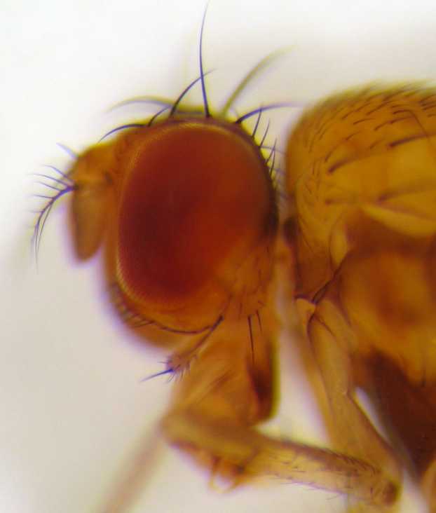
''Drosophila'' (), from Ancient Greek δρόσος (''drósos''), meaning "dew", and φίλος (''phílos''), meaning "loving", is a genus of fly, belonging to the family Drosophilidae, whose members are often called "small fruit flies" or pomace flies, vinegar flies, or wine flies, a reference to the characteristic of many species to linger around overripe or rotting fruit. They should not be confused with the Tephritidae, a related family, which are also called fruit flies (sometimes referred to as "true fruit flies"); tephritids feed primarily on unripe or ripe fruit, with many species being regarded as destructive agricultural pests, especially the Mediterranean fruit fly. One species of ''Drosophila'' in particular, ''Drosophila melanogaster'', has been heavily used in research in genetics and is a common model organism in developmental biology. The terms "fruit fly" and "''Drosophila''" are often used synonymously with ''D. melanogaster'' in modern biological literatur ... [...More Info...] [...Related Items...] OR: [Wikipedia] [Google] [Baidu] |
Mitochondrial Fission
Mitochondrial fission is the process by which mitochondria divide or segregate into two separate mitochondrial organelles. Mitochondrial fission is counteracted by mitochondrial fusion, where two mitochondria fuse together to form a larger one. Fusion can result in elongated mitochondrial networks. In healthy cells, mitochondrial fission and fusion are balanced, and disruptions to these processes are linked to various diseases. Mitochondrial fission is coordinated with the mitochondrial DNA replication process. Some of the proteins involved in mitochondrial fission have been identified, and mutations in some of these proteins are associated with mitochondrial diseases. Mitochondrial fission plays a role in the cellular stress response and in apoptosis (programmed cell death). Mechanism Drp1 The Drp1 protein, a member of the dynamin family of large GTPases, is transcribed from the ''DNM1L'' gene. Alternative splicing produces at least ten isoforms of Drp1, which regulate tissue-s ... [...More Info...] [...Related Items...] OR: [Wikipedia] [Google] [Baidu] |
PINK1 With Healthy Mitochondria
PTEN-induced kinase 1 (PINK1) is a mitochondrial serine/threonine-protein kinase encoded by the ''PINK1'' gene. It is thought to protect cells from stress-induced mitochondrial dysfunction. PINK1 activity causes the parkin protein to bind to depolarized mitochondria to induce autophagy of those mitochondria. PINK1 is processed by healthy mitochondria and released to trigger neuron differentiation. Mutations in this gene cause one form of autosomal recessive early-onset Parkinson's disease. Structure PINK1 is synthesized as a 63000 Da protein which is often cleaved by PARL, between the 103-Alanine and the 104-Phenylalanine residues, into a 53000 Da fragment. PINK1 contains an N-terminal mitochondrial localization sequence, a putative transmembrane sequence, a Ser/Thr kinase domain, and a C-terminal regulatory sequence. The protein has been found to localize to the outer membrane of mitochondria, but can also be found throughout the cytosol. Experiments suggest the Ser/Thr ... [...More Info...] [...Related Items...] OR: [Wikipedia] [Google] [Baidu] |
Degradation Of Damaged Mitochondria With PINK1
Degradation may refer to: Science * Degradation (geology), lowering of a fluvial surface by erosion * Degradation (telecommunications), of an electronic signal * Biodegradation of organic substances by living organisms * Environmental degradation in ecology * Land degradation, a process in which the value of the biophysical environment is affected by a combination of human-induced processes acting upon the land * Polymer degradation, as plastics age Other * Elegant degradation, gradual rather than sudden * Graceful degradation, in a fault-tolerant system * Degradation (knighthood), revocation of knighthood * Cashiering, whereby a military officer is dismissed for misconduct * Reduction in rank, whereby a military officer is reduced to a lower rank for misconduct * Degradation, the former ceremony of defrocking a disgraced priest * ''Degradation'', a song by the Violent Femmes, from '' Add It Up (1981–1993)'' See also * '' Dégradé'', 2015 Palestinian film * Humiliation ... [...More Info...] [...Related Items...] OR: [Wikipedia] [Google] [Baidu] |
Parkin (protein)
Parkin is a 465-amino acid residue E3 ubiquitin ligase, a protein that in humans and mice is encoded by the ''PARK2'' gene. Parkin plays a critical role in ubiquitination – the process whereby molecules are covalently labelled with ubiquitin (Ub) and directed towards degradation in proteasomes or lysosomes. Ubiquitination involves the sequential action of three enzymes. First, an E1 ubiquitin-activating enzyme binds to inactive Ub in eukaryotic cells via a thioester bond and mobilises it in an ATP-dependent process. Ub is then transferred to an E2 ubiquitin-conjugating enzyme before being conjugated to the target protein via an E3 ubiquitin ligase. There exists a multitude of E3 ligases, which differ in structure and substrate specificity to allow selective targeting of proteins to intracellular degradation. In particular, parkin recognises proteins on the outer membrane of mitochondria upon cellular insult and mediates the clearance of damaged mitochondria via autophagy an ... [...More Info...] [...Related Items...] OR: [Wikipedia] [Google] [Baidu] |
Mitochondrial Inner Membrane
The inner mitochondrial membrane (IMM) is the mitochondrial membrane which separates the mitochondrial matrix from the intermembrane space. Structure The structure of the inner mitochondrial membrane is extensively folded and compartmentalized. The numerous invaginations of the membrane are called cristae, separated by crista junctions from the inner boundary membrane juxtaposed to the outer membrane. Cristae significantly increase the total membrane surface area compared to a smooth inner membrane and thereby the available working space for oxidative phosphorylation. The inner membrane creates two compartments. The region between the inner and outer membrane, called the intermembrane space, is largely continuous with the cytosol, while the more sequestered space inside the inner membrane is called the matrix. Cristae For typical liver mitochondria, the area of the inner membrane is about 5 times as large as the outer membrane due to cristae. This ratio is variable and mitocho ... [...More Info...] [...Related Items...] OR: [Wikipedia] [Google] [Baidu] |


