|
Oxyphil Cell (parathyroid)
Parathyroid oxyphil cells, also named oncocytes, are one out of the two types of cells found in the parathyroid gland, the other being parathyroid chief cell. Oxyphil cells are only found in a select few number of species and humans are one of them. These cells can be found in clusters in the center of the section and at the periphery. Oxyphil cells appear at the onset of puberty, but have no known function. It is perceived that oxyphil cells may be derived from chief cells at puberty, as they are not present at birth like chief cells. Oxyphil cells increase in number with age. Although the terms oncocyte, oxyphil cell, and Hürthle cell are used interchangeably, "Hürthle cell" is used only to indicate cells of thyroid follicular origin.Cannon, J. (2011). The Significance of Hurthle Cells in Thyroid Disease. The Oncologist. doi:10.1634/theoncologist.2010-0253 Structure Oxyphil cells may be binucleated and proteins found within their cytoplasms are basic, resulting in acidophi ... [...More Info...] [...Related Items...] OR: [Wikipedia] [Google] [Baidu] |
Micrograph
A micrograph is an image, captured photographically or digitally, taken through a microscope or similar device to show a magnify, magnified image of an object. This is opposed to a macrograph or photomacrograph, an image which is also taken on a microscope but is only slightly magnified, usually less than 10 times. Micrography is the practice or art of using microscopes to make photographs. A photographic micrograph is a photomicrograph, and one taken with an electron microscope is an electron micrograph. A micrograph contains extensive details of microstructure. A wealth of information can be obtained from a simple micrograph like behavior of the material under different conditions, the phases found in the system, failure analysis, grain size estimation, elemental analysis and so on. Micrographs are widely used in all fields of microscopy. Types Photomicrograph A light micrograph or photomicrograph is a micrograph prepared using an optical microscope, a process referred to ... [...More Info...] [...Related Items...] OR: [Wikipedia] [Google] [Baidu] |
Enzyme Activity
Enzyme assays are laboratory methods for measuring enzyme, enzymatic activity. They are vital for the study of enzyme kinetics and enzyme inhibitor, enzyme inhibition. Enzyme units The quantity or concentration of an enzyme can be expressed in Mole (unit), molar amounts, as with any other chemical, or in terms of activity in enzyme units. Enzyme activity Enzyme activity is a measure of the quantity of active enzyme present and is thus dependent on various physical conditions, ''which should be specified''. It is calculated using the following formula: :\mathrm=\mathrm_\text=\mathrm\times\mathrm where :\mathrm = Enzyme activity :\mathrm_\text = Moles of substrate converted per unit time :\mathrm = Rate of the reaction :\mathrm = Reaction volume The SI unit is the katal, 1 katal = 1 mole (unit), mol s−1 (mole per second), but this is an excessively large unit. A more practical and commonly used value is enzyme unit (U) = 1 μmol min−1 (micromole per minute). 1 U correspon ... [...More Info...] [...Related Items...] OR: [Wikipedia] [Google] [Baidu] |
Endocrine Cells
The endocrine system is a messenger system in an organism comprising feedback loops of hormones that are released by internal glands directly into the circulatory system and that target and regulate distant organs. In vertebrates, the hypothalamus is the neural control center for all endocrine systems. In humans, the major endocrine glands are the thyroid, parathyroid, pituitary, pineal, and adrenal glands, and the (male) testis and (female) ovaries. The hypothalamus, pancreas, and thymus also function as endocrine glands, among other functions. (The hypothalamus and pituitary glands are organs of the neuroendocrine system. One of the most important functions of the hypothalamusit is located in the brain adjacent to the pituitary glandis to link the endocrine system to the nervous system via the pituitary gland.) Other organs, such as the kidneys, also have roles within the endocrine system by secreting certain hormones. The study of the endocrine system and its disorders ... [...More Info...] [...Related Items...] OR: [Wikipedia] [Google] [Baidu] |
Neuroendocrine Cell
Neuroendocrine cells are cells that receive neuronal input (through neurotransmitters released by nerve cells or neurosecretory cells) and, as a consequence of this input, release messenger molecules (hormones) into the blood. In this way they bring about an integration between the nervous system and the endocrine system, a process known as neuroendocrine integration. An example of a neuroendocrine cell is a cell of the adrenal medulla (innermost part of the adrenal gland), which releases adrenaline to the blood. The adrenal medullary cells are controlled by the sympathetic division of the autonomic nervous system. These cells are modified postganglionic neurons. Autonomic nerve fibers lead directly to them from the central nervous system. The adrenal medullary hormones are kept in vesicles much in the same way neurotransmitters are kept in neuronal vesicles. Hormonal effects can last up to ten times longer than those of neurotransmitters. Sympathetic nerve fiber impulses stimulat ... [...More Info...] [...Related Items...] OR: [Wikipedia] [Google] [Baidu] |
Pituitary Gland
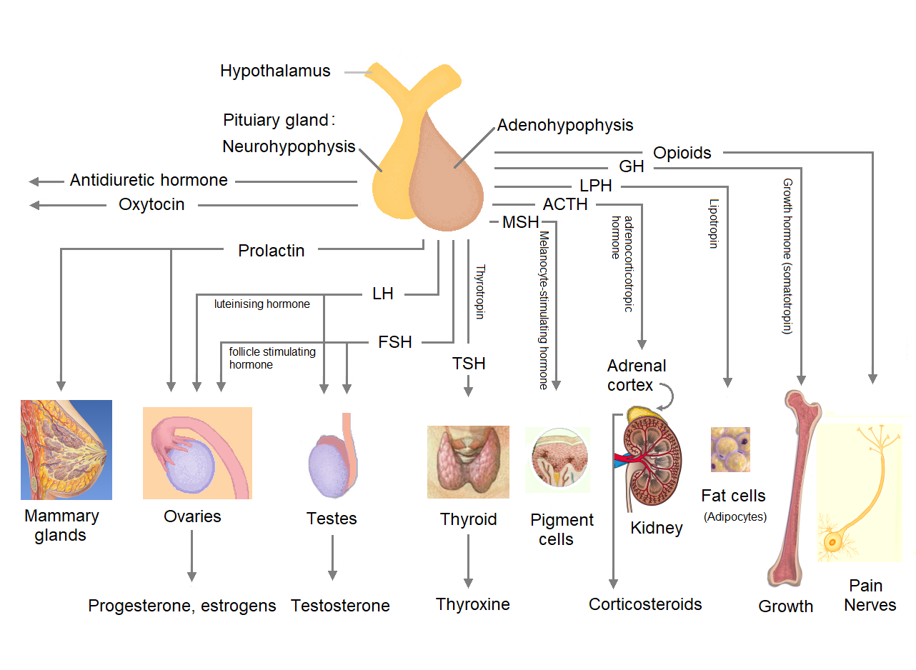
The pituitary gland or hypophysis is an endocrine gland in vertebrates. In humans, the pituitary gland is located at the base of the human brain, brain, protruding off the bottom of the hypothalamus. The pituitary gland and the hypothalamus control much of the body's endocrine system. It is seated in part of the sella turcica a fossa (anatomy), depression in the sphenoid bone, known as the hypophyseal fossa. The human pituitary gland is ovoid, oval shaped, about 1 cm in diameter, in weight on average, and about the size of a kidney bean. Digital version. There are two main lobes of the pituitary, an anterior pituitary, anterior lobe, and a posterior pituitary, posterior lobe joined and separated by a small intermediate lobe. The anterior lobe (adenohypophysis) is the glandular part that produces and secretes several hormones. The posterior lobe (neurohypophysis) secretes neurohypophysial hormones produced in the hypothalamus. Both lobes have different origins and they are both co ... [...More Info...] [...Related Items...] OR: [Wikipedia] [Google] [Baidu] |
Basophil Cell
An anterior pituitary basophil is a type of cell in the anterior pituitary which manufactures hormones. It is called a basophil because it is basophilic (readily takes up bases), and typically stains a relatively deep blue or purple. These basophils are further classified by the hormones they produce. (It is usually not possible to distinguish between these cell types using standard staining techniques.) *Produced only in pregnancy by the developing embryo. See also * Chromophobe cell :* Melanotroph * Chromophil :* Acidophil cell * Oxyphil cell * Oxyphil cell (parathyroid) * Pituitary gland * Neuroendocrine cell * Basophilic Basophilic is a technical term used by pathologists. It describes the appearance of cells, tissues and cellular structures as seen through the microscope after a histological section has been stained with a basic dye. The most common such dye ... References External links * {{Authority control Histology ... [...More Info...] [...Related Items...] OR: [Wikipedia] [Google] [Baidu] |
Acidophil Cell
In the anterior pituitary, the term "acidophil" is used to describe two different types of cells which stain well with acidic dyes. * somatotrophs, which secrete growth hormone (a peptide hormone) * lactotrophs, which secrete prolactin (a peptide hormone) When using standard staining techniques, they cannot be distinguished from each other (though they can be distinguished from basophils and chromophobes), and are therefore identified simply as "acidophils". See also * Eosinophilic * Acidophile (histology) * Basophilic * Chromophobe cell :* Melanotroph * Chromophil :* Basophil cell * Oxyphil cell * Oxyphil cell (parathyroid) * Pituitary gland * Neuroendocrine cell Neuroendocrine cells are cells that receive neuronal input (through neurotransmitters released by nerve cells or neurosecretory cells) and, as a consequence of this input, release messenger molecules (hormones) into the blood. In this way they bri ... References {{Authority control Histology ... [...More Info...] [...Related Items...] OR: [Wikipedia] [Google] [Baidu] |
Chromophil
A chromophil is a cell which is easily stainable by absorbing chromium salts used in histology to increase the visual contrast of samples for microscopy. Function Chromophil cells are mostly hormone-producing cells containing so-called chromaffin granules. In these subcellular structures, amino acid precursors to certain hormones are accumulated and subsequently decarboxylated to the corresponding amines, for example epinephrine, norepinephrine, dopamine or serotonin. Chromophil cells therefore belong to the group of APUD (amine precursor uptake and decarboxylation) cells. Location These cells are scattered throughout the whole body, but particularly in glands such as the hypothalamus, hypophysis, thyroid, parathyroid and pancreas. In adult animals, chromophil cells make up the largest portion of the stria in the cochlear duct. See also * Chromophobe cell * Melanotroph * Acidophil cell * Basophil cell * Oxyphil cell (pathology) * Oxyphil cell (parathyroid) * Pituitary glan ... [...More Info...] [...Related Items...] OR: [Wikipedia] [Google] [Baidu] |
Melanotroph
A melanotroph (or melanotrope) is a cell in the pituitary gland that generates melanocyte-stimulating hormone (α‐MSH) from its precursor pro-opiomelanocortin. Chronic stress can induce the secretion of α‐MSH in melanotrophs and lead to their subsequent degeneration. See also * Chromophobe cell * Chromophil :* Acidophil cell :* Basophil cell * Oxyphil cell :* Oxyphil cell (parathyroid) * Pituitary gland * Neuroendocrine cell * List of distinct cell types in the adult human body The list of human cell types provides an enumeration and description of the various specialized cells found within the human body, highlighting their distinct functions, characteristics, and contributions to overall physiological processes. Cell ... References Endocrine system {{Biochemistry-stub ... [...More Info...] [...Related Items...] OR: [Wikipedia] [Google] [Baidu] |
Chromophobe Cell
A chromophobe cell is a cell that does not stain readily, and thus appears relatively pale under the microscope. It is contrasted with a chromophil cell that does stain easily. Chromophobe cells are one of three cell stain types present in the anterior and intermediate lobes of the pituitary gland, the others being basophilic and acidophilic. One type of chromophobe cell is known as amphophilic. Amphophils are epithelial cells found in the anterior and intermediate lobes of the pituitary. Together, these epithelial cells are responsible for producing the hormones of the anterior pituitary and releasing them into the bloodstream. Melanotrophs (also, Melanotropes) are another type of chromophobe which secrete melanocyte-stimulating hormone (MSH). Clinical significance "Chromophobe" also refers to a type of renal cell carcinoma Renal cell carcinoma (RCC) is a kidney cancer that originates in the lining of the Proximal tubule, proximal convoluted tubule, a part of the very smal ... [...More Info...] [...Related Items...] OR: [Wikipedia] [Google] [Baidu] |
List Of Human Cell Types Derived From The Germ Layers
This is a list of Cell (biology), cells in humans derived from the three embryonic germ layers – ectoderm, mesoderm, and endoderm. Cells derived from ectoderm Surface ectoderm Skin * Trichocyte (human), Trichocyte * Keratinocyte Anterior pituitary * Gonadotropic cell, Gonadotrope * Corticotropic cell, Corticotrope * Thyrotropic cell, Thyrotrope * Somatotropic cell, Somatotrope * Prolactin cell, Lactotroph Tooth enamel * Ameloblast Neural crest Peripheral nervous system * Neuron * Neuroglia, Glia ** Schwann cell ** Satellite glial cell Neuroendocrine system * Chromaffin cell * Glomus cell Skin * Melanocyte ** Nevus cell * Merkel cell Teeth * Odontoblast * Cementoblast Eyes * Corneal keratocyte Smooth muscle Neural tube Central nervous system * Neuron * Glia ** Astrocyte ** Ependyma, Ependymocytes ** Müller glia (retina) ** Oligodendrocyte ** Oligodendrocyte progenitor cell ** Pituicyte (posterior pituitary) Pineal gland * Pinealocyte Cells derived from mesoderm ... [...More Info...] [...Related Items...] OR: [Wikipedia] [Google] [Baidu] |
Calcium-sensing Receptor
The calcium-sensing receptor (CaSR) is a Class C G-protein coupled receptor which senses extracellular levels of calcium ions. It is primarily expressed in the parathyroid gland, the renal tubules of the kidney, pancreatic islets and the brain. In the parathyroid gland, it controls calcium homeostasis by regulating the release of parathyroid hormone (PTH). In the kidney, it has an inhibitory effect on the re-absorption of calcium, potassium, sodium, and water depending on which segment of the tubule is being activated. CaSR has regulatory role in insulin secretion, adhesion and beta-cell proliferation in pancreatic islets. Since the initial review of CaSR, there has been in-depth analysis of its role related to parathyroid disease and other roles related to tissues and organs in the body. 1993, Brown et al. isolated a clone named BoPCaR (bovine parathyroid calcium receptor) which replicated the effect when introduced to polyvalent cations. Because of this, the ability to ... [...More Info...] [...Related Items...] OR: [Wikipedia] [Google] [Baidu] |

