|
Opsismodysplasia
Opsismodysplasia is a type of skeletal dysplasia (a bone disease that interferes with bone development) first described by Zonana and associates in 1977, and designated under its current name by Maroteaux (1984). Derived from the Greek ''opsismos'' ("late"), the name "opsismodysplasia" describes a delay in bone maturation. In addition to this delay, the disorder is characterized by micromelia (short or undersized bones), particularly of the hands and feet, delay of ossification (bone cell formation), platyspondyly (flattened vertebrae), irregular metaphyses, an array of facial aberrations and respiratory distress related to chronic infection. Opsismodysplasia is congenital, being apparent at birth. It has a variable mortality, with some affected individuals living to adulthood. The disorder is rare, with an incidence of less than 1 per 1,000,000 worldwide. It is inherited in an autosomal recessive pattern, which means the defective (mutated) gene that causes the disorder is located ... [...More Info...] [...Related Items...] OR: [Wikipedia] [Google] [Baidu] |
Osteochondrodysplasia
An osteochondrodysplasia,Etymology: . or skeletal dysplasia, is a disorder of the development of bone and cartilage. Osteochondrodysplasias are rare diseases. About 1 in 5,000 babies are born with some type of skeletal dysplasia. Nonetheless, if taken collectively, genetic skeletal dysplasias or osteochondrodysplasias comprise a recognizable group of genetically determined disorders with generalized skeletal affection. These disorders lead to disproportionate short stature and bone abnormalities, particularly in the arms, legs, and spine. Skeletal dysplasia can result in marked functional limitation and even mortality. Osteochondrodysplasias or skeletal dysplasia subtypes can overlap in clinical aspects, therefore plain radiography is absolutely necessary to establish an accurate diagnosis. Magnetic resonance imaging can provide further diagnostic insights and guide treatment strategies especially in cases of spinal involvement. As some disorders that cause skeletal dysplasia have ... [...More Info...] [...Related Items...] OR: [Wikipedia] [Google] [Baidu] |
Cartilage
Cartilage is a resilient and smooth type of connective tissue. Semi-transparent and non-porous, it is usually covered by a tough and fibrous membrane called perichondrium. In tetrapods, it covers and protects the ends of long bones at the joints as articular cartilage, and is a structural component of many body parts including the rib cage, the neck and the bronchial tubes, and the intervertebral discs. In other taxa, such as chondrichthyans and cyclostomes, it constitutes a much greater proportion of the skeleton. It is not as hard and rigid as bone, but it is much stiffer and much less flexible than muscle. The matrix of cartilage is made up of glycosaminoglycans, proteoglycans, collagen fibers and, sometimes, elastin. It usually grows quicker than bone. Because of its rigidity, cartilage often serves the purpose of holding tubes open in the body. Examples include the rings of the trachea, such as the cricoid cartilage and carina. Cartilage is composed of specialized c ... [...More Info...] [...Related Items...] OR: [Wikipedia] [Google] [Baidu] |
Genetic Carrier
A hereditary carrier (genetic carrier or just carrier), is a person or other organism that has inherited a recessive allele for a genetic trait or mutation but usually does not display that trait or show symptoms of the disease. Carriers are, however, able to pass the allele onto their offspring, who may then express the genetic trait. Carriers in autosomal inheritances Autosomal dominant-recessive inheritance is made possible by the fact that the individuals of most species (including all higher animals and plants) have two alleles of most hereditary predispositions because the chromosomes in the cell nucleus are usually present in pairs (diploid). Carriers can be female or male as the autosomes are homologous independently from the sex. In carriers the expression of a certain characteristic is recessive. The individual has both a genetic predisposition for the dominant trait and a genetic predisposition for the recessive trait, and the dominant expression prevails in th ... [...More Info...] [...Related Items...] OR: [Wikipedia] [Google] [Baidu] |
Autosomal Recessive - En
An autosome is any chromosome that is not a sex chromosome. The members of an autosome pair in a diploid cell have the same morphology, unlike those in allosomal (sex chromosome) pairs, which may have different structures. The DNA in autosomes is collectively known as atDNA or auDNA. For example, humans have a diploid genome that usually contains 22 pairs of autosomes and one allosome pair (46 chromosomes total). The autosome pairs are labeled with numbers (1–22 in humans) roughly in order of their sizes in base pairs, while allosomes are labelled with their letters. By contrast, the allosome pair consists of two X chromosomes in females or one X and one Y chromosome in males. Unusual combinations XYY, XXY, XXX, XXXX, XXXXX or XXYY, among other irregular combinations, are known to occur and usually cause developmental abnormalities. Autosomes still contain sexual determination genes even though they are not sex chromosomes. For example, the SRY gene on the Y chromosom ... [...More Info...] [...Related Items...] OR: [Wikipedia] [Google] [Baidu] |
Tibia
The tibia (; : tibiae or tibias), also known as the shinbone or shankbone, is the larger, stronger, and anterior (frontal) of the two Leg bones, bones in the leg below the knee in vertebrates (the other being the fibula, behind and to the outside of the tibia); it connects the knee with the ankle bones, ankle. The tibia is found on the anatomical terms of location#Medial, medial side of the leg next to the fibula and closer to the median plane. The tibia is connected to the fibula by the interosseous membrane of leg, forming a type of fibrous joint called a syndesmosis with very little movement. The tibia is named for the flute ''aulos, tibia''. It is the second largest bone in the human body, after the femur. The leg bones are the strongest long bones as they support the rest of the body. Structure In human anatomy, the tibia is the second largest bone next to the femur. As in other vertebrates the tibia is one of two bones in the lower leg, the other being the fibula, and is a ... [...More Info...] [...Related Items...] OR: [Wikipedia] [Google] [Baidu] |
Femur
The femur (; : femurs or femora ), or thigh bone is the only long bone, bone in the thigh — the region of the lower limb between the hip and the knee. In many quadrupeds, four-legged animals the femur is the upper bone of the hindleg. The Femoral head, top of the femur fits into a socket in the pelvis called the hip joint, and the bottom of the femur connects to the shinbone (tibia) and kneecap (patella) to form the knee. In humans the femur is the largest and thickest bone in the body. Structure The femur is the only bone in the upper Human leg, leg. The two femurs converge Anatomical terms of location, medially toward the knees, where they articulate with the Anatomical terms of location, proximal ends of the tibiae. The angle at which the femora converge is an important factor in determining the femoral-tibial angle. In females, thicker pelvic bones cause the femora to converge more than in males. In the condition genu valgum, ''genu valgum'' (knock knee), the femurs conve ... [...More Info...] [...Related Items...] OR: [Wikipedia] [Google] [Baidu] |
Tarsus (skeleton)
In the human body, the tarsus (: tarsi) is a cluster of seven articulating bones in each foot situated between the lower end of the tibia and the fibula of the lower leg and the metatarsus. It is made up of the midfoot (Cuboid bone, cuboid, medial, intermediate, and lateral cuneiform bone, cuneiform, and navicular) and hindfoot (Talus bone, talus and calcaneus). The tarsus articulates with the bones of the metatarsus, which in turn articulate with the proximal phalanges of the toes. The joint between the tibia and fibula above and the tarsus below is referred to as the ankle, ankle joint proper. In humans the largest bone in the tarsus is the calcaneus, which is the weight-bearing bone within the heel of the foot. Human anatomy Bones The talus bone or ankle bone is connected superiorly to the two bones of the lower leg, the tibia and fibula, to form the ankle, ankle joint or talocrural joint; inferiorly, at the subtalar joint, to the calcaneus or heel bone. Together, the ... [...More Info...] [...Related Items...] OR: [Wikipedia] [Google] [Baidu] |
Carpal Bones
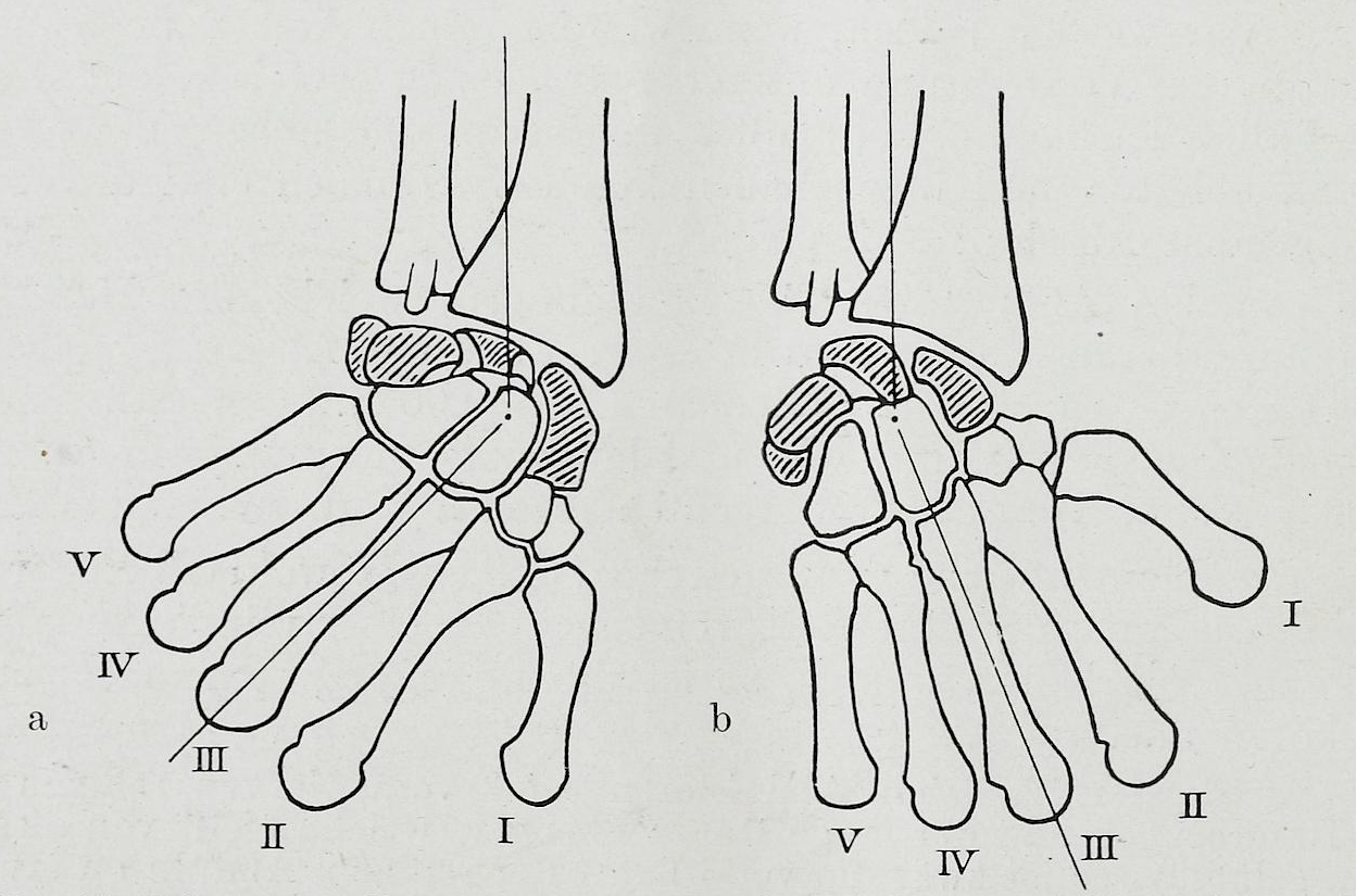
The carpal bones are the eight small bones that make up the wrist (carpus) that connects the hand to the forearm. The terms "carpus" and "carpal" are derived from the Latin wikt:carpus#Latin, carpus and the Greek language, Greek wikt:καρπός#Ancient Greek, καρπός (karpós), meaning "wrist". In human anatomy, the main role of the carpal bones is to joint, articulate with the radius (bone), radial and ulnar heads to form a highly mobile condyloid joint (i.e. wrist joint),Kingston 2000, pp 126-127 to provide attachments for thenar and hypothenar muscles, and to form part of the rigid carpal tunnel which allows the median nerve and tendons of the anterior compartment of the forearm, anterior forearm muscles to be transmitted to the hand and fingers. In tetrapods, the carpus is the sole cluster of bones in the wrist between the radius (bone), radius and ulna and the metacarpus. The bones of the carpus do not belong to individual fingers (or toes in quadrupeds), whereas those ... [...More Info...] [...Related Items...] OR: [Wikipedia] [Google] [Baidu] |
Ossification Center
An ossification center is a point where ossification of the hyaline cartilage begins. The first step in ossification is that the chondrocytes at this point become hypertrophic and arrange themselves in rows. The matrix in which they are imbedded increases in quantity, so that the cells become further separated from each other. A deposit of calcareous material now takes place in this matrix, between the rows of cells, so that they become separated from each other by longitudinal columns of calcified matrix, presenting a granular and opaque appearance. Here and there the matrix between two cells of the same row also becomes calcified, and transverse bars of calcified substance stretch across from one calcareous column to another. Thus, there are longitudinal groups of the cartilage cells enclosed in oblong cavities, the walls of which are formed of calcified matrix which cuts off all nutrition from the cells; the cells, in consequence, atrophy, leaving spaces called the primary ... [...More Info...] [...Related Items...] OR: [Wikipedia] [Google] [Baidu] |
Wrist
In human anatomy, the wrist is variously defined as (1) the carpus or carpal bones, the complex of eight bones forming the proximal skeletal segment of the hand; "The wrist contains eight bones, roughly aligned in two rows, known as the carpal bones." (2) the wrist joint or radiocarpal joint, the joint between the radius and the carpus and; (3) the anatomical region surrounding the carpus including the distal parts of the bones of the forearm and the proximal parts of the metacarpus or five metacarpal bones and the series of joints between these bones, thus referred to as ''wrist joints''. "With the large number of bones composing the wrist (ulna, radius, eight carpas, and five metacarpals), it makes sense that there are many, many joints that make up the structure known as the wrist." This region also includes the carpal tunnel, the anatomical snuff box, bracelet lines, the flexor retinaculum, and the extensor retinaculum. As a consequence of these various definitions, f ... [...More Info...] [...Related Items...] OR: [Wikipedia] [Google] [Baidu] |
Hand
A hand is a prehensile, multi-fingered appendage located at the end of the forearm or forelimb of primates such as humans, chimpanzees, monkeys, and lemurs. A few other vertebrates such as the Koala#Characteristics, koala (which has two thumb#Opposition and apposition, opposable thumbs on each "hand" and fingerprints extremely similar to human fingerprints) are often described as having "hands" instead of paws on their front limbs. The raccoon is usually described as having "hands" though opposable thumbs are lacking. Some evolutionary anatomists use the term ''hand'' to refer to the appendage of digits on the forelimb more generally—for example, in the context of whether the three Digit (anatomy), digits of the bird hand involved the same Homology (biology), homologous loss of two digits as in the dinosaur hand. The human hand usually has five digits: Finger numbering#Four-finger system, four fingers plus one thumb; however, these are often referred to collectively as Finger ... [...More Info...] [...Related Items...] OR: [Wikipedia] [Google] [Baidu] |
Carpal
The carpal bones are the eight small bones that make up the wrist (carpus) that connects the hand to the forearm. The terms "carpus" and "carpal" are derived from the Latin carpus and the Greek καρπός (karpós), meaning "wrist". In human anatomy, the main role of the carpal bones is to articulate with the radial and ulnar heads to form a highly mobile condyloid joint (i.e. wrist joint),Kingston 2000, pp 126-127 to provide attachments for thenar and hypothenar muscles, and to form part of the rigid carpal tunnel which allows the median nerve and tendons of the anterior forearm muscles to be transmitted to the hand and fingers. In tetrapods, the carpus is the sole cluster of bones in the wrist between the radius and ulna and the metacarpus. The bones of the carpus do not belong to individual fingers (or toes in quadrupeds), whereas those of the metacarpus do. The corresponding part of the foot is the tarsus. The carpal bones allow the wrist to move and rotate vertic ... [...More Info...] [...Related Items...] OR: [Wikipedia] [Google] [Baidu] |


