|
Occipitofrontalis Muscle
The occipitofrontalis muscle (epicranius muscle) is a muscle which covers parts of the skull. It consists of two parts or bellies: the occipital belly, near the occipital bone, and the frontal belly, near the frontal bone. It is supplied by the supraorbital artery, the supratrochlear artery, and the occipital artery. It is innervated by the facial nerve. In humans, the occipitofrontalis helps to create facial expressions. Structure The occipitofrontalis muscle consists of two parts or bellies: * the occipital belly, near the occipital bone. It originates on the lateral two-thirds of the highest nuchal line, and on the mastoid process of the temporal bone. It inserts into the epicranial aponeurosis. * the frontal belly, near the frontal bone. It originates from an intermediate tendon that connects to the occipital belly. It inserts in the fascia of the facial muscles and in the skin above the eyes and nose. Some sources consider the occipital and frontal bellies to b ... [...More Info...] [...Related Items...] OR: [Wikipedia] [Google] [Baidu] |
Epicranial Aponeurosis
The epicranial aponeurosis (aponeurosis epicranialis, galea aponeurotica) is an aponeurosis (a tough layer of dense fibrous tissue). It covers the upper part of the skull in humans and many other animals. Structure In humans, the epicranial aponeurosis originates from the external occipital protuberance and highest nuchal lines of the occipital bone. It merges with the occipitofrontalis muscle. In front, it forms a short and narrow prolongation between its union with the frontalis muscle (the frontal part of the occipitofrontalis muscle). On either side, the epicranial aponeurosis attaches to the anterior auricular muscles and the superior auricular muscles. Here it is less aponeurotic, and is continued over the temporal fascia to the zygomatic arch as a layer of laminated areolar tissue. It is closely connected to the integument by the firm, dense, fibro-fatty layer which forms the superficial fascia of the scalp. It is attached to the pericranium by loose cellular tis ... [...More Info...] [...Related Items...] OR: [Wikipedia] [Google] [Baidu] |
Mastoid Process
The mastoid part of the temporal bone is the posterior (back) part of the temporal bone, one of the bones of the skull. Its rough surface gives attachment to various muscles (via tendons) and it has openings for blood vessels. From its borders, the mastoid part articulates with two other bones. Etymology The word "mastoid" is derived from the Greek word for "breast", a reference to the shape of this bone. Surfaces Outer surface Its outer surface is rough and gives attachment to the occipitalis and posterior auricular muscles. It is perforated by numerous foramina (holes); for example, the mastoid foramen is situated near the posterior border and transmits a vein to the transverse sinus and a small branch of the occipital artery to the dura mater. The position and size of this foramen are very variable; it is not always present; sometimes it is situated in the occipital bone, or in the suture between the temporal and the occipital. Mastoid process The mastoid process ... [...More Info...] [...Related Items...] OR: [Wikipedia] [Google] [Baidu] |
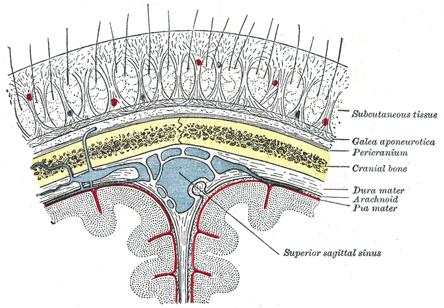
Scalp
The scalp is the area of the head where head hair grows. It is made up of skin, layers of connective and fibrous tissues, and the membrane of the skull. Anatomically, the scalp is part of the epicranium, a collection of structures covering the cranium. The scalp is bordered by the face at the front, and by the neck at the sides and back. The scientific study of hair and scalp is called trichology. Structure Layers The scalp is usually described as having five layers, which can be remembered using the mnemonic 'SCALP': * S: Skin. The skin of the scalp contains numerous hair follicles and sebaceous glands. * C: Connective tissue. A dense subcutaneous layer of fat and fibrous tissue that lies beneath the skin, containing the nerves and vessels of the scalp. * A: Aponeurosis. The epicranial aponeurosis or galea aponeurotica is a tough layer of dense fibrous tissue which anchors the above layers in place. It runs from the frontalis muscle anteriorly to the occipitalis ... [...More Info...] [...Related Items...] OR: [Wikipedia] [Google] [Baidu] |
Edinburgh
Edinburgh is the capital city of Scotland and one of its 32 Council areas of Scotland, council areas. The city is located in southeast Scotland and is bounded to the north by the Firth of Forth and to the south by the Pentland Hills. Edinburgh had a population of in , making it the List of towns and cities in Scotland by population, second-most populous city in Scotland and the List of cities in the United Kingdom, seventh-most populous in the United Kingdom. The Functional urban area, wider metropolitan area had a population of 912,490 in the same year. Recognised as the capital of Scotland since at least the 15th century, Edinburgh is the seat of the Scottish Government, the Scottish Parliament, the Courts of Scotland, highest courts in Scotland, and the Palace of Holyroodhouse, the official residence of the Monarchy of the United Kingdom, British monarch in Scotland. It is also the annual venue of the General Assembly of the Church of Scotland. The city has long been a cent ... [...More Info...] [...Related Items...] OR: [Wikipedia] [Google] [Baidu] |
Churchill Livingstone
Churchill Livingstone is an academic publisher. It was formed in 1971 from the merger of Longman's medical list, E & S Livingstone (Edinburgh, Scotland) and J & A Churchill (London, England) and was owned by Pearson. Harcourt acquired Churchill Livingstone in 1997. It is now integrated as an imprint in Elsevier's health science The following outline is provided as an overview of and topical guide to health sciences: Health sciences – those sciences that focus on health, or health care, as core parts of their subject matter. Health sciences relate to multiple ... division after Elsevier acquired Harcourt in 2001. In the past it published a number of classic medical texts, including Sir William Osler's textbook '' The Principles and Practice of Medicine, Gray's Anatomy,'' and '' Myles' Textbook for Midwives.'' In the 1980s, in addition to new texts in all areas of clinical medicine, it published an extensive list of medical and nursing textbooks in low-cost e ... [...More Info...] [...Related Items...] OR: [Wikipedia] [Google] [Baidu] |
Lambdoid Suture
The lambdoid suture, or lambdoidal suture, is a dense, fibrous connective tissue joint on the posterior aspect of the skull that connects the parietal bones with the occipital bone. It is continuous with the occipitomastoid suture. Structure The lambdoid suture is between the paired parietal bones and the occipital bone of the skull. It runs from the asterion on each side. Nerve supply The lambdoid suture may be supplied by a branch of the supraorbital nerve, a branch of the frontal branch of the trigeminal nerve. Clinical significance At birth, the bones of the skull do not meet. If certain bones of the skull grow too fast, then craniosynostosis (premature closure of the sutures) may occur. This can result in skull deformities. If the lambdoid suture closes too soon on one side, the skull will appear twisted and asymmetrical, a condition called "plagiocephaly". Plagiocephaly refers to the shape and not the condition. The condition is craniosynostosis. The lambdoid s ... [...More Info...] [...Related Items...] OR: [Wikipedia] [Google] [Baidu] |
Supraorbital Nerve
The supraorbital nerve is one of two terminal branches - the other being the supratrochlear nerve - of the frontal nerve (itself a branch of the ophthalmic nerve (CN V1)). It exits the orbit via the supraorbital foramen/notch before splitting into a medial branch and a lateral branch. It innervates the skin of the forehead, upper eyelid, and the root of the nose. Structure Origin The supraorbital nerve branches from the frontal nerve midway between the base and apex of the orbit. Course It travels anteriorly superior to the levator palpebrae superioris muscle. It exits the orbit through the supraorbital foramen/notch in the superior margin orbit, exiting it lateral to the supratrochlear nerve. It then ascends onto the forehead deep to the corrugator supercilii muscle and frontalis muscles. Fate It divides into a medial branch and lateral branch - usually after emerging from the orbit, but sometimes already within the orbit. Distribution The supraorbital nerv ... [...More Info...] [...Related Items...] OR: [Wikipedia] [Google] [Baidu] |
Artery
An artery () is a blood vessel in humans and most other animals that takes oxygenated blood away from the heart in the systemic circulation to one or more parts of the body. Exceptions that carry deoxygenated blood are the pulmonary arteries in the pulmonary circulation that carry blood to the lungs for oxygenation, and the umbilical arteries in the fetal circulation that carry deoxygenated blood to the placenta. It consists of a multi-layered artery wall wrapped into a tube-shaped channel. Arteries contrast with veins, which carry deoxygenated blood back towards the heart; or in the pulmonary and fetal circulations carry oxygenated blood to the lungs and fetus respectively. Structure The anatomy of arteries can be separated into gross anatomy, at the macroscopic scale, macroscopic level, and histology, microanatomy, which must be studied with a microscope. The arterial system of the human body is divided into systemic circulation, systemic arteries, carrying blood from the ... [...More Info...] [...Related Items...] OR: [Wikipedia] [Google] [Baidu] |
Temporoparietalis Muscle
The temporoparietalis muscle is a distinct muscle of the head. It lies above the superior auricular muscle, auricularis superior muscle. It lies just inferior to the epicranial aponeurosis of the occipitofrontalis muscle. The temporoparietalis muscle may be used in Reconstructive surgery, reconstructive Otorhinolaryngology, ear surgery. References Muscles of the head and neck {{Anatomy-stub ... [...More Info...] [...Related Items...] OR: [Wikipedia] [Google] [Baidu] |
Terminologia Anatomica
''Terminologia Anatomica'' (commonly abbreviated TA) is the international standard for human anatomy, human anatomical terminology. It is developed by the Federative International Programme on Anatomical Terminology (FIPAT) a program of the International Federation of Associations of Anatomists (IFAA). History The sixth edition of the previous standard, ''Nomina Anatomica'', was released in 1989. The first edition of ''Terminologia Anatomica'', superseding Nomina Anatomica, was developed by the Federative Committee on Anatomical Terminology (FCAT) and the International Federation of Associations of Anatomists (IFAA) and released in 1998. In April 2011, this edition was published online by the Federative International Programme on Anatomical Terminologies (FIPAT), the successor of FCAT. The first edition contained 7635 Latin items. The second edition was released online by FIPAT in 2019 and approved and adopted by the IFAA General Assembly in 2020. The latest errata is dated Au ... [...More Info...] [...Related Items...] OR: [Wikipedia] [Google] [Baidu] |
Nose
A nose is a sensory organ and respiratory structure in vertebrates. It consists of a nasal cavity inside the head, and an external nose on the face. The external nose houses the nostrils, or nares, a pair of tubes providing airflow through the nose for Respiration (physiology), respiration. Where the nostrils pass through the nasal cavity they widen, are known as nasal fossae, and contain nasal concha, turbinates and olfactory mucosa. The nasal cavity also connects to the paranasal sinuses (dead-end air cavities for pressure buffering and humidification). From the nasal cavity, the nostrils continue into the pharynx, a switch track valve connecting the respiratory system, respiratory and digestive systems. In humans, the nose is located centrally on the face and serves as an alternative respiratory passage especially during suckling for infants. The protruding nose that is completely separate from the mouth part is a characteristic found only in theria, therian mammals. It has b ... [...More Info...] [...Related Items...] OR: [Wikipedia] [Google] [Baidu] |
Facial Muscles
The facial muscles are a group of striated skeletal muscles supplied by the facial nerve (cranial nerve VII) that, among other things, control facial expression. These muscles are also called mimetic muscles. They are only found in mammals, although they derive from neural crest cells found in all vertebrates. They are the only muscles that attach to the dermis. Structure The facial muscles are just under the skin ( subcutaneous) muscles that control facial expression. They generally originate from the surface of the skull bone (rarely the fascia), and insert on the skin of the face. When they contract, the skin moves. These muscles also cause wrinkles at right angles to the muscles’ action line. Nerve supply The facial muscles are supplied by the facial nerve (cranial nerve VII), with each nerve serving one side of the face. In contrast, the nearby masticatory muscles are supplied by the mandibular nerve, a branch of the trigeminal nerve (cranial nerve V). List of muscl ... [...More Info...] [...Related Items...] OR: [Wikipedia] [Google] [Baidu] |


