|
Neurofilaments
Neurofilaments (NF) are classed as type IV intermediate filaments found in the cytoplasm of neurons. They are protein polymers measuring 10 nm in diameter and many micrometers in length. Together with microtubules (~25 nm) and microfilaments (7 nm), they form the neuronal cytoskeleton. They are believed to function primarily to provide structural support for axons and to regulate axon diameter, which influences nerve conduction velocity. The proteins that form neurofilaments are members of the intermediate filament protein family, which is divided into six types based on their gene organization and protein structure. Types I and II are the keratins which are expressed in epithelia. Type III contains the proteins vimentin, desmin, peripherin and glial fibrillary acidic protein (GFAP). Type IV consists of the neurofilament proteins NF-L, NF-M, NF-H and α-internexin. Type V consists of the nuclear lamins, and type VI consists of the protein nestin. The ty ... [...More Info...] [...Related Items...] OR: [Wikipedia] [Google] [Baidu] |
NEFL
Neurofilament light polypeptide is a protein that in humans is encoded by the NEFL gene. Structure Neurofilament light polypeptide is a member of the intermediate filament protein family. This protein family consists of over 50 human proteins divided into 5 major classes, the Class I and II keratins, Class III vimentin, Glial fibrillary acidic protein, GFAP, desmin and the others, the Class IV neurofilaments and the Class V nuclear lamins. There are four major neurofilament subunits, NF-L, NF-M, NF-H and α-internexin. These form heteropolymers which assemble to produce 10 nm neurofilaments which are only expressed in neurons where they are major structural proteins, particularly concentrated in large projection axons. The NF-L protein is encoded by the ''NEFL'' gene. Function These neurofilament heteropolymers assemble into the cytoskeleton of axons, where they provide structural support and help regulate axonal diameter and conduction velocity. Axons are particularly sens ... [...More Info...] [...Related Items...] OR: [Wikipedia] [Google] [Baidu] |
Peripherin
Peripherin is a type III intermediate filament protein expressed mainly in neurons of the peripheral nervous system. It is also found in neurons of the central nervous system that have projections toward peripheral structures, such as spinal motor neurons. Its size, structure, and sequence/location of protein motifs is similar to other type III intermediate filament proteins such as desmin, vimentin and glial fibrillary acidic protein. Like these proteins, peripherin can self-assemble to form homopolymeric filamentous networks (networks formed from peripherin protein dimers), but it can also heteropolymerize with neurofilaments in several neuronal types. This protein in humans is encoded by the ''PRPH'' gene. Peripherin is thought to play a role in neurite elongation during development and axonal regeneration after injury, but its exact function is unknown. It is also associated with some of the major neuropathologies that characterize amyotropic lateral sclerosis (ALS), but desp ... [...More Info...] [...Related Items...] OR: [Wikipedia] [Google] [Baidu] |
Nestin (protein)
Nestin is a protein that in humans is encoded by the NES gene. Nestin (acronym for neuroepithelial stem cell protein) is a type VI intermediate filament (IF) protein. These intermediate filament proteins are expressed mostly in nerve cells where they are implicated in the radial growth of the axon. Seven genes encode for the heavy (NF-H), medium (NF-M) and light neurofilament (NF-L) proteins, nestin and α-internexin in nerve cells, synemin α and desmuslin/synemin β (two alternative transcripts of the DMN gene) in muscle cells, and syncoilin (also in muscle cells). Members of this group mostly preferentially coassemble as heteropolymers in tissues. Steinert et al. has shown that nestin forms homodimers and homotetramers but does not form IF by itself in vitro. In mixtures, nestin preferentially co-assembles with purified vimentin or the type IV IF protein internexin to form heterodimer coiled-coil molecules. Gene Structurally, nestin has the shortest head domain (N-termin ... [...More Info...] [...Related Items...] OR: [Wikipedia] [Google] [Baidu] |
Internexin
Internexin, alpha-internexin, is a Class IV intermediate filament approximately 66 kDa. The protein was originally purified from rat optic nerve and spinal cord.Levavasseur F, Zhu Q, and JP Julien. No requirement of alpha-internexin for nervous system development and for radial growth of axons. Molecular Brain Research. 69:104-112. (1999). The protein copurifies with other neurofilament subunits, as it was originally discovered, however in some mature neurons it can be the only neurofilament expressed. The protein is present in developing neuroblasts and in the central nervous system of adults. The protein is a major component of the intermediate filament network in small interneurons and cerebellar granule cells, where it is present in the parallel fibers. Structure Alpha-internexin has a homologous central rod domain of approximately 310 amino acid residues that form a highly conserved alpha helical region. The central rod domain is responsible for coiled-coil structure an ... [...More Info...] [...Related Items...] OR: [Wikipedia] [Google] [Baidu] |
Internexin
Internexin, alpha-internexin, is a Class IV intermediate filament approximately 66 kDa. The protein was originally purified from rat optic nerve and spinal cord.Levavasseur F, Zhu Q, and JP Julien. No requirement of alpha-internexin for nervous system development and for radial growth of axons. Molecular Brain Research. 69:104-112. (1999). The protein copurifies with other neurofilament subunits, as it was originally discovered, however in some mature neurons it can be the only neurofilament expressed. The protein is present in developing neuroblasts and in the central nervous system of adults. The protein is a major component of the intermediate filament network in small interneurons and cerebellar granule cells, where it is present in the parallel fibers. Structure Alpha-internexin has a homologous central rod domain of approximately 310 amino acid residues that form a highly conserved alpha helical region. The central rod domain is responsible for coiled-coil structure and ... [...More Info...] [...Related Items...] OR: [Wikipedia] [Google] [Baidu] |
NEFM
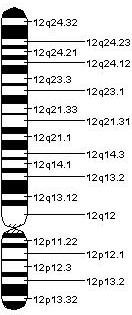
Neurofilament medium polypeptide (NF-M) is a protein that in humans is encoded by the ''NEFM'' gene. Function Neurofilaments are type IV intermediate filament heteropolymers composed of light (NEFL), medium (this protein), and heavy ( NEFH) chains. Neurofilaments comprise the exoskeleton and functionally maintain neuronal caliber. They may also play a role in intracellular transport to axons and dendrites. This gene encodes the medium neurofilament protein. This protein is commonly used as a biomarker In biomedical contexts, a biomarker, or biological marker, is a measurable indicator of some biological state or condition. Biomarkers are often measured and evaluated using blood, urine, or soft tissues to examine normal biological processes, ... of neuronal damage. References Further reading * * * * * * * * * * * * * * * * * * * Human proteins {{gene-8-stub ... [...More Info...] [...Related Items...] OR: [Wikipedia] [Google] [Baidu] |
NEFH
Neurofilament, heavy polypeptide (NEFH) is a protein that in humans is encoded by the ''NEFH'' gene. It is the gene for a heavy protein subunit that is combined with medium and light subunits to make neurofilaments, which form the framework for nerve cells. Mutations in the NEFH gene are associated with Charcot-Marie-Tooth disease. References Genes on human chromosome 22 {{Gene-22-stub ... [...More Info...] [...Related Items...] OR: [Wikipedia] [Google] [Baidu] |
Lysine
Lysine (symbol Lys or K) is an α-amino acid that is a precursor to many proteins. Lysine contains an α-amino group (which is in the protonated form when the lysine is dissolved in water at physiological pH), an α-carboxylic acid group (which is in the deprotonated form when the lysine is dissolved in water at physiological pH), and a side chain (which is partially protonated when the lysine is dissolved in water at physiological pH), and so it is classified as a basic, charged (in water at physiological pH), aliphatic amino acid. It is encoded by the codons AAA and AAG. Like almost all other amino acids, the α-carbon is chiral and lysine may refer to either enantiomer or a racemic mixture of both. For the purpose of this article, lysine will refer to the biologically active enantiomer L-lysine, where the α-carbon is in the ''S'' configuration. The human body cannot synthesize lysine. It is essential in humans and must therefore be obtained from the diet. In orga ... [...More Info...] [...Related Items...] OR: [Wikipedia] [Google] [Baidu] |
Glutamic Acid
Glutamic acid (symbol Glu or E; known as glutamate in its anionic form) is an α- amino acid that is used by almost all living beings in the biosynthesis of proteins. It is a non-essential nutrient for humans, meaning that the human body can synthesize enough for its use. It is also the most abundant excitatory neurotransmitter in the vertebrate nervous system. It serves as the precursor for the synthesis of the inhibitory gamma-aminobutyric acid (GABA) in GABAergic neurons. Its molecular formula is . Glutamic acid exists in two optically isomeric forms; the dextrorotatory -form is usually obtained by hydrolysis of gluten or from the waste waters of beet-sugar manufacture or by fermentation.Webster's Third New International Dictionary of the English Language Unabridged, Third Edition, 1971. Its molecular structure could be idealized as HOOC−CH()−()2−COOH, with two carboxyl groups −COOH and one amino group −. However, in the solid state and mildly acidic water s ... [...More Info...] [...Related Items...] OR: [Wikipedia] [Google] [Baidu] |
SDS-PAGE
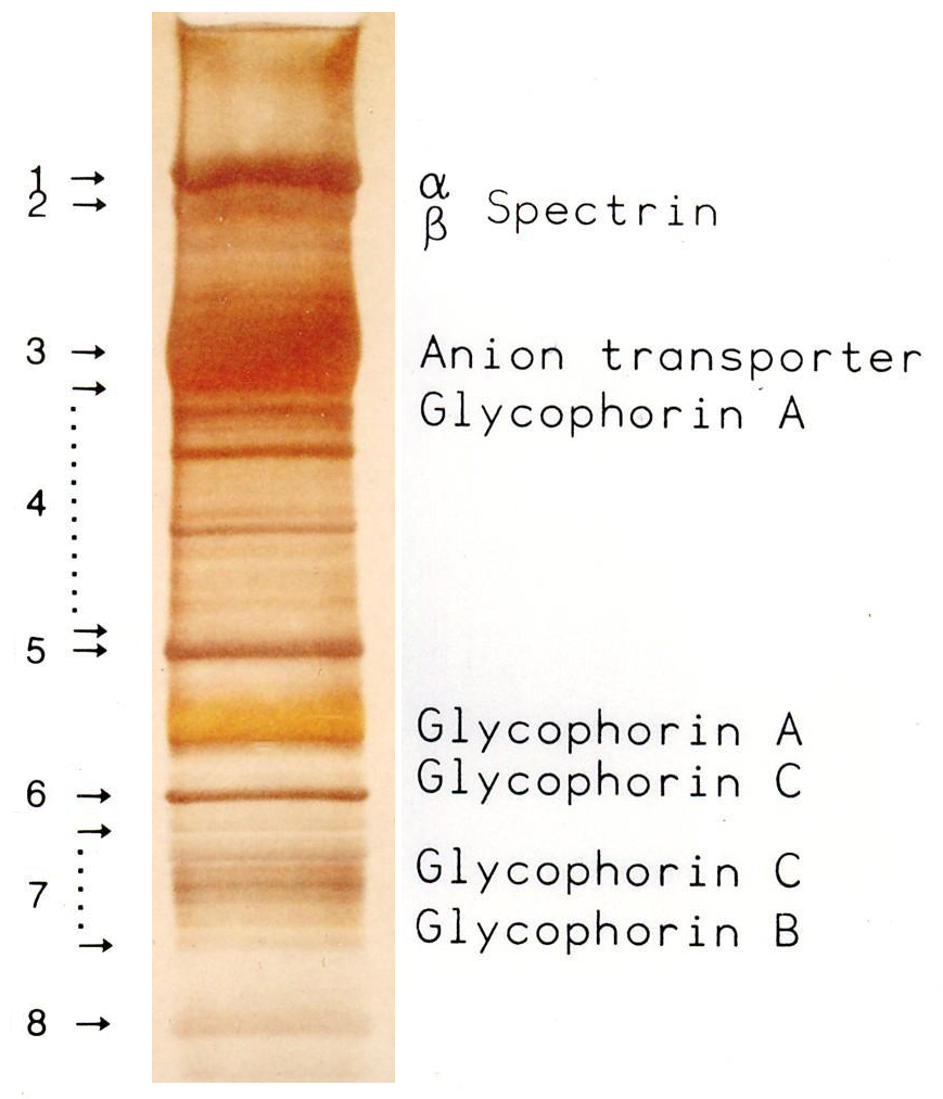
SDS-PAGE (sodium dodecyl sulfate–polyacrylamide gel electrophoresis) is a Discontinuous electrophoresis, discontinuous electrophoretic system developed by Ulrich K. Laemmli which is commonly used as a method to separate proteins with molecular masses between 5 and 250 Kilodalton, kDa. The combined use of sodium dodecyl sulfate (SDS, also known as sodium lauryl sulfate) and polyacrylamide gel eliminates the influence of structure and charge, and proteins are separated by differences in their size. At least up to 2012, the publication describing it was the most frequently cited paper by a single author, and the second most cited overall. Properties SDS-PAGE is an electrophoresis method that allows protein separation by mass. The medium (also referred to as ′matrix′) is a polyacrylamide-based discontinuous gel. The polyacrylamide-gel is typically sandwiched between two glass plates in a slab gel. Although tube gels (in glass cylinders) were used historically, they were rapid ... [...More Info...] [...Related Items...] OR: [Wikipedia] [Google] [Baidu] |
Molecular Mass
The molecular mass () is the mass of a given molecule, often expressed in units of daltons (Da). Different molecules of the same compound may have different molecular masses because they contain different isotopes of an element. The derived quantity relative molecular mass is the unitless ratio of the mass of a molecule to the atomic mass constant (which is equal to one dalton). The molecular mass and relative molecular mass are distinct from but related to the ''molar mass''. The molar mass is defined as the mass of a given substance divided by the amount of the substance, and is expressed in grams per mole (g/mol). That makes the molar mass an ''average'' of many particles or molecules (weighted by abundance of the isotopes), and the molecular mass the mass of one specific particle or molecule. The molar mass is usually the more appropriate quantity when dealing with macroscopic (weigh-able) quantities of a substance. The definition of molecular weight is most authoritat ... [...More Info...] [...Related Items...] OR: [Wikipedia] [Google] [Baidu] |
Horizontal Neurons
Horizontal cells are the laterally interconnecting neurons having cell bodies in the inner nuclear layer of the retina of vertebrate eyes. They help integrate and regulate the input from multiple photoreceptor cells. Among their functions, horizontal cells are believed to be responsible for increasing contrast via lateral inhibition and adapting both to bright and dim light conditions. Horizontal cells provide inhibitory feedback to rod and cone photoreceptors. They are thought to be important for the antagonistic center-surround property of the receptive fields of many types of retinal ganglion cells. Other retinal neurons include photoreceptor cells, bipolar cells, amacrine cells, and retinal ganglion cells. Structure Depending on the species, there are typically one or two classes of horizontal cells, with a third type sometimes proposed. Horizontal cells span across photoreceptors and summate inputs before synapsing onto photoreceptor cells. Horizontal cells may also sy ... [...More Info...] [...Related Items...] OR: [Wikipedia] [Google] [Baidu] |


