|
Nasopharyngeal Angiofibroma
Nasopharyngeal angiofibroma is an angiofibroma also known as juvenile nasal angiofibroma, fibromatous hamartoma, and angiofibromatous hamartoma of the nasal cavity. It is a benign but locally aggressive vascular tumor of the nasopharynx that arises from the superior margin of the sphenopalatine foramen and grows in the back of the nasal cavity. It most commonly affects adolescent males. Though it is a benign tumor, it is locally invasive and can invade the nose, cheek, orbit (frog face deformity), or brain. Features The tumor is highly vascular, meaning that it has a rich blood supply. Clinically, an individual with one may present with nosebleeds, followed by nasal obstruction and then mucus discharge from the nose. Grossly, it is a firm mass that may be yellow, dark red, or even black. Histologically, it presents with several vascular spaces of varying sizes and wall thicknesses as well as fibrous or collagenous Stroma (tissue), stroma with fibroblasts. Mast cells are common ... [...More Info...] [...Related Items...] OR: [Wikipedia] [Google] [Baidu] |
Micrograph
A micrograph is an image, captured photographically or digitally, taken through a microscope or similar device to show a magnify, magnified image of an object. This is opposed to a macrograph or photomacrograph, an image which is also taken on a microscope but is only slightly magnified, usually less than 10 times. Micrography is the practice or art of using microscopes to make photographs. A photographic micrograph is a photomicrograph, and one taken with an electron microscope is an electron micrograph. A micrograph contains extensive details of microstructure. A wealth of information can be obtained from a simple micrograph like behavior of the material under different conditions, the phases found in the system, failure analysis, grain size estimation, elemental analysis and so on. Micrographs are widely used in all fields of microscopy. Types Photomicrograph A light micrograph or photomicrograph is a micrograph prepared using an optical microscope, a process referred to ... [...More Info...] [...Related Items...] OR: [Wikipedia] [Google] [Baidu] |
H&E Stain
Hematoxylin and eosin stain ( or haematoxylin and eosin stain or hematoxylin–eosin stain; often abbreviated as H&E stain or HE stain) is one of the principal tissue stains used in histology. It is the most widely used stain in medical diagnosis and is often the ''gold standard.'' For example, when a pathologist looks at a biopsy of a suspected cancer, the histological section is likely to be stained with H&E. H&E is the combination of two histological stains: hematoxylin and eosin. The hematoxylin stains cell nuclei a purplish blue, and eosin stains the extracellular matrix and cytoplasm pink, with other structures taking on different shades, hues, and combinations of these colors. Hence a pathologist can easily differentiate between the nuclear and cytoplasmic parts of a cell, and additionally, the overall patterns of coloration from the stain show the general layout and distribution of cells and provides a general overview of a tissue sample's structure. Thus, patte ... [...More Info...] [...Related Items...] OR: [Wikipedia] [Google] [Baidu] |
ENT Surgery
Otorhinolaryngology ( , abbreviated ORL and also known as otolaryngology, otolaryngology–head and neck surgery (ORL–H&N or OHNS), or ear, nose, and throat (ENT)) is a surgical subspecialty within medicine that deals with the surgical and medical management of conditions of the head and neck. Doctors who specialize in this area are called otorhinolaryngologists, otolaryngologists, head and neck surgeons, or ENT surgeons or physicians. Patients seek treatment from an otorhinolaryngologist for diseases of the ear, Human nose, nose, throat, base of skull, base of the skull, head, and neck. These commonly include functional diseases that affect the senses and activities of eating, drinking, speaking, breathing, swallowing, and hearing. In addition, ENT surgery encompasses the surgical management of cancers and benign tumors and reconstruction of the head and neck as well as plastic surgery of the face, scalp, and neck. Etymology The term is a combination of Neo-Latin classic ... [...More Info...] [...Related Items...] OR: [Wikipedia] [Google] [Baidu] |
Angiofibroma
Angiofibroma (AGF) is a descriptive term for a wide range of benign skin or mucous membrane (i.e. the outer membrane lining body cavities such as the mouth and nose) lesions in which individuals have: # benign papules, i.e. pinhead-sized elevations that lack visible evidence of containing fluid; # nodule (medicine), nodules, i.e. small firm lumps usually > 1 mm in diameter; and/or # tumors, i.e. masses often regarded as ~8 mm or larger. Diagnosis AGF lesions share common Macroscopic scale, macroscopic (i.e. gross) and microscopic appearances. Grossly, AGF lesions consist of multiple papules, one or more skin-colored to erythematous, dome-shaped nodules, or usually just a single tumor. Microscopically, they consist of spindle-shaped and stellate-shaped cells centered around dilated and thin-walled blood vessels in a background of coarse bundles of collagen (i.e. the main fibrous component of connective tissue). Angiofibromas have been divided into different types but commonly a spe ... [...More Info...] [...Related Items...] OR: [Wikipedia] [Google] [Baidu] |
Benign
Malignancy () is the tendency of a medical condition to become progressively worse; the term is most familiar as a characterization of cancer. A ''malignant'' tumor contrasts with a non-cancerous benign tumor, ''benign'' tumor in that a malignancy is not self-limited in its growth, is capable of invading into adjacent tissues, and may be capable of spreading to distant tissues. A benign tumor has none of those properties, but may still be harmful to health. The term benign in more general medical use characterizes a condition or growth that is not cancerous, i.e. does not spread to other parts of the body or invade nearby tissue. Sometimes the term is used to suggest that a condition is not dangerous or serious. Malignancy in cancers is characterized by anaplasia, invasiveness, and metastasis. Malignant tumors are also characterized by genome instability, so that cancers, as assessed by whole genome sequencing, frequently have between 10,000 and 100,000 mutations in their ent ... [...More Info...] [...Related Items...] OR: [Wikipedia] [Google] [Baidu] |
Vascular Tumor
A vascular tumor is a vascular anomaly where a tumor forms from cells that make blood or lymph vessels; a soft tissue growth that can be either benign or malignant. Examples of vascular tumors include hemangiomas, hemangioendotheliomas, Kaposi's sarcomas, angiosarcomas, and hemangioblastomas. An angioma refers to any type of benign vascular tumor. Some vascular tumors can be associated with serious blood-clotting disorders, making correct diagnosis critical. A vascular tumor may be described in terms of being ''highly vascularized'', or ''poorly vascularized'', referring to the degree of blood supply to the tumor. Classification Vascular tumors make up one of the classifications of vascular anomalies. The other grouping is vascular malformations. Vascular tumors can be further subclassified as being benign, borderline or aggressive, and malignant. Vascular tumors are described as ''proliferative'', and vascular malformations as ''nonproliferative''. Types A vascular t ... [...More Info...] [...Related Items...] OR: [Wikipedia] [Google] [Baidu] |
Nasopharynx
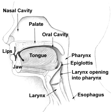
The pharynx (: pharynges) is the part of the throat behind the mouth and nasal cavity, and above the esophagus and trachea (the tubes going down to the stomach and the lungs respectively). It is found in vertebrates and invertebrates, though its structure varies across species. The pharynx carries food to the esophagus and air to the larynx. The flap of cartilage called the epiglottis stops food from entering the larynx. In humans, the pharynx is part of the digestive system and the conducting zone of the respiratory system. (The conducting zone—which also includes the nostrils of the nose, the larynx, trachea, bronchi, and bronchioles—filters, warms, and moistens air and conducts it into the lungs). The human pharynx is conventionally divided into three sections: the nasopharynx, oropharynx, and laryngopharynx (hypopharynx). In humans, two sets of pharyngeal muscles form the pharynx and determine the shape of its lumen. They are arranged as an inner layer of longitudina ... [...More Info...] [...Related Items...] OR: [Wikipedia] [Google] [Baidu] |
Sphenopalatine Foramen
The sphenopalatine foramen is a foramen of the skull that connects the nasal cavity and the pterygopalatine fossa. It gives passage to the sphenopalatine artery, nasopalatine nerve, and the superior nasal nerve (all passing from the pterygopalatine fossa into the nasal cavity). Structure The processes of the superior border of the palatine bone are separated by the ''sphenopalatine notch'', which is converted into the sphenopalatine foramen by the under surface of the body of the sphenoid. The sphenopalatine foramen is situated posterior to the middle nasal meatus orbital process of palatine bone, anterior to the sphenoidal process of palatine bone, inferior to the body and of the sphenoid bone, and superior to the superior margin of the perpendicular plate of palatine bone. Relations The ethmoid crest (a reliable surgical landmark A landmark is a recognizable natural or artificial feature used for navigation, a feature that stands out from its near environment and ... [...More Info...] [...Related Items...] OR: [Wikipedia] [Google] [Baidu] |
Nasal Cavity
The nasal cavity is a large, air-filled space above and behind the nose in the middle of the face. The nasal septum divides the cavity into two cavities, also known as fossae. Each cavity is the continuation of one of the two nostrils. The nasal cavity is the uppermost part of the respiratory system and provides the nasal passage for inhaled air from the nostrils to the nasopharynx and rest of the respiratory tract. The paranasal sinuses surround and drain into the nasal cavity. Structure The term "nasal cavity" can refer to each of the two cavities of the nose, or to the two sides combined. The lateral wall of each nasal cavity mainly consists of the maxilla. However, there is a deficiency that is compensated for by the perpendicular plate of the palatine bone, the medial pterygoid plate, the labyrinth of ethmoid and the inferior concha. The paranasal sinuses are connected to the nasal cavity through small orifices called ostia. Most of these ostia communicat ... [...More Info...] [...Related Items...] OR: [Wikipedia] [Google] [Baidu] |
Stroma (tissue)
Stroma () is the part of a tissue (biology), tissue or organ (anatomy), organ with a structural or connective role. It is made up of all the parts without specific functions of the organ - for example, connective tissue, blood vessels, ducts, etc. The other part, the parenchyma, consists of the cells that perform the function of the tissue or organ. There are multiple ways of classifying tissues: one classification scheme is based on tissue functions and another analyzes their cellular components. Stromal tissue falls into the "functional" class that contributes to the body's support and movement. Stromal cell, The cells which make up stroma tissues serve as a matrix in which the other cells are embedded. Stroma is made of various types of stromal cells. Examples of stroma include: * stroma of iris * stroma of cornea * stroma of ovary * stroma of thyroid gland * stroma of thymus * stroma of bone marrow * lymph node stromal cell *Mesenchymal stem cell, multipotent stromal cell (me ... [...More Info...] [...Related Items...] OR: [Wikipedia] [Google] [Baidu] |
Arteriovenous Malformation
An arteriovenous malformation (AVM) is an abnormal connection between arteries and veins, bypassing the capillary system. Usually congenital, this vascular anomaly is widely known because of its occurrence in the central nervous system (usually as a cerebral AVM), but can appear anywhere in the body. The symptoms of AVMs can range from none at all to intense pain or bleeding, and they can lead to other serious medical problems. Signs and symptoms Symptoms of AVMs vary according to their location. Most neurological AVMs produce few to no symptoms. Often the malformation is discovered as part of an autopsy or during treatment of an unrelated disorder (an " incidental finding"); in rare cases, its expansion or a micro-bleed from an AVM in the brain can cause epilepsy, neurological deficit, or pain. The most general symptoms of a cerebral AVM include headaches and epileptic seizures, with more specific symptoms that normally depend on its location and the individual, in ... [...More Info...] [...Related Items...] OR: [Wikipedia] [Google] [Baidu] |


