|
NUT Carcinoma
NUT carcinoma (NC; formerly NUT midline carcinoma (NMC)) is a rare genetically defined, very aggressive squamous-cell carcinoma, squamous cell epithelium, epithelial cancer that usually arises in the midline of the body and is characterized by a chromosomal rearrangement in the nuclear protein in testis gene (i.e. ''NUTM1'' gene). In approximately 75% of cases, the coding sequence of ''NUTM1'' in Karyotype#Types of banding, band 14 on the Locus (genetics)#Nomenclature, long (or "q") arm of chromosome 15 is fused to ''BRD4'' or ''BRD3'', which creates a chimeric gene that encodes the ''BRD-NUT'' fusion protein. The remaining cases, the fusion of NUTM1 is to an unknown partner gene, usually called ''NUT''-variant. The name NUT carcinoma was introduced as the carcinoma does not only occur in the body midline; therefore, World Health Organization, WHO also changed the name in 2015 in the WHO Classification of Tumours of the Lung, Pleura, Thymus and Heart. Signs and symptoms The number ... [...More Info...] [...Related Items...] OR: [Wikipedia] [Google] [Baidu] |
Micrograph
A micrograph is an image, captured photographically or digitally, taken through a microscope or similar device to show a magnify, magnified image of an object. This is opposed to a macrograph or photomacrograph, an image which is also taken on a microscope but is only slightly magnified, usually less than 10 times. Micrography is the practice or art of using microscopes to make photographs. A photographic micrograph is a photomicrograph, and one taken with an electron microscope is an electron micrograph. A micrograph contains extensive details of microstructure. A wealth of information can be obtained from a simple micrograph like behavior of the material under different conditions, the phases found in the system, failure analysis, grain size estimation, elemental analysis and so on. Micrographs are widely used in all fields of microscopy. Types Photomicrograph A light micrograph or photomicrograph is a micrograph prepared using an optical microscope, a process referred to ... [...More Info...] [...Related Items...] OR: [Wikipedia] [Google] [Baidu] |
Head
A head is the part of an organism which usually includes the ears, brain, forehead, cheeks, chin, eyes, nose, and mouth, each of which aid in various sensory functions such as sight, hearing, smell, and taste. Some very simple animals may not have a head, but many bilaterally symmetric forms do, regardless of size. Heads develop in animals by an evolutionary trend known as cephalization. In bilaterally symmetrical animals, nervous tissue concentrate at the anterior region, forming structures responsible for information processing. Through biological evolution, sense organs and feeding structures also concentrate into the anterior region; these collectively form the head. Human head The human head is an anatomical unit that consists of the skull, hyoid bone and cervical vertebrae. The skull consists of the brain case which encloses the cranial cavity, and the facial skeleton, which includes the mandible. There are eight bones in the brain case ... [...More Info...] [...Related Items...] OR: [Wikipedia] [Google] [Baidu] |
Histone Deacetylase Inhibitor
Histone deacetylase inhibitors (HDAC inhibitors, HDACi, HDIs) are chemical compounds that enzyme inhibitor, inhibit histone deacetylases. Since acetylation of histones, deacetylation of histones produces transcriptionally silenced heterochromatin, HDIs can render chromatin more transcriptionally active and induce epigenomic changes. HDIs have a long history of use in psychiatry and neurology as mood stabilizers and anti-epileptics, such as valproic acid. Since at least 2003 they have been investigated as possible treatments for cancers, parasitic and inflammatory diseases. Cellular biochemistry/pharmacology To carry out gene expression, a cell must control the coiling and uncoiling of DNA around histones. This is accomplished with the assistance of histone acetyltransferase, histone acetyl transferases (HAT), which acetylation of histones , acetylate the lysine residues in core histones leading to a less compact and more transcriptionally active euchromatin, and, on the conv ... [...More Info...] [...Related Items...] OR: [Wikipedia] [Google] [Baidu] |
BET Inhibitors
BET inhibitors are a class of drugs that reversibly bind the bromodomains of Bromodomain and Extra-Terminal motif (BET) proteins BRD2, BRD3, BRD4, and BRDT, and prevent protein-protein interaction between BET proteins and acetylated histones and transcription factors. Discovery and development Thienodiazepine BET inhibitors were discovered in a phenotypic drug screen by scientists at Yoshitomi Pharmaceuticals (now Mitsubishi Tanabe Pharma) in the early 1990s, and their potential both as anti-inflammatories and anti-cancer agents noted. OncoEthix (acquired by Merck in 2014) in-licensed OTX-015 from Mitsubishi and in 2012 initiated the first BET inhibitor clinical trial for oncology (ClinicalTrials.gov Identifier: NCT01713582). BET inhibitors were also independently discovered in phenotypic screens for small molecule inducers of Apolipoprotein A-I by both GSK and Resverlogix. In 2010, the use of JQ1, a tert-butyl synthetic precursor of OTX-015, was published having activity in ... [...More Info...] [...Related Items...] OR: [Wikipedia] [Google] [Baidu] |
NUT Gene
Nut often refers to: * Nut (fruit), fruit composed of a hard shell and a seed * Nut (food), a dry and edible fruit or seed, including but not limited to true nuts * Nut (hardware), fastener used with a bolt Nut, NUT or Nuts may also refer to: Arts, entertainment, and media Comics * ''Nuts'', comic in the National Lampoon by Gahan Wilson (1970s) * ''Nuts'', comic strip in alternative newspapers by M. Wartella (1990s) Fictional characters * Nut (Marvel Comics), fictional character evoking the Egyptian sky goddess * Nut (movie character), character portrayed by Shing Fui-On in two late 20th-century Hong Kong crime films Films * ''Nuts'' (1987 film), American drama * ''Nuts'' (2012 film), French comedy * ''Nuts!'' (film), animated documentary on John R. Brinkley Television *NBC Universal Television Studio, or NUTS, former name of television arm of NBCUniversal / Universal Television * Nuts TV, British television channel related to ''Nuts'' magazine Other uses in arts, entert ... [...More Info...] [...Related Items...] OR: [Wikipedia] [Google] [Baidu] |
Chromosomal Translocation
In genetics, chromosome translocation is a phenomenon that results in unusual rearrangement of chromosomes. This includes "balanced" and "unbalanced" translocation, with three main types: "reciprocal", "nonreciprocal" and "Robertsonian" translocation. Reciprocal translocation is a chromosome abnormality caused by exchange of parts between non-homologous chromosomes. Two detached fragments of two different chromosomes are switched. Robertsonian translocation occurs when two non-homologous chromosomes get attached, meaning that given two healthy pairs of chromosomes, one of each pair "sticks" and blends together homogeneously. Each type of chromosomal translocation can result in disorders for growth, function and the development of an individuals body, often resulting from a change in their genome. A gene fusion may be created when the translocation joins two otherwise-separated genes. It is detected on cytogenetics or a karyotype of affected cells. Translocations can be bala ... [...More Info...] [...Related Items...] OR: [Wikipedia] [Google] [Baidu] |
Immunohistochemistry
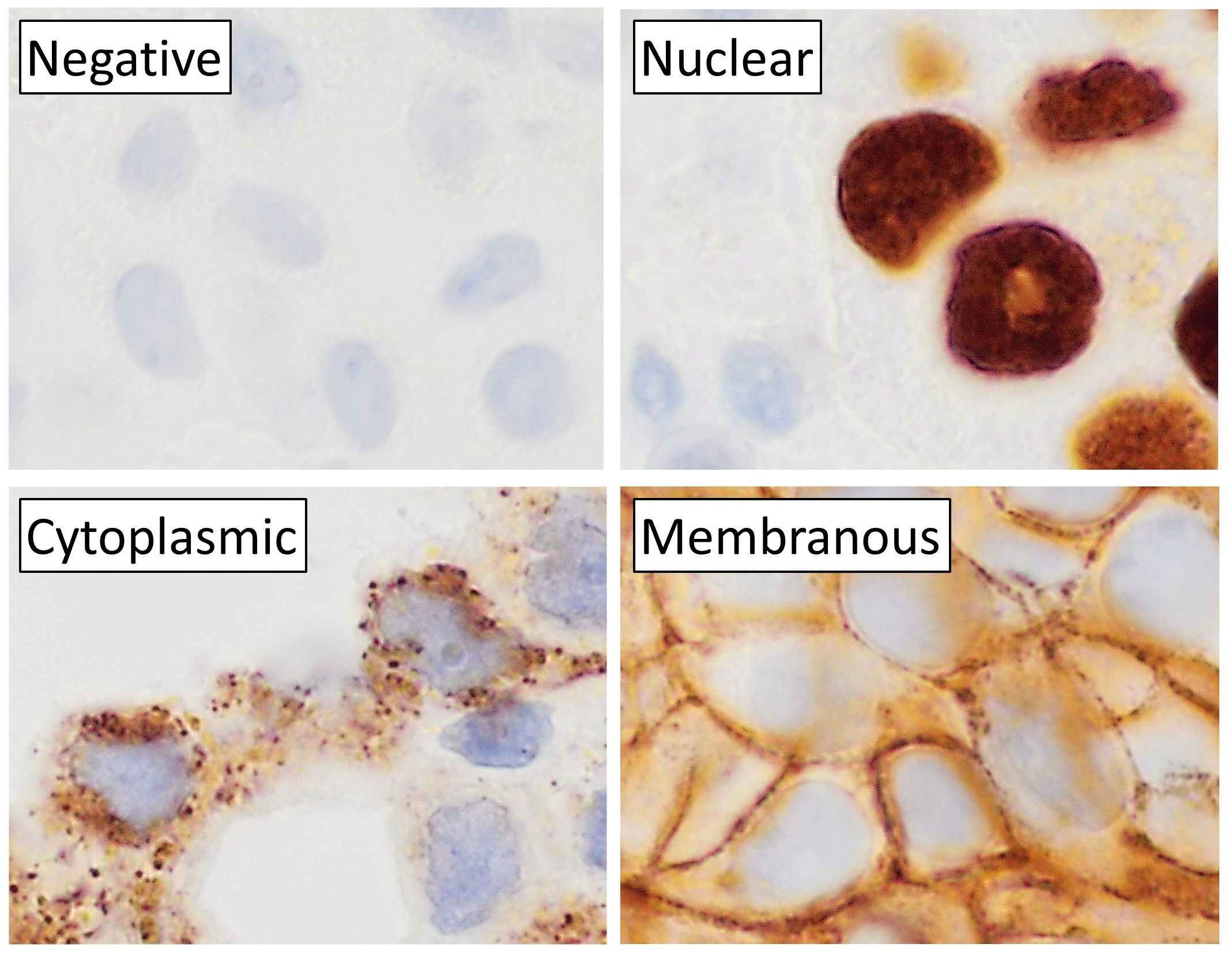
Immunohistochemistry is a form of immunostaining. It involves the process of selectively identifying antigens in cells and tissue, by exploiting the principle of Antibody, antibodies binding specifically to antigens in biological tissues. Albert Coons, Albert Hewett Coons, Ernst Berliner, Ernest Berliner, Norman Jones and Hugh J Creech was the first to develop immunofluorescence in 1941. This led to the later development of immunohistochemistry. Immunohistochemical staining is widely used in the diagnosis of abnormal cells such as those found in cancerous tumors. In some cancer cells certain tumor antigens are expressed which make it possible to detect. Immunohistochemistry is also widely used in basic research, to understand the distribution and localization of biomarkers and differentially expressed proteins in different parts of a biological tissue. Sample preparation Immunohistochemistry can be performed on tissue that has been fixed and embedded in Paraffin wax, paraffin, ... [...More Info...] [...Related Items...] OR: [Wikipedia] [Google] [Baidu] |
CD34
CD34 is a transmembrane phosphoglycoprotein protein encoded by the CD34 gene in humans, mice, rats and other species. CD34 derives its name from the cluster of differentiation protocol that identifies cell surface antigens. CD34 was first described on hematopoietic stem cells independently by Civin et al. and Tindle et al. as a cell surface glycoprotein and functions as a cell-cell adhesion factor. It may also mediate the attachment of hematopoietic stem cells to bone marrow extracellular matrix or directly to stromal cells. Clinically, it is associated with the selection and enrichment of hematopoietic stem cells for bone marrow transplants. Due to these historical and clinical associations, CD34 expression is almost ubiquitously related to hematopoietic cells; however, it is actually found on many other cell types as well. Function The CD34 protein is a member of a family of single-pass transmembrane sialomucin proteins that show expression on early haematopoietic ... [...More Info...] [...Related Items...] OR: [Wikipedia] [Google] [Baidu] |
Carcinoembryonic Antigen
Carcinoembryonic antigen (CEA) describes a set of highly-related glycoproteins involved in cell adhesion. CEA is normally produced in gastrointestinal tissue during fetal development, but the production stops before birth. Consequently, CEA is usually present at very low levels in the blood of healthy adults (about 2–4 ng/mL). However, the serum levels are raised in some types of cancer, which means that it can be used as a tumor marker in clinical tests. Serum levels can also be elevated in heavy smoking, smokers. CEA are Glycosylphosphatidylinositol, glycosyl phosphatidyl inositol (GPI) cell-surface-anchored glycoproteins whose specialized Sialyl-Lewis X, sialo Fucosylation, fucosylated glycoforms serve as functional colon carcinoma L-selectin and E-selectin ligands, which may be critical to the metastatic dissemination of colon carcinoma cells. Immunologically they are characterized as members of the CD66 cluster of differentiation. The proteins include CD66a, CD66b, CD6 ... [...More Info...] [...Related Items...] OR: [Wikipedia] [Google] [Baidu] |
TP63
Tumor protein p63, typically referred to as p63, also known as transformation-related protein 63, is a protein that in humans is encoded by the ''TP63'' (also known as the '' p63'') gene. The ''TP63'' gene was discovered 20 years after the discovery of the ''p53'' tumor suppressor gene and along with ''p73'' constitutes the ''p53'' gene family based on their structural similarity. Despite being discovered significantly later than ''p53'', phylogenetic analysis of ''p53'', ''p63'' and ''p73'', suggest that ''p63'' was the original member of the family from which ''p53'' and ''p73'' evolved. Function Tumor protein p63 is a member of the p53 family of transcription factors. p63 -/- mice have several developmental defects which include the lack of limbs and other tissues, such as teeth and mammary glands, which develop as a result of interactions between mesenchyme and epithelium. TP63 encodes for two main isoforms by alternative promoters (TAp63 and ΔNp63). ΔNp63 is involved in ... [...More Info...] [...Related Items...] OR: [Wikipedia] [Google] [Baidu] |
Cytokeratin
Cytokeratins are keratin proteins found in the intracytoplasmic cytoskeleton of epithelial tissue. They are an important component of intermediate filaments, which help cells resist mechanical stress. Expression of these cytokeratins within epithelial cells is largely specific to particular organs or tissues. Thus they are used clinically to identify the cell of origin of various human tumors. Naming The term ''cytokeratin'' began to be used in the late 1970s, when the protein subunits of keratin intermediate filaments inside cells were first being identified and characterized. In 2006 a new systematic nomenclature for mammalian keratins was created, and the proteins previously called ''cytokeratins'' are simply called ''keratins'' (human epithelial category). For example, cytokeratin-4 (CK-4) has been renamed keratin-4 (K4). However, they are still commonly referred to as cytokeratins in clinical practice. Types There are two categories of cytokeratins: the acidic type I ... [...More Info...] [...Related Items...] OR: [Wikipedia] [Google] [Baidu] |
Mediastinum
The mediastinum (from ;: mediastina) is the central compartment of the thoracic cavity. Surrounded by loose connective tissue, it is a region that contains vital organs and structures within the thorax, mainly the heart and its vessels, the esophagus, the trachea, the vagus nerve, vagus, phrenic nerve, phrenic and cardiac nerves, the thoracic duct, the thymus and the lymph nodes of the central chest. Anatomy The mediastinum lies within the thorax and is enclosed on the right and left by pulmonary pleurae, pleurae. It is surrounded by the chest wall in front, the lungs to the sides and the Spine (anatomy), spine at the back. It extends from the sternum in front to the vertebral column behind. It contains all the organs of the thorax except the lungs. It is continuous with the loose connective tissue of the neck. The mediastinum can be divided into an upper (or superior) and lower (or inferior) part: * The superior mediastinum starts at the superior thoracic aperture and ends ... [...More Info...] [...Related Items...] OR: [Wikipedia] [Google] [Baidu] |


