|
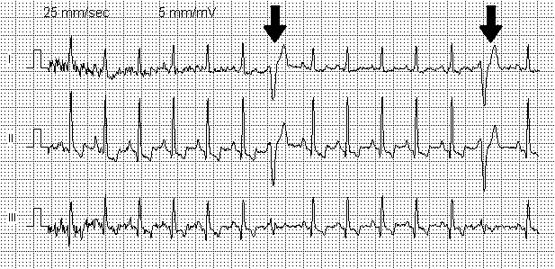
Myocardial Contusion
A blunt cardiac injury is an injury to the heart as the result of blunt trauma, typically to the anterior chest wall. It can result in a variety of specific injuries to the heart, the most common of which is a myocardial contusion, which is a term for a bruise (contusion) to the heart after an injury. Other injuries which can result include septal defects and valvular failures. The right ventricle is thought to be most commonly affected due to its anatomic location as the most anterior surface of the heart. Myocardial contusion is not a specific diagnosis and the extent of the injury can vary greatly. Usually, there are other chest injuries seen with a myocardial contusion such as rib fractures, pneumothorax, and heart valve injury. When a myocardial contusion is suspected, consideration must be given to any other chest injuries, which will likely be determined by clinical signs, tests, and imaging. The signs and symptoms of a myocardial contusion can manifest in different ways in ... [...More Info...] [...Related Items...] OR: [Wikipedia] [Google] [Baidu] |
Emergency Medicine
Emergency medicine is the medical specialty concerned with the care of illnesses or injuries requiring immediate medical attention. Emergency physicians (or "ER doctors") specialize in providing care for unscheduled and undifferentiated patients of all ages. As frontline providers, in coordination with emergency medical services, they are responsible for initiating resuscitation, stabilization, and early interventions during the acute phase of a medical condition. Emergency physicians generally practice in hospital emergency departments, pre-hospital settings via emergency medical services, and intensive care units. Still, they may also work in primary care settings such as urgent care clinics. Sub-specialties of emergency medicine include disaster medicine, medical toxicology, point-of-care ultrasonography, critical care medicine, emergency medical services, hyperbaric medicine, sports medicine, palliative care, or aerospace medicine. Various models for emerg ... [...More Info...] [...Related Items...] OR: [Wikipedia] [Google] [Baidu] |
Bundle Branch Block
A bundle branch block is a partial or complete interruption in the flow of electrical impulses in either of the bundle branches of the heart's electrical system. Anatomy and physiology The heart's electrical activity begins in the sinoatrial node (the heart's natural pacemaker), which is situated on the upper right atrium. The impulse travels next through the left and right atria and summates at the atrioventricular node. From the AV node the electrical impulse travels down the bundle of His and divides into the right and left bundle branches. The right bundle branch contains one fascicle. The left bundle branch subdivides into two fascicles: the left anterior fascicle, and the left posterior fascicle. Other sources divide the left bundle branch into three fascicles: the left anterior, the left posterior, and the left septal fascicle. The thicker left posterior fascicle bifurcates, with one fascicle being in the septal aspect. Ultimately, the fascicles divide into mill ... [...More Info...] [...Related Items...] OR: [Wikipedia] [Google] [Baidu] |
Commotio Cordis
Commotio cordis (Latin, "agitation / disruption of the heart") is a rare disruption of heart rhythm that occurs as a result of a blow to the area directly over the heart (the precordial region) at a critical instant during the cycle of a heartbeat. The condition is 97% fatal if not treated within three minutes. This sudden rise in intracavitary pressure leads to disruption of normal heart electrical activity, followed instantly by ventricular fibrillation, complete disorganization of the heart's pumping function, and cardiac arrest. It is not caused by mechanical damage to the heart muscle or surrounding organs and is not the result of heart disease. Its incidence in the United States is fewer than 20 cases per year, often occurring in boys participating in sports, most commonly in baseball when a ball strikes a player in the chest. Commotio cordis can occur only upon impact within a narrow window of about 40 milliseconds in the cardiac electrical cycle, explaining why it is ... [...More Info...] [...Related Items...] OR: [Wikipedia] [Google] [Baidu] |
Constrictive Pericarditis
Constrictive pericarditis is a condition characterized by a thickened, fibrotic pericardium, limiting the heart's ability to function normally. In many cases, the condition continues to be difficult to diagnose and therefore benefits from a good understanding of the underlying cause. Signs and symptoms Signs and symptoms of constrictive pericarditis are consistent with the following: fatigue, swollen abdomen, difficulty breathing (dyspnea), swelling of legs and general weakness. Related conditions are bacterial pericarditis, pericarditis and pericarditis after a heart attack. Causes The cause of constrictive pericarditis in the developing world are idiopathic in origin, though likely infectious in nature. In regions where tuberculosis is common, it is the cause in a large portion of cases. Causes of constrictive pericarditis include: * Tuberculosis * Incomplete drainage of purulent pericarditis * Fungal and parasitic infections * Chronic pericarditis * Postviral pericarditis * ... [...More Info...] [...Related Items...] OR: [Wikipedia] [Google] [Baidu] |
Pericardial Effusion
A pericardial effusion is an abnormal accumulation of fluid in the pericardial cavity. The pericardium is a two-part membrane surrounding the heart: the outer fibrous Connective tissue, connective membrane and an inner two-layered serous membrane. The two layers of the serous membrane enclose the pericardial cavity (the potential space) between them. This pericardial space contains a small amount of pericardial fluid, normally 15-50 mL in volume. The pericardium, specifically the pericardial fluid provides lubrication, maintains the anatomic position of the heart in the chest (levocardia), and also serves as a barrier to protect the heart from infection and inflammation in adjacent tissues and organs. By definition, a pericardial effusion occurs when the volume of fluid in the cavity exceeds the normal limit. If large enough, it can compress the heart, causing cardiac tamponade and obstructive shock. Some of the presenting symptoms are shortness of breath, chest pain, chest pressu ... [...More Info...] [...Related Items...] OR: [Wikipedia] [Google] [Baidu] |
Heart Failure
Heart failure (HF), also known as congestive heart failure (CHF), is a syndrome caused by an impairment in the heart's ability to Cardiac cycle, fill with and pump blood. Although symptoms vary based on which side of the heart is affected, HF typically presents with shortness of breath, Fatigue (medical), excessive fatigue, and bilateral peripheral edema, leg swelling. The severity of the heart failure is mainly decided based on ejection fraction and also measured by the severity of symptoms. Other conditions that have symptoms similar to heart failure include obesity, kidney failure, liver disease, anemia, and thyroid disease. Common causes of heart failure include coronary artery disease, heart attack, hypertension, high blood pressure, atrial fibrillation, valvular heart disease, alcohol use disorder, excessive alcohol consumption, infection, and cardiomyopathy. These cause heart failure by altering the structure or the function of the heart or in some cases both. There are ... [...More Info...] [...Related Items...] OR: [Wikipedia] [Google] [Baidu] |
Pericardiocentesis
Pericardiocentesis (PCC), also called pericardial tap, is a medical procedure where fluid is aspirated from the pericardium (the sac enveloping the heart). Anatomy and physiology The pericardium is a fibrous sac surrounding the heart composed of two layers: an inner visceral pericardium and an outer parietal pericardium. The area between these two layers is known as the pericardial space and normally contains 15 to 50 mL of serous fluid. This fluid protects the heart by serving as a shock absorber and provides lubrication to the heart during contraction. The elastic nature of the pericardium allows it to accommodate a small amount of extra fluid, roughly 80 to 120 mL, in the acute setting. However, once a critical volume is reached, even small amounts of extra fluid can rapidly increase pressure within the pericardium. This pressure can significantly hinder the ability of the heart to contract, leading to cardiac tamponade. If accumulation of fluid is slow and occurs over ... [...More Info...] [...Related Items...] OR: [Wikipedia] [Google] [Baidu] |
Tamponade
Tamponade () is the closure or blockage (as of a wound or body cavity) by or as if by a tampon, especially to stop bleeding. Tamponade is a useful method of stopping a hemorrhage. This can be achieved by applying an absorbent dressing directly into a wound, thereby absorbing excess blood and creating a blockage, or by applying direct pressure with a hand or a tourniquet. Not to be confused with a tamponade that occurs as a result of health problems. For example: cardiac tamponade is a condition where fluid collects in the pericardial sac increasing pressure within the pericardium which in turn prevents the ventricles from expanding fully significantly reducing the efficiency of the heart. It is considered a medical emergency and if left unchecked is fatal. Bladder tamponade is obstruction of the urinary bladder outlet due to heavy blood clot formation within it. [...More Info...] [...Related Items...] OR: [Wikipedia] [Google] [Baidu] |
Echocardiography
Echocardiography, also known as cardiac ultrasound, is the use of ultrasound to examine the heart. It is a type of medical imaging, using standard ultrasound or Doppler ultrasound. The visual image formed using this technique is called an echocardiogram, a cardiac echo, or simply an echo. Echocardiography is routinely used in the diagnosis, management, and follow-up of patients with any suspected or known heart diseases. It is one of the most widely used diagnostic imaging modalities in cardiology. It can provide a wealth of helpful information, including the size and shape of the heart (internal chamber size quantification), pumping capacity, location and extent of any tissue damage, and assessment of valves. An echocardiogram can also give physicians other estimates of heart function, such as a calculation of the cardiac output, ejection fraction, and diastolic function (how well the heart relaxes). Echocardiography is an important tool in assessing wall motion abnorma ... [...More Info...] [...Related Items...] OR: [Wikipedia] [Google] [Baidu] |
Premature Ventricular Contraction
A premature ventricular contraction (PVC) is a common event where the heartbeat is initiated by Purkinje fibers in the ventricles rather than by the sinoatrial node. PVCs may cause no symptoms or may be perceived as a "skipped beat" or felt as palpitations in the chest. PVCs do not usually pose any danger. The electrical events of the heart detected by the electrocardiogram (ECG) allow a PVC to be easily distinguished from a normal heart beat. However, very frequent PVCs can be symptomatic of an underlying heart condition (such as arrhythmogenic right ventricular cardiomyopathy). Furthermore, very frequent (over 20% of all heartbeats) PVCs are considered a risk factor for arrhythmia-induced cardiomyopathy, in which the heart muscle becomes less effective and symptoms of heart failure may develop. Ultrasound of the heart is therefore recommended in people with frequent PVCs. If PVCs are frequent or troublesome, medication (beta blockers or certain calcium channel blockers) ... [...More Info...] [...Related Items...] OR: [Wikipedia] [Google] [Baidu] |
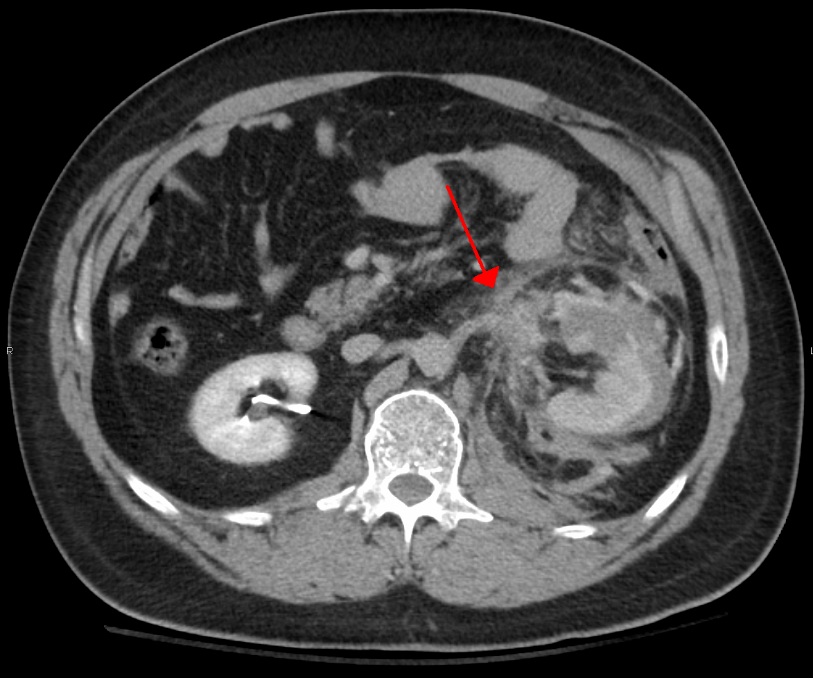
Blunt Trauma
A blunt trauma, also known as a blunt force trauma or non-penetrating trauma, is a physical trauma due to a forceful impact without penetration of the body's surface. Blunt trauma stands in contrast with penetrating trauma, which occurs when an object pierces the skin, enters body tissue, and creates an open wound. Blunt trauma occurs due to direct physical trauma or impactful force to a body part. Such incidents often occur with road traffic collisions, assaults, and sports-related injuries, and are notably common among the elderly who experience falls. Blunt trauma can lead to a wide range of injuries including contusions, concussions, abrasions, lacerations, internal or external hemorrhages, and bone fractures. The severity of these injuries depends on factors such as the force of the impact, the area of the body affected, and the underlying comorbidities of the affected individual. In some cases, blunt force trauma can be life-threatening and may require immedia ... [...More Info...] [...Related Items...] OR: [Wikipedia] [Google] [Baidu] |
Ventricular Fibrillation
Ventricular fibrillation (V-fib or VF) is an abnormal heart rhythm in which the Ventricle (heart), ventricles of the heart Fibrillation, quiver. It is due to disorganized electrical conduction system of the heart, electrical activity. Ventricular fibrillation results in cardiac arrest with loss of consciousness and no pulse. This is followed by sudden cardiac death in the absence of treatment. Ventricular fibrillation is initially found in about 10% of people with cardiac arrest. Ventricular fibrillation can occur due to coronary heart disease, valvular heart disease, cardiomyopathy, Brugada syndrome, long QT syndrome, electric shock, or intracranial hemorrhage. Diagnosis is by an electrocardiogram (ECG) showing irregular unformed QRS complexes without any clear P wave (electrocardiography), P waves. An important differential diagnosis is torsades de pointes. Treatment is with cardiopulmonary resuscitation (CPR) and defibrillation. Defibrillation#Change to a biphasic waveform, ... [...More Info...] [...Related Items...] OR: [Wikipedia] [Google] [Baidu] |


