|
Muscular Branches Of The Radial Nerve
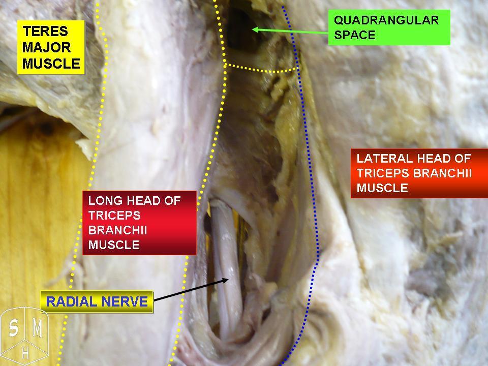
The muscular branches of the radial nerve supply the triceps brachii, anconæus, brachioradialis, and extensor carpi radialis longus, and are grouped as medial, posterior, and lateral. Medial The medial muscular branches supply the medial head of the triceps brachii. That to the medial head is a long, slender filament, which lies close to the ulnar nerve as far as the lower third of the arm, and is therefore frequently spoken of as the ''ulnar collateral nerve''. Posterior The posterior muscular branch, of large size, arises from the nerve in the groove between the triceps brachii and the humerus. It divides into filaments, which supply the medial and lateral heads of the triceps brachii and the anconæus muscles. The branch for the latter muscle is a long, slender filament, which descends in the substance of the medial head of the triceps brachii. Lateral The lateral muscular branches supply the brachioradialis, extensor carpi radialis longus, and the lateral part of the bra ... [...More Info...] [...Related Items...] OR: [Wikipedia] [Google] [Baidu] |
Posterior Compartment Of The Arm
Posterior may refer to: * Posterior (anatomy), the end of an organism opposite to anterior ** Buttocks, as a euphemism * Posterior horn (other) * Posterior probability The posterior probability is a type of conditional probability that results from updating the prior probability with information summarized by the likelihood via an application of Bayes' rule. From an epistemological perspective, the posteri ..., the conditional probability that is assigned when the relevant evidence is taken into account * Posterior tense, a relative future tense {{disambiguation ... [...More Info...] [...Related Items...] OR: [Wikipedia] [Google] [Baidu] |
Mobile Wad
The mobile wad (or mobile wad of Henry) is a group of the following three muscles found in the lateral compartment of the forearm:Baek 2004, pp 508–509 * brachioradialis * extensor carpi radialis brevis * extensor carpi radialis longus It is also sometimes known as the "wad of three",Biel 2019, p138 "lateral compartment",DuParc 2003, 55–030–A-10 or "radial group"Platzer 2004, p 164 of the forearm. Function These three muscles act as flexors at the elbow joint.Note: The extensor carpi muscles are so named because they extend at the carpus, not at the elbow. The extensor carpi radialis brevis and longus are both weak flexors at the elbow joint. Brevis moves the arm from ulnar abduction to its mid-position and flexes dorsally. Longus is a weak pronator in the flexed arm and a supinator in the outstretched arm. At the carpal joints longus acts in dorsiflexion with the extensor carpi ulnaris and in radial abduction with the flexor carpi radialis In anatomy, flexor carpi ... [...More Info...] [...Related Items...] OR: [Wikipedia] [Google] [Baidu] |
Radial Nerve
The radial nerve is a nerve in the human body that supplies the posterior portion of the upper limb. It innervates the medial and lateral heads of the triceps brachii muscle of the arm, as well as all 12 muscles in the Posterior compartment of the forearm, posterior osteofascial compartment of the forearm and the associated joints and overlying skin. It originates from the brachial plexus, carrying fibers from the posterior roots of spinal nerves C5, C6, C7, C8 and T1. The radial nerve and its branches provide Motor neuron, motor innervation to the dorsal arm muscles (the triceps brachii and the anconeus) and the extrinsic extensors of the wrists and hands; it also provides cutaneous Nerve supply to the skin, sensory innervation to most of the back of the hand, except for the back of the little finger and adjacent half of the ring finger (which are innervated by the ulnar nerve). The radial nerve divides into a deep branch, which becomes the posterior interosseous nerve, and a su ... [...More Info...] [...Related Items...] OR: [Wikipedia] [Google] [Baidu] |
Triceps Brachii
The triceps, or triceps brachii (Latin for "three-headed muscle of the arm"), is a large muscle on the back of the upper limb of many vertebrates. It consists of three parts: the medial, lateral, and long head. All three heads cross the elbow joint. However, the long head also crosses the shoulder joint. The triceps muscle contracts when the elbow is straightened and expands when the elbow is bent. The long head gets a further contraction when the arm is behind the torso due to how it crosses the shoulder joint. It is the muscle principally responsible for extension of the elbow joint (straightening of the arm). Structure * The long head arises from the infraglenoid tubercle of the scapula. It extends distally anterior to the teres minor and posterior to the teres major. * The medial head arises proximally in the humerus, just inferior to the groove of the radial nerve; from the dorsal (back) surface of the humerus; from the medial intermuscular septum; and its distal ... [...More Info...] [...Related Items...] OR: [Wikipedia] [Google] [Baidu] |
Anconæus
The anconeus muscle (or anconaeus/anconæus) is a small muscle on the posterior aspect of the elbow joint. Some consider anconeus to be a continuation of the triceps brachii muscle. Some sources consider it to be part of the posterior compartment of the arm, while others consider it part of the posterior compartment of the forearm. The anconeus muscle can easily be palpated just lateral to the olecranon process of the ulna. Structure Anconeus originates on the posterior surface of the lateral epicondyle of the humerus and inserts distally on the superior posterior surface of the ulna and the lateral aspect of the olecranon. Innervation Anconeus is innervated by a branch of the radial nerve (cervical roots 7 and 8) from the posterior cord of the brachial plexus called the nerve to the anconeus. The somatomotor portion of radial nerve innervating anconeus bifurcates from the main branch in the radial groove of the humerus. This innervation pattern follows the rules of innervation ... [...More Info...] [...Related Items...] OR: [Wikipedia] [Google] [Baidu] |
Brachioradialis
The brachioradialis is a muscle of the forearm that flexes the forearm at the elbow. It is also capable of both pronation and supination, depending on the position of the forearm. It is attached to the distal styloid process of the radius by way of the brachioradialis tendon, and to the lateral supracondylar ridge of the humerus. Structure The brachioradialis is a superficial, fusiform muscle on the lateral side of the forearm. It originates proximally on the lateral supracondylar ridge of the humerus. It inserts distally on the radius, at the base of its styloid process. Near the elbow, it forms the lateral limit of the cubital fossa, or elbow pit. Nerve supply Despite the bulk of the muscle body being visible from the anterior aspect of the forearm, the brachioradialis is a posterior compartment muscle and consequently is innervated by the radial nerve. Of the muscles that receive innervation from the radial nerve, it is one of only four that receive input directly from the ... [...More Info...] [...Related Items...] OR: [Wikipedia] [Google] [Baidu] |
Extensor Carpi Radialis Longus
The extensor carpi radialis longus is one of the five main muscles that control movements at the wrist. This muscle is quite long, starting on the lateral side of the humerus, and attaching to the base of the second metacarpal bone (metacarpal of the index finger). Structure It originates from the lateral supracondylar ridge of the humerus, from the lateral intermuscular septum, and by a few fibers from the lateral epicondyle of the humerus. The fibers end at the upper third of the forearm in a flat tendon, which runs along the lateral border of the radius, beneath the abductor pollicis longus and extensor pollicis brevis; it then passes beneath the dorsal carpal ligament, where it lies in a groove on the back of the radius common to it and the extensor carpi radialis brevis, immediately behind the styloid process. One of the three muscles of the radial forearm group, it initially lies beside the brachioradialis, but becomes mostly tendon early on. Passing between the brachi ... [...More Info...] [...Related Items...] OR: [Wikipedia] [Google] [Baidu] |
Humerus
The humerus (; : humeri) is a long bone in the arm that runs from the shoulder to the elbow. It connects the scapula and the two bones of the lower arm, the radius (bone), radius and ulna, and consists of three sections. The humeral upper extremity of humerus, upper extremity consists of a rounded head, a narrow neck, and two short processes (tubercles, sometimes called tuberosities). The body of humerus, body is cylindrical in its upper portion, and more prism (geometry), prismatic below. The lower extremity of humerus, lower extremity consists of 2 epicondyles, 2 processes (trochlea of the humerus, trochlea and capitulum of the humerus, capitulum), and 3 fossae (radial fossa, coronoid fossa, and olecranon fossa). As well as its true anatomical neck, the constriction below the greater and lesser tubercles of the humerus is referred to as its Surgical neck of the humerus, surgical neck due to its tendency to fracture, thus often becoming the focus of surgeons. Etymology The word ... [...More Info...] [...Related Items...] OR: [Wikipedia] [Google] [Baidu] |
Brachialis
The brachialis (brachialis anticus) is a muscle in the upper arm that flexes the elbow. It lies beneath the biceps brachii, and makes up part of the floor of the region known as the cubital fossa (elbow pit). It originates from the anterior aspect of the distal humerus; it inserts onto the tuberosity of the ulna. It is innervated by the musculocutaneous nerve, and commonly also receives additional innervation from the radial nerve."Brachialis Muscle." Kenhub. Kenhub, Aug. 2001 The brachialis is the prime mover of elbow flexion generating about 50% more power than the biceps.Saladin, Kenneth S, Stephen J. Sullivan, and Christina A. Gan. Anatomy & Physiology: The Unity of Form and Function. 2015. Print. Structure Origin The brachialis originates from the anterior surface of the distal half of the humerus, near the insertion of the deltoid muscle, which it embraces by two angular processes. Its origin extends below to within 2.5 cm of the margin of the articular surface of t ... [...More Info...] [...Related Items...] OR: [Wikipedia] [Google] [Baidu] |

