|
Multifocal Micronodular Pneumocyte Hyperplasia
Multifocal micronodular pneumocyte hyperplasia (MMPH) is a subtype of pneumocytic hyperplasia (hyperplasia of pneumocytes lining pulmonary alveoli). Several synonymous terms have been done for this entity: adenomatoid proliferation of alveolar epithelium, papillary alveolar hamartoma, multifocal alveolar hyperplasia, multinodular pneumocyte hyperplasia. These multifocal lesions are observed in tuberous sclerosis, and can be associated with lymphangioleiomyomatosis and perivascular epithelioid cell tumour (PEComa or clear cell "sugar tumor")). It can be diagnosed through lung biopsy using thoracoscopy. Microscopy * Well-demarcated, nodular lesions ranging 2–5 mm in pulmonary parenchyma. * Type II pneumocytes without nuclear atypia lined thickened alveolar septa and proliferated papillary structures. * Enlarged cuboidal cells lining mildly thickened alveolar septa. * Enlarged cuboidal cells have abundant, eosinophilic cytoplasm and large, round nuclei. * Papillary pat ... [...More Info...] [...Related Items...] OR: [Wikipedia] [Google] [Baidu] |
Multifocal Micronodular Pneumocyte Hyperplasia - Tuberous Sclerosis - A3 -- High Mag
Progressive lenses, also called multifocal lenses, progressive addition lenses (PAL), varifocal lenses, progressive power lenses, graduated prescription lenses, or progressive spectacle lenses are corrective lenses used in eyeglasses to correct presbyopia and other disorders of accommodation. They are characterised by a gradient of increasing lens power, added to the wearer's correction for the other refractive errors. The gradient starts at the wearer's distance prescription at the top of the lens and reaches a maximum addition power, or the full reading addition, at the bottom of the lens. The length of the progressive power gradient on the lens surface depends on the design of the lens, with a final addition power between 0.75 and 3.50 dioptres. The addition value prescribed depends on the level of presbyopia of the patient. In general the older the patient, the higher the addition. History The first patent for a PAL was British Patent 15,735, granted to Owen Aves with ... [...More Info...] [...Related Items...] OR: [Wikipedia] [Google] [Baidu] |
Pneumocytic Hyperplasia
Pneumocytic hyperplasia is an hyperplasia of pneumocytes lining pulmonary alveoli A pulmonary alveolus (plural: alveoli, from Latin ''alveolus'', "little cavity"), also known as an air sac or air space, is one of millions of hollow, distensible cup-shaped cavities in the lungs where oxygen is exchanged for carbon dioxide. Al .... Types *Pulmonary atypical adenomatous hyperplasia * Multifocal micronodular pneumocyte hyperplasia References Pulmonary lesion {{med-sign-stub ... [...More Info...] [...Related Items...] OR: [Wikipedia] [Google] [Baidu] |
Pneumocyte
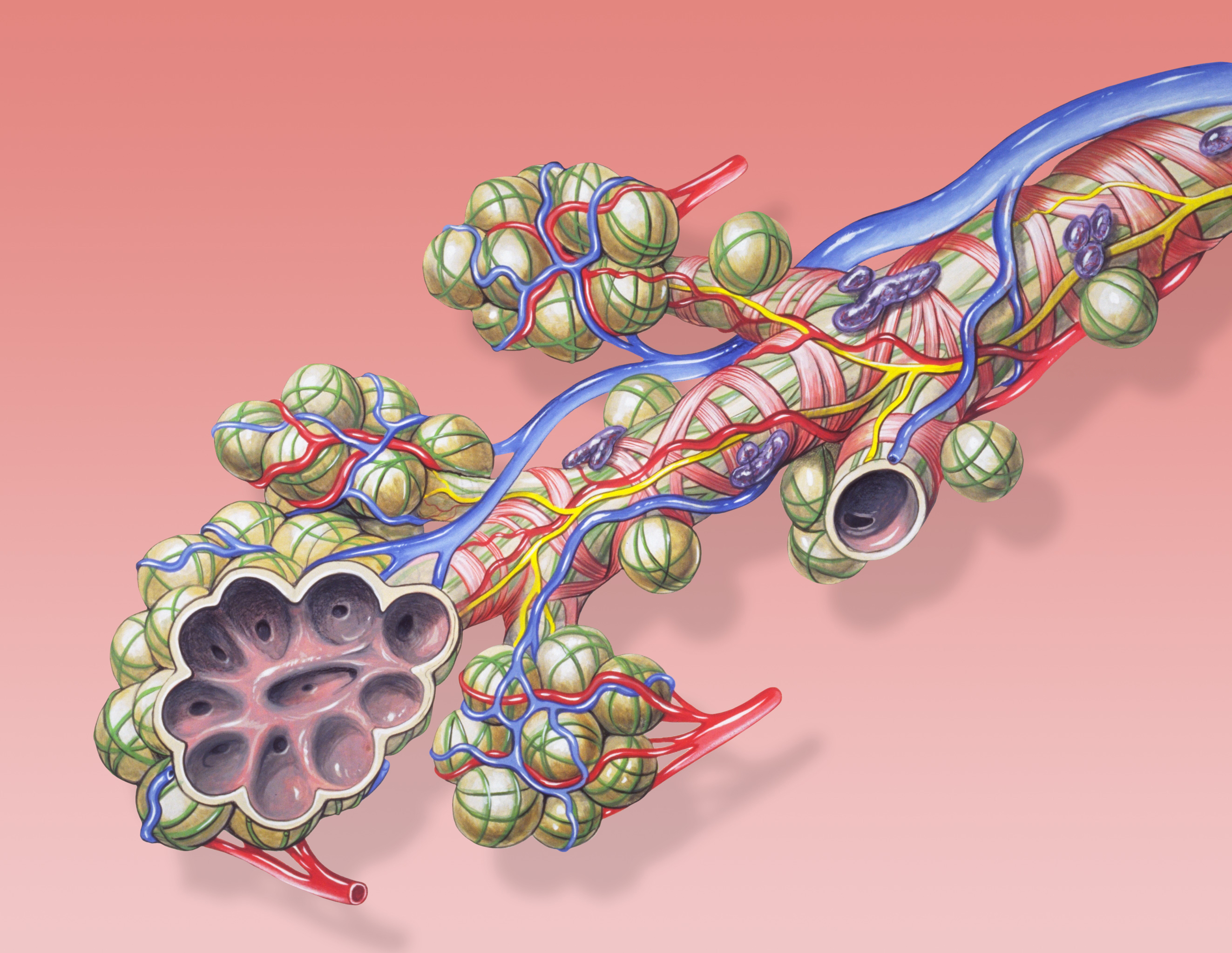
A pulmonary alveolus (plural: alveoli, from Latin ''alveolus'', "little cavity"), also known as an air sac or air space, is one of millions of hollow, distensible cup-shaped cavities in the lungs where oxygen is exchanged for carbon dioxide. Alveoli make up the functional tissue of the mammalian lungs known as the lung parenchyma, which takes up 90 percent of the total lung volume. Alveoli are first located in the respiratory bronchioles that mark the beginning of the respiratory zone. They are located sparsely in these bronchioles, line the walls of the alveolar ducts, and are more numerous in the blind-ended alveolar sacs. The acini are the basic units of respiration, with gas exchange taking place in all the alveoli present. The alveolar membrane is the gas exchange surface, surrounded by a network of capillaries. Across the membrane oxygen is diffused into the capillaries and carbon dioxide released from the capillaries into the alveoli to be breathed out. Alveoli ar ... [...More Info...] [...Related Items...] OR: [Wikipedia] [Google] [Baidu] |
Pulmonary Alveoli
A pulmonary alveolus (plural: alveoli, from Latin ''alveolus'', "little cavity"), also known as an air sac or air space, is one of millions of hollow, distensible cup-shaped cavities in the lungs where oxygen is exchanged for carbon dioxide. Alveoli make up the functional tissue of the mammalian lungs known as the lung parenchyma, which takes up 90 percent of the total lung volume. Alveoli are first located in the respiratory bronchioles that mark the beginning of the respiratory zone. They are located sparsely in these bronchioles, line the walls of the alveolar ducts, and are more numerous in the blind-ended alveolar sacs. The acini are the basic units of respiration, with gas exchange taking place in all the alveoli present. The alveolar membrane is the gas exchange surface, surrounded by a network of capillaries. Across the membrane oxygen is diffused into the capillaries and carbon dioxide released from the capillaries into the alveoli to be breathed out. Alveoli a ... [...More Info...] [...Related Items...] OR: [Wikipedia] [Google] [Baidu] |
Tuberous Sclerosis
Tuberous sclerosis complex (TSC) is a rare multisystem autosomal dominant genetic disease that causes non-cancerous tumours to grow in the brain and on other vital organs such as the kidneys, heart, liver, eyes, lungs and skin. A combination of symptoms may include seizures, intellectual disability, developmental delay, behavioral problems, skin abnormalities, lung disease, and kidney disease. TSC is caused by a mutation of either of two genes, '' TSC1'' and '' TSC2'', which code for the proteins hamartin and tuberin, respectively, with ''TSC2'' mutations accounting for the majority and tending to cause more severe symptoms. These proteins act as tumor growth suppressors, agents that regulate cell proliferation and differentiation. Prognosis is highly variable and depends on the symptoms, but life expectancy is normal for many. The prevalence of the disease is estimated to be 7 to 12 in 100,000. The disease is often abbreviated to tuberous sclerosis, which refers to ... [...More Info...] [...Related Items...] OR: [Wikipedia] [Google] [Baidu] |
Lymphangioleiomyomatosis
Lymphangioleiomyomatosis (LAM) is a rare, progressive and systemic disease that typically results in cystic lung destruction. It predominantly affects women, especially during childbearing years. The term sporadic LAM is used for patients with LAM not associated with tuberous sclerosis complex (TSC), while TSC-LAM refers to LAM that is associated with TSC. Signs and symptoms The average age of onset is the early to mid 30s. Exertional dyspnea (shortness of breath) and spontaneous pneumothorax (lung collapse) have been reported as the initial presentation of the disease in 49% and 46% of patients, respectively. Diagnosis is typically delayed 5 to 6 years. The condition is often misdiagnosed as asthma or chronic obstructive pulmonary disease. The first pneumothorax, or lung collapse, precedes the diagnosis of LAM in 82% of patients. The consensus clinical definition of LAM includes multiple symptoms: * Fatigue * Cough * Coughing up blood (rarely massive) * Chest pain * Chylous com ... [...More Info...] [...Related Items...] OR: [Wikipedia] [Google] [Baidu] |
Perivascular Epithelioid Cell Tumour
Perivascular epithelioid cell tumour, also known as PEComa or PEC tumour, is a family of mesenchymal tumours consisting of perivascular epithelioid cells (PECs). These are rare tumours that can occur in any part of the human body. The cell type from which these tumours originate remains unknown. Normally, no perivascular epitheloid cells exist; the name refers to the characteristics of the tumour when examined under the microscope. Establishing the malignant potential of these tumours remains challenging although criteria have been suggested; some PEComas display malignant features whereas others can cautiously be labeled as having 'uncertain malignant potential'. The most common tumours in the PEComa family are renal angiomyolipoma and pulmonary lymphangioleiomyomatosis, both of which are more common in patients with tuberous sclerosis complex. The genes responsible for this multi-system genetic disease have also been implicated in other PEComas. Many PEComa types shows a femal ... [...More Info...] [...Related Items...] OR: [Wikipedia] [Google] [Baidu] |
Sugar Tumor
Perivascular epithelioid cell tumour, also known as PEComa or PEC tumour, is a family of mesenchymal tumours consisting of perivascular epithelioid cells (PECs). These are rare tumours that can occur in any part of the human body. The cell type from which these tumours originate remains unknown. Normally, no perivascular epitheloid cells exist; the name refers to the characteristics of the tumour when examined under the microscope. Establishing the malignant potential of these tumours remains challenging although criteria have been suggested; some PEComas display malignant features whereas others can cautiously be labeled as having 'uncertain malignant potential'. The most common tumours in the PEComa family are renal angiomyolipoma and pulmonary lymphangioleiomyomatosis, both of which are more common in patients with tuberous sclerosis complex. The genes responsible for this multi-system genetic disease have also been implicated in other PEComas. Many PEComa types shows a ... [...More Info...] [...Related Items...] OR: [Wikipedia] [Google] [Baidu] |
Lung Biopsy
A lung biopsy is an interventional procedure performed to diagnose lung pathology by obtaining a small piece of lung which is examined under a microscope. Beyond microscopic examination for cellular morphology and architecture, special stains and cultures can be performed on the tissue obtained. Types A lung biopsy can be performed percutaneously (through the skin, typically guided by a CT Scan), via bronchoscopy with ultrasound guidance, or by surgery, either open or by video-assisted thoracoscopic surgery (VATS). Reasons to perform A lung biopsy is performed when a lung lesion is suspicious for lung cancer, or when cancer cannot be distinguished from another disease, such as aspergillosis. Lung biopsy also plays a role in the diagnosis of interstitial lung disease. Risks Any approach to lung biopsy risks causing a pneumothorax. Careful technique can limit this risk, which ranges from less than 1% to about 10%. The precise risk of pneumothorax depends on technique and on u ... [...More Info...] [...Related Items...] OR: [Wikipedia] [Google] [Baidu] |
Thoracoscopy
Thoracoscopy is a medical procedure involving internal examination, biopsy and/or resection/ drainage of disease or masses within the pleural cavity, usually with video assistance. Thoracoscopy may be performed either under general anaesthesia or under sedation with local anaesthetic. History Thoracoscopy was first performed by Sir Francis Cruise of the Mater Misericordiae Hospital in Dublin in conjunction with Dr Samuel Gordon in 1865. It was further developed by Hans Christian Jacobaeus, a Swedish internist in 1910 for the treatment of tuberculous intra-thoracic adhesions. He used a cystoscope to examine the thoracic cavity, developing his technique over the next twenty years. Today, thoracoscopy is performed using specialized thoracoscopes. These instruments include a light source and a lens for viewing and may have ports through which other instruments may be inserted for the purpose of tissue removal and manipulation. Video-assisted thoracoscopic surgery Video-ass ... [...More Info...] [...Related Items...] OR: [Wikipedia] [Google] [Baidu] |
Atypical Adenomatous Hyperplasia
Atypical adenomatous hyperplasia is a subtype of pneumocytic hyperplasia in the lung. It can be a precursor lesion of in situ adenocarcinoma of the lung (bronchioloalveolar carcinoma). In prostate tissue biopsy, it can be confused for adenocarcinoma of the prostate. The needle biopsy rate is less than 1%. Pathology Morphological differential diagnosis * Multifocal micronodular pneumocyte hyperplasia (MMPH) * in situ pulmonary adenocarcinoma (bronchioloalveolar carcinoma – BAC) Variants * multiple atypical adenomatous hyperplasia * disseminated AAH Histopathological images Image:Pulmonary_adeocaricnoma_(1)_localized_noninvasive_type.jpg Image:Pulmonary adeocaricnoma (2) localized noninvasive type.jpg Image:Pulmonary adeocaricnoma (3) localized noninvasive type.jpg Image:Pulmonary_adeocaricnoma_(4)_localized_noninvasive_type.jpg See also * EGFR * KRAS ''KRAS'' ( Kirsten rat sarcoma virus) is a gene that provides instructions for making a protein called K-Ras, ... [...More Info...] [...Related Items...] OR: [Wikipedia] [Google] [Baidu] |


