|
Mixed Connective Tissue Disease
Mixed connective tissue disease (MCTD) is a systemic autoimmune disease that shares characteristics with at least two other systemic autoimmune diseases, including Systemic scleroderma, systemic sclerosis (Ssc), systemic lupus erythematosus (SLE), polymyositis/dermatomyositis (PM/DM), and rheumatoid arthritis. The idea behind the "mixed" disease is that this specific autoantibody is also present in other autoimmune diseases such as systemic lupus erythematosus, polymyositis, scleroderma, etc. MCTD was characterized as an individual disease in 1972 by Sharp et al., and the term was introduced by Leroy in 1980. Some experts consider MCTD to be the same as undifferentiated connective tissue disease, but other experts specifically reject this idea because undifferentiated connective tissue disease is not necessarily associated with serum antibodies directed against the U1-RNP. Furthermore, MCTD is associated with a more clearly defined set of signs and symptoms. Signs and symptoms T ... [...More Info...] [...Related Items...] OR: [Wikipedia] [Google] [Baidu] |
Autoimmune Disease
An autoimmune disease is a condition that results from an anomalous response of the adaptive immune system, wherein it mistakenly targets and attacks healthy, functioning parts of the body as if they were foreign organisms. It is estimated that there are more than 80 recognized autoimmune diseases, with recent scientific evidence suggesting the existence of potentially more than 100 distinct conditions. Nearly any body part can be involved. Autoimmune diseases are a separate class from autoinflammatory diseases. Both are characterized by an immune system malfunction which may cause similar symptoms, such as rash, swelling, or fatigue, but the cardinal cause or mechanism of the diseases is different. A key difference is a malfunction of the innate immune system in autoinflammatory diseases, whereas in autoimmune diseases there is a malfunction of the adaptive immune system. Symptoms of autoimmune diseases can significantly vary, primarily based on the specific type of the d ... [...More Info...] [...Related Items...] OR: [Wikipedia] [Google] [Baidu] |
Sclerodactyly
Sclerodactyly is a localized thickening and tightness of the skin of the fingers or toes that yields a characteristic claw-like appearance and spindle shape of the affected digits, and renders them immobile or of limited mobility. The thickened, discolored patches of skin are called morphea, and may involve connective tissue below the skin, as well as muscle and other tissues. Sclerodactyly is often preceded by months or even years by Raynaud's phenomenon when it is part of systemic scleroderma. The term "sclerodactyly" comes . It is generally associated with systemic scleroderma and mixed connective tissue disease, and auto-immune disorders. Sclerodactyly is one component of the limited cutaneous form of systemic sclerosis (lcSSc), also known as CREST syndrome (CREST is an acronym that stands for calcinosis, Raynaud's phenomenon, esophageal dysmotility, sclerodactyly, and telangiectasia.) Sclerodactyly is also one component of Huriez Syndrome, along with palmoplantar ... [...More Info...] [...Related Items...] OR: [Wikipedia] [Google] [Baidu] |
Pleural Effusion
A pleural effusion is accumulation of excessive fluid in the pleural space, the potential space that surrounds each lung. Under normal conditions, pleural fluid is secreted by the parietal pleural capillaries at a rate of 0.6 millilitre per kilogram weight per hour, and is cleared by lymphatic absorption leaving behind only 5–15 millilitres of fluid, which helps to maintain a functional vacuum between the parietal and visceral pleurae. Excess fluid within the pleural space can impair inhalation, inspiration by upsetting the functional vacuum and hydrostatically increasing the resistance against lung expansion, resulting in a fully or partially collapsed lung. Various kinds of fluid can accumulate in the pleural space, such as serous fluid (hydrothorax), blood (hemothorax), pus (pyothorax, more commonly known as pleural empyema), chyle (chylothorax), or very rarely urine (urinothorax) or feces (coprothorax). When unspecified, the term "pleural effusion" normally refers to hydro ... [...More Info...] [...Related Items...] OR: [Wikipedia] [Google] [Baidu] |
Pulmonary Hypertension
Pulmonary hypertension (PH or PHTN) is a condition of increased blood pressure in the pulmonary artery, arteries of the lungs. Symptoms include dypsnea, shortness of breath, Syncope (medicine), fainting, tiredness, chest pain, pedal edema, swelling of the legs, and a fast heartbeat. The condition may make it difficult to exercise. Onset is typically gradual. According to the definition at the 6th World Symposium of Pulmonary Hypertension in 2018, a patient is deemed to have pulmonary hypertension if the pulmonary mean arterial pressure is greater than 20mmHg at rest, revised down from a purely arbitrary 25mmHg, and pulmonary vascular resistance (PVR) greater than 3 Wood units. The cause is often unknown. Risk factors include a family history, prior pulmonary embolism (blood clots in the lungs), HIV/AIDS, sickle cell disease, cocaine use, chronic obstructive pulmonary disease, sleep apnea, living at high altitudes, and problems with the mitral valve. The underlying mechanism typ ... [...More Info...] [...Related Items...] OR: [Wikipedia] [Google] [Baidu] |
Interstitial Lung Disease
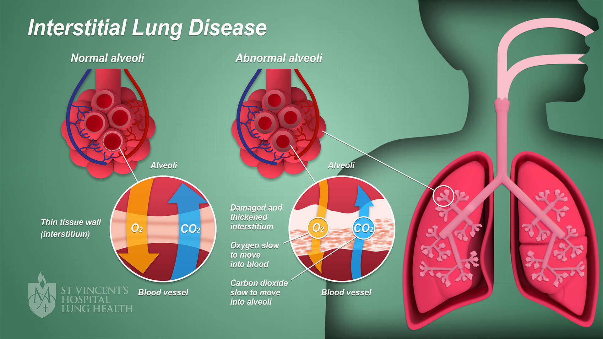
Interstitial lung disease (ILD), or diffuse parenchymal lung disease (DPLD), is a group of respiratory diseases affecting the interstitium (the tissue) and space around the alveoli (air sacs) of the lungs. It concerns alveolar epithelium, pulmonary capillary endothelium, basement membrane, and perivascular and perilymphatic tissues. It may occur when an injury to the lungs triggers an abnormal healing response. Ordinarily, the body generates just the right amount of tissue to repair damage, but in interstitial lung disease, the repair process is disrupted, and the tissue around the air sacs (alveoli) becomes scarred and thickened. This makes it more difficult for oxygen to pass into the bloodstream. The disease presents itself with the following symptoms: shortness of breath, nonproductive coughing, fatigue, and weight loss, which tend to develop slowly, over several months. The average rate of survival for someone with this disease is between three and five years. The term IL ... [...More Info...] [...Related Items...] OR: [Wikipedia] [Google] [Baidu] |
Arthritis Mutilans
Arthritis mutilans is a rare medical condition involving severe inflammation damaging the joints of the hands and feet, and resulting in deformation and problems with moving the affected areas; it can also affect the spine. As an uncommon arthropathy, arthritis mutilans was originally described as affecting the hands, feet, fingers, and/or toes, but can refer in general to severe derangement of any joint damaged by arthropathy. First described in modern medical literature by Marie and Leri in 1913, in the hands, arthritis mutilans is also known as opera glass hand (''la main en lorgnette'' in French), or chronic absorptive arthritis. Sometimes there is foot involvement in which toes shorten and on which painful calluses develop in a condition known as opera glass foot, or ''pied en lorgnette''. Signs and symptoms For a person with arthritis mutilans in the hands, the fingers become shortened by arthritis, and the shortening may become severe enough that the hand looks paw-like, w ... [...More Info...] [...Related Items...] OR: [Wikipedia] [Google] [Baidu] |
Swan Neck Deformity
Swan neck deformity is a deformed position of the finger, in which the joint closest to the fingertip is permanently bent toward the palm while the nearest joint to the palm is bent away from it ( DIP flexion with PIP hyperextension). It is commonly caused by injury, hypermobility or inflammatory conditions like rheumatoid arthritis or sometimes familial (congenital, like Ehlers–Danlos syndrome). Pathophysiology Swan neck deformity has many of possible causes arising from the DIP, PIP, or even the MCP joints. In all cases, there is a stretching of the volar plate at the PIP joint to allow hyperextension, plus some damage to the attachment of the extensor tendon to the base of the distal phalanx that produces a hyperflexed mallet finger. Duck bill deformity is a similar condition affecting the thumb (which cannot have true swan neck deformity because it does not have enough joints). Diagnosis Diagnosis of swan neck deformity is mainly clinical. MRI of the hand may sugges ... [...More Info...] [...Related Items...] OR: [Wikipedia] [Google] [Baidu] |
Boutonniere Deformity
Boutonniere deformity is a deformed position of the fingers or toes, in which the joint nearest the knuckle (the proximal interphalangeal joint, or PIP) is permanently bent toward the palm while the farthest joint (the distal interphalangeal joint, or DIP) is bent back away ( PIP flexion with DIP hyperextension). Causes include injury, inflammatory conditions like rheumatoid arthritis, psoriatic arthritis, and genetic conditions like Ehlers-Danlos syndrome. Pathophysiology This flexion deformity of the proximal interphalangeal joint is due to interruption of the central slip of the extensor tendon such that the lateral slips separate and the head of the proximal phalanx pops through the gap like a finger through a button hole (thus the name, from French ''boutonnière'' "button hole"). The distal joint is subsequently drawn into hyperextension because the two peripheral slips of the extensor tendon are stretched by the head of the proximal phalanx (note that the two periphera ... [...More Info...] [...Related Items...] OR: [Wikipedia] [Google] [Baidu] |
Arthritis
Arthritis is a general medical term used to describe a disorder that affects joints. Symptoms generally include joint pain and stiffness. Other symptoms may include redness, warmth, Joint effusion, swelling, and decreased range of motion of the affected joints. In certain types of arthritis, other organs such as the skin are also affected. Onset can be gradual or sudden. There are several types of arthritis. The most common forms are osteoarthritis (most commonly seen in weightbearing joints) and rheumatoid arthritis. Osteoarthritis usually occurs as an individual ages and often affects the hips, knees, shoulders, and fingers. Rheumatoid arthritis is an autoimmune disorder that often affects the hands and feet. Other types of arthritis include gout, lupus, and septic arthritis. These are inflammatory based types of rheumatic disease. Early treatment for arthritis commonly includes resting the affected joint and conservative measures such as heating or icing. Weight Weight ... [...More Info...] [...Related Items...] OR: [Wikipedia] [Google] [Baidu] |
Calcinosis Cutis
Calcinosis cutis is an uncommon condition marked by calcium buildup in the skin and subcutaneous tissues. Calcinosis cutis can range in intensity from little nodules in one area of the body to huge, crippling lesions affecting a vast portion of the body. Five kinds of the condition are typically distinguished: calciphylaxis, idiopathic calcification, iatrogenic calcification, dystrophic calcification, and metastatic calcification. Tumors, inflammation, varicose veins, infections, connective tissue disease, hyperphosphatemia, and hypercalcemia can all lead to calcinosis. Systemic sclerosis is linked to calcineuris cutis. Calcinosis is seen in Limited Cutaneous Systemic Sclerosis, also known as CREST syndrome (the "C" in CREST). Signs and symptoms Lesions might be more severe and widespread, or they can develop gradually and show no symptoms. The nodules may cause pain and hinder function in addition to having a variety of sizes and shapes. The underlying condition determine ... [...More Info...] [...Related Items...] OR: [Wikipedia] [Google] [Baidu] |
Telangiectasia
Telangiectasias (), also known as spider veins, are small dilated blood vessels that can occur near the surface of the skin or mucous membranes, measuring between 0.5 and 1 millimeter in diameter. These dilated blood vessels can develop anywhere on the body, but are commonly seen on the face around the nose, cheeks and chin. Dilated blood vessels can also develop on the legs, although when they occur on the legs, they often have underlying venous reflux or "hidden varicose veins" (see Venous hypertension section below). When found on the legs, they are found specifically on the upper thigh, below the knee joint and around the ankles. Many patients with spider veins seek the assistance of physicians who specialize in vein care or peripheral vascular disease. These physicians are called vascular surgeons or phlebologists. More recently, interventional radiologists have started treating venous problems. Some telangiectasias are due to developmental abnormalities that can close ... [...More Info...] [...Related Items...] OR: [Wikipedia] [Google] [Baidu] |
Discoid Lupus Erythematosus
Discoid lupus erythematosus is the most common type of chronic cutaneous lupus (CCLE), an autoimmune skin condition on the lupus erythematosus spectrum of illnesses. It presents with red, painful, inflamed and coin-shaped patches of skin with a scaly and crusty appearance, most often on the scalp, cheeks, and ears. Hair loss may occur if the lesions are on the scalp.James, William; Berger, Timothy; Elston, Dirk (2005). ''Andrews' Diseases of the Skin: Clinical Dermatology''. (10th ed.) Saunders. Chapter 8. . The lesions can then develop severe scarring, and the centre areas may appear lighter in color with a rim darker than the normal skin. These lesions can last for years without treatment. Patients with systemic lupus erythematous develop discoid lupus lesions with some frequency. However, patients who present initially with discoid lupus infrequently develop systemic lupus. Discoid lupus can be divided into localized, generalized, and childhood discoid lupus. The lesions are d ... [...More Info...] [...Related Items...] OR: [Wikipedia] [Google] [Baidu] |


