|
Minimal Change Nephropathy
Minimal change disease (MCD), also known as lipoid nephrosis or nil disease, among others, is a disease affecting the kidneys which causes nephrotic syndrome. Nephrotic syndrome leads to the loss of significant amounts of protein to the urine (proteinuria), which causes the widespread edema (soft tissue swelling) and impaired kidney function commonly experienced by those affected by the disease. It is most common in children and has a peak incidence at 2 to 6 years of age. MCD is responsible for 10–25% of nephrotic syndrome cases in adults. It is also the most common cause of nephrotic syndrome of unclear cause (idiopathic) in children. Signs and symptoms The clinical signs of minimal change disease are proteinuria (abnormal excretion of proteins, mainly albumin, into the urine), edema (swelling of soft tissues as a consequence of water retention), weight gain, and hypoalbuminemia (low serum albumin). These signs are referred to collectively as nephrotic syndrome. The firs ... [...More Info...] [...Related Items...] OR: [Wikipedia] [Google] [Baidu] |
Podocyte
Podocytes are cells in Bowman's capsule in the kidneys that wrap around capillaries of the glomerulus. Podocytes make up the epithelial lining of Bowman's capsule, the third layer through which filtration of blood takes place. Bowman's capsule filters the blood, retaining large molecules such as proteins while smaller molecules such as water, salts, and sugars are filtered as the first step in the formation of urine. Although various viscera have epithelial layers, the name visceral epithelial cells usually refers specifically to podocytes, which are specialized epithelial cells that reside in the visceral layer of the capsule. The podocytes have long primary processes called ''trabeculae'' that form secondary processes known as ''pedicels'' or foot processes (for which the cells are named '' podo-'' + '' -cyte''). The pedicels wrap around the capillaries and leave slits between them. Blood is filtered through these slits, each known as a filtration slit, slit diaphragm, or sl ... [...More Info...] [...Related Items...] OR: [Wikipedia] [Google] [Baidu] |
Periorbital Edema
The periorbita is the area around the orbit In celestial mechanics, an orbit (also known as orbital revolution) is the curved trajectory of an object such as the trajectory of a planet around a star, or of a natural satellite around a planet, or of an artificial satellite around an .... Sometimes it refers specifically to the layer of tissue surrounding the orbit that consists of periosteum. However, it may refer to anything that is around the orbit, such as in periorbital cellulitis. References {{Authority control Tissues (biology) Human eye anatomy ... [...More Info...] [...Related Items...] OR: [Wikipedia] [Google] [Baidu] |
Lymphokine
Lymphokines are a subset of cytokines that are produced by a type of immune cell known as a lymphocyte. They are protein mediators typically produced by T cells to direct the immune system response by signaling between its cells. Lymphokines have many roles, including the attraction of other immune cells, including macrophages and other lymphocytes, to an infected site and their subsequent activation to prepare them to mount an immune response. Circulating lymphocytes can detect a very small concentration of lymphokine and then move up the concentration gradient towards where the immune response is required. Lymphokines aid B cells to produce antibodies. Important lymphokines secreted by the T helper cell include:Guyton, AC; Hall, JE. (2006) Medical Physiology, Elsevier Saunders. 11th edition, pp.447. * Interleukin 2 Interleukin-2 (IL-2) is an interleukin, which is a type of cytokine signaling molecule forming part of the immune system. It is a 15.5–16 Dalton (unit) ... [...More Info...] [...Related Items...] OR: [Wikipedia] [Google] [Baidu] |
Complement (biology)
The complement system, also known as complement cascade, is a part of the humoral, innate immune system and enhances (complements) the ability of antibodies and phagocytic cells to clear microbes and damaged cells from an organism, promote inflammation, and attack the pathogen's cell membrane. Despite being part of the innate immune system, the complement system can be recruited and brought into action by antibodies generated by the adaptive immune system. The complement system consists of a number of small, inactive, liver synthesized protein precursors circulating in the blood. When stimulated by one of several triggers, proteases in the system cleave specific proteins to release cytokines and initiate an amplifying cascade of further cleavages. The end result of this ''complement activation'' or ''complement fixation'' cascade is stimulation of phagocytes to clear foreign and damaged material, inflammation to attract additional phagocytes, and activation of the cell-killing ... [...More Info...] [...Related Items...] OR: [Wikipedia] [Google] [Baidu] |
Electron Microscopy
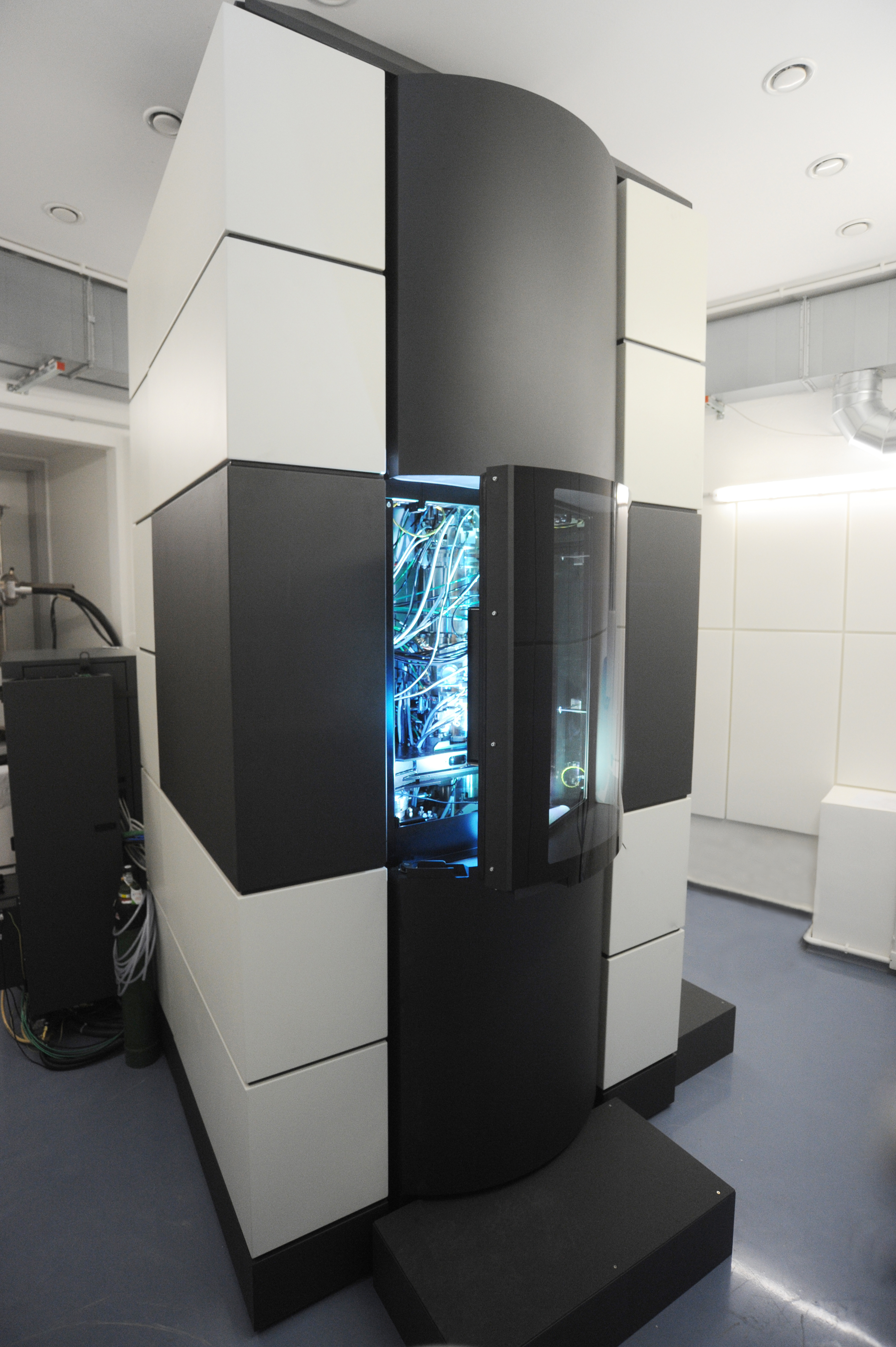
An electron microscope is a microscope that uses a beam of electrons as a source of illumination. It uses electron optics that are analogous to the glass lenses of an optical light microscope to control the electron beam, for instance focusing it to produce magnified images or electron diffraction patterns. As the wavelength of an electron can be up to 100,000 times smaller than that of visible light, electron microscopes have a much higher resolution of about 0.1 nm, which compares to about 200 nm for light microscopes. ''Electron microscope'' may refer to: * Transmission electron microscope (TEM) where swift electrons go through a thin sample * Scanning transmission electron microscope (STEM) which is similar to TEM with a scanned electron probe * Scanning electron microscope (SEM) which is similar to STEM, but with thick samples * Electron microprobe similar to a SEM, but more for chemical analysis * Low-energy electron microscope (LEEM), used to image surfaces * ... [...More Info...] [...Related Items...] OR: [Wikipedia] [Google] [Baidu] |
Immunoglobulins
An antibody (Ab) or immunoglobulin (Ig) is a large, Y-shaped protein belonging to the immunoglobulin superfamily which is used by the immune system to identify and neutralize antigens such as bacteria and viruses, including those that cause disease. Each individual antibody recognizes one or more specific antigens, and antigens of virtually any size and chemical composition can be recognized. Antigen literally means "antibody generator", as it is the presence of an antigen that drives the formation of an antigen-specific antibody. Each of the branching chains comprising the "Y" of an antibody contains a paratope that specifically binds to one particular epitope on an antigen, allowing the two molecules to bind together with precision. Using this mechanism, antibodies can effectively "tag" the antigen (or a microbe or an infected cell bearing such an antigen) for attack by cells of the immune system, or can neutralize it directly (for example, by blocking a part of a virus that i ... [...More Info...] [...Related Items...] OR: [Wikipedia] [Google] [Baidu] |
Immunofluorescence
Immunofluorescence (IF) is a light microscopy-based technique that allows detection and localization of a wide variety of target biomolecules within a cell or tissue at a quantitative level. The technique utilizes the binding specificity of antibodies and antigens. The specific region an antibody recognizes on an antigen is called an epitope. Several antibodies can recognize the same epitope but differ in their binding affinity. The antibody with the higher affinity for a specific epitope will surpass antibodies with a lower affinity for the same epitope. By conjugating the antibody to a fluorophore, the position of the target biomolecule is visualized by exciting the fluorophore and measuring the emission of light in a specific predefined wavelength using a fluorescence microscope. It is imperative that the binding of the fluorophore to the antibody itself does not interfere with the immunological specificity of the antibody or the binding capacity of its antigen. Immunofluore ... [...More Info...] [...Related Items...] OR: [Wikipedia] [Google] [Baidu] |
Glomerulus (kidney)
The glomerulus (: glomeruli) is a network of small blood vessels (capillaries) known as a ''tuft'', located at the beginning of a nephron in the kidney. Each of the two kidneys contains about one million nephrons. The tuft is structurally supported by the mesangium (the space between the blood vessels), composed of Intraglomerular mesangial cell, intraglomerular mesangial cells. The blood is filtered across the capillary walls of this tuft through the glomerular filtration barrier, which yields its Ultrafiltration (renal), filtrate of water and soluble substances to a cup-like sac known as Bowman's capsule. The filtrate then enters the Nephron#Renal tubule, renal tubule of the nephron. The glomerulus receives its blood supply from an afferent arteriole of the renal arterial circulation. Unlike most capillary beds, the glomerular capillaries exit into efferent arterioles rather than venules. The resistance of the efferent arterioles causes sufficient hydrostatic pressure within th ... [...More Info...] [...Related Items...] OR: [Wikipedia] [Google] [Baidu] |
Pathognomonic
Pathognomonic (synonym ''pathognomic'') is a term, often used in medicine, that means "characteristic for a particular disease". A pathognomonic sign is a particular sign whose presence means that a particular disease is present beyond any doubt. The absence of a pathognomonic sign does not rule out the disease. Labelling a sign or symptom "pathognomonic" represents a marked intensification of a "diagnostic" sign or symptom. The word is an adjective of Greek origin derived from πάθος ''pathos'' 'disease' and γνώμων ''gnomon'' 'indicator' (from γιγνώσκω ''gignosko'' 'I know, I recognize'). Practical use While some findings may be classic, typical or highly suggestive in a certain condition, they may not occur ''uniquely'' in this condition and therefore may not directly imply a specific diagnosis. A pathognomonic sign or symptom has very high positive predictive value and high specificity but does not need to have high sensitivity: for example it can som ... [...More Info...] [...Related Items...] OR: [Wikipedia] [Google] [Baidu] |
Mesangial
The glomerulus (: glomeruli) is a network of small blood vessels (capillaries) known as a ''tuft'', located at the beginning of a nephron in the kidney. Each of the two kidneys contains about one million nephrons. The tuft is structurally supported by the mesangium (the space between the blood vessels), composed of intraglomerular mesangial cells. The blood is filtered across the capillary walls of this tuft through the glomerular filtration barrier, which yields its filtrate of water and soluble substances to a cup-like sac known as Bowman's capsule. The filtrate then enters the renal tubule of the nephron. The glomerulus receives its blood supply from an afferent arteriole of the renal arterial circulation. Unlike most capillary beds, the glomerular capillaries exit into efferent arterioles rather than venules. The resistance of the efferent arterioles causes sufficient hydrostatic pressure within the glomerulus to provide the force for ultrafiltration. The glomerulus and its ... [...More Info...] [...Related Items...] OR: [Wikipedia] [Google] [Baidu] |
Light Microscope
The optical microscope, also referred to as a light microscope, is a type of microscope that commonly uses visible spectrum, visible light and a system of lens (optics), lenses to generate magnified images of small objects. Optical microscopes are the oldest design of microscope and were possibly invented in their present compound form in the 17th century. Basic optical microscopes can be very simple, although many complex designs aim to improve optical resolution, resolution and sample contrast (vision), contrast. The object is placed on a stage and may be directly viewed through one or two eyepieces on the microscope. In high-power microscopes, both eyepieces typically show the same image, but with a stereo microscope, slightly different images are used to create a 3-D effect. A camera is typically used to capture the image (micrograph). The sample can be lit in a variety of ways. Transparent objects can be lit from below and solid objects can be lit with light coming through ... [...More Info...] [...Related Items...] OR: [Wikipedia] [Google] [Baidu] |
Blood Clots
A thrombus ( thrombi) is a solid or semisolid aggregate from constituents of the blood (platelets, fibrin, red blood cells, white blood cells) within the circulatory system during life. A blood clot is the final product of the blood coagulation step in hemostasis in or out of the circulatory system. There are two components to a thrombus: aggregated platelets and red blood cells that form a plug, and a mesh of cross-linked fibrin protein. The substance making up a thrombus is sometimes called cruor. A thrombus is a healthy response to injury intended to stop and prevent further bleeding, but can be harmful in thrombosis, when a clot obstructs blood flow through a healthy blood vessel in the circulatory system. In the microcirculation consisting of the very small and smallest blood vessels the capillaries, tiny thrombi known as microclots can obstruct the flow of blood in the capillaries. This can cause a number of problems particularly affecting the alveoli in the lungs ... [...More Info...] [...Related Items...] OR: [Wikipedia] [Google] [Baidu] |


