|
Minicell
In bacteriology, minicells are bacterial cells that are smaller than usual. The first minicells reported were from a strain of ''Escherichia coli'' that had a mutation in the Min System that lead to mis-localization of the septum during cell division and the production of cells of random sizes. Generation of minicells The first report of minicells in the scientific literature dates to 1930., but the first use of the name "minicell" dates to 1967 Minicells of a variety of gram negative and gram positive bacteria, including ''Escherichia coli'' and ''Salmonella enterica'', have been reported, but in principle, minicells could be generated for any bacterial species that can be genetically edited. Minicells can not reproduce because they do not contain a full copy of the genome. Normal role of minicells in bacteriology Scientists hypothesize that minicells are produced by normal bacteria in times of stress so that damaged areas of the cell can be expelled. Applications of ... [...More Info...] [...Related Items...] OR: [Wikipedia] [Google] [Baidu] |
Min System
The Min System is a mechanism composed of three proteins MinC, MinD, and MinE used by ''E. coli'' as a means of properly localizing the septum prior to cell division. Each component participates in generating a dynamic oscillation of FtsZ protein inhibition between the two bacterial poles to precisely specify the mid-zone of the cell, allowing the cell to accurately divide in two. This system is known to function in conjunction with a second negative regulatory system, the nucleoid occlusion system (NO), to ensure proper spatial and temporal regulation of chromosomal segregation and division. History The initial discovery of this family of proteins is attributed to Adler et al. (1967). First identified as '' E. coli'' mutants that could not produce a properly localized septum, resulting in the generation of minicells due to mislocalized cell division occurring near the bacterial poles. This caused miniature vesicles to pinch off, void of essential molecular constituents per ... [...More Info...] [...Related Items...] OR: [Wikipedia] [Google] [Baidu] |
Bacterial Strain
In biology, a strain is a genetic variant, a subtype or a culture within a biological species. Strains are often seen as inherently artificial concepts, characterized by a specific intent for genetic isolation. This is most easily observed in microbiology where strains are derived from a single cell colony and are typically quarantined by the physical constraints of a Petri dish. Strains are also commonly referred to within virology, botany, and with rodents used in experimental studies. Microbiology and virology It has been said that "there is no universally accepted definition for the terms 'strain', ' variant', and 'isolate' in the virology community, and most virologists simply copy the usage of terms from others". A strain is a genetic variant or subtype of a microorganism (e.g., a virus, bacterium or fungus). For example, a "flu strain" is a certain biological form of the influenza or "flu" virus. These flu strains are characterized by their differing isoforms of surfa ... [...More Info...] [...Related Items...] OR: [Wikipedia] [Google] [Baidu] |
Escherichia Coli
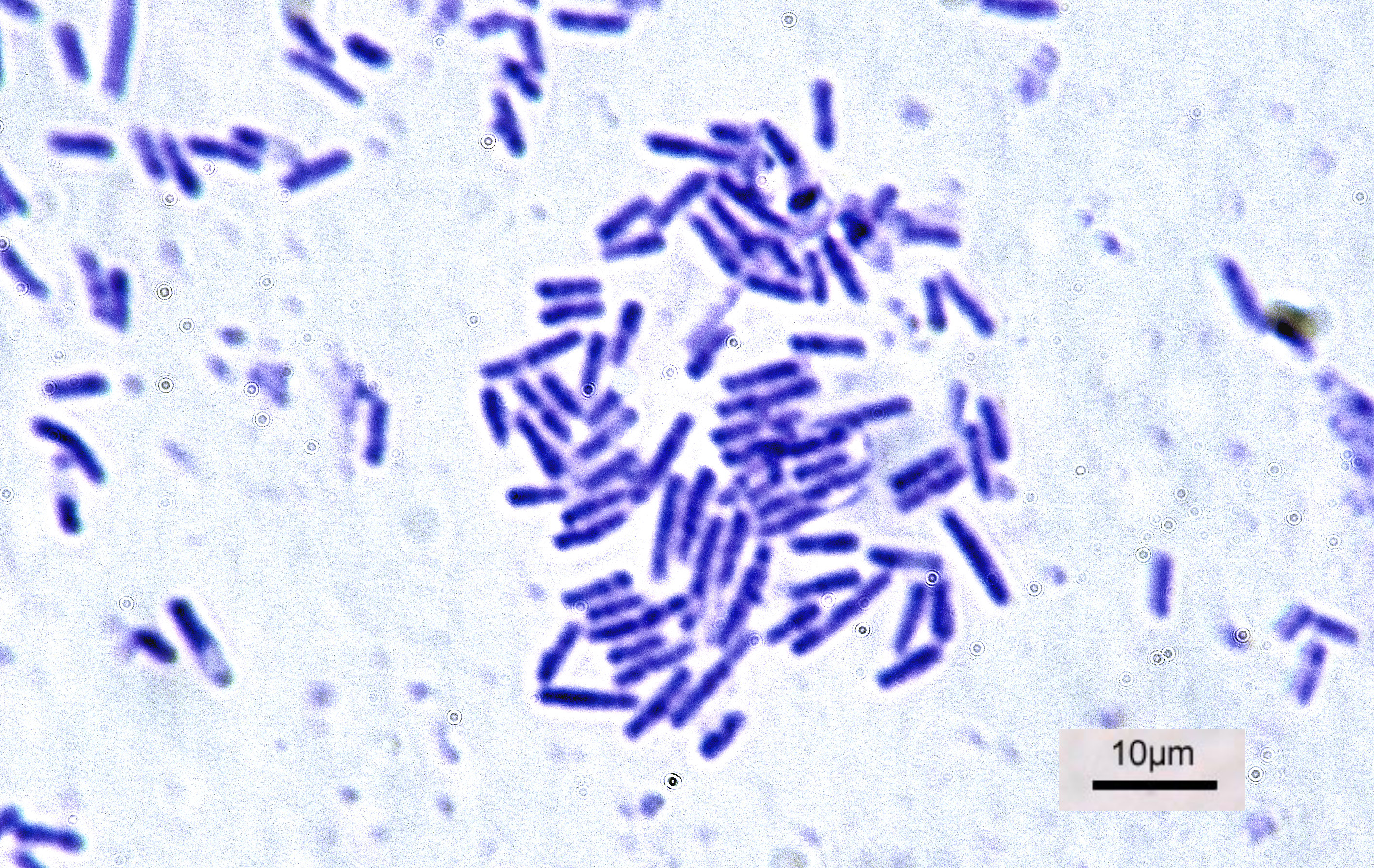
''Escherichia coli'' (),Wells, J. C. (2000) Longman Pronunciation Dictionary. Harlow ngland Pearson Education Ltd. also known as ''E. coli'' (), is a Gram-negative, facultative anaerobic, rod-shaped, coliform bacterium of the genus '' Escherichia'' that is commonly found in the lower intestine of warm-blooded organisms. Most ''E. coli'' strains are harmless, but some serotypes ( EPEC, ETEC etc.) can cause serious food poisoning in their hosts, and are occasionally responsible for food contamination incidents that prompt product recalls. Most strains do not cause disease in humans and are part of the normal microbiota of the gut; such strains are harmless or even beneficial to humans (although these strains tend to be less studied than the pathogenic ones). For example, some strains of ''E. coli'' benefit their hosts by producing vitamin K2 or by preventing the colonization of the intestine by pathogenic bacteria. These mutually beneficial relationships between ''E. co ... [...More Info...] [...Related Items...] OR: [Wikipedia] [Google] [Baidu] |
Septum
In biology, a septum (Latin for ''something that encloses''; plural septa) is a wall, dividing a cavity or structure into smaller ones. A cavity or structure divided in this way may be referred to as septate. Examples Human anatomy * Interatrial septum, the wall of tissue that is a sectional part of the left and right atria of the heart * Interventricular septum, the wall separating the left and right ventricles of the heart * Lingual septum, a vertical layer of fibrous tissue that separates the halves of the tongue. * Nasal septum: the cartilage wall separating the nostrils of the nose * Alveolar septum: the thin wall which separates the alveoli from each other in the lungs * Orbital septum, a palpebral ligament in the upper and lower eyelids * Septum pellucidum or septum lucidum, a thin structure separating two fluid pockets in the brain * Uterine septum, a malformation of the uterus * Vaginal septum, a lateral or transverse partition inside the vagina * Interm ... [...More Info...] [...Related Items...] OR: [Wikipedia] [Google] [Baidu] |
Gram Negative
The gram (originally gramme; SI unit symbol g) is a unit of mass in the International System of Units (SI) equal to one one thousandth of a kilogram. Originally defined as of 1795 as "the absolute weight of a volume of pure water equal to the cube of the hundredth part of a metre melting ice", the defining temperature (~0 °C) was later changed to 4 °C, the temperature of maximum density of water. However, by the late 19th century, there was an effort to make the Base unit (measurement), base unit the kilogram and the gram a derived unit. In 1960, the new International System of Units defined a ''gram'' as one one-thousandth of a kilogram (i.e., one gram is Scientific notation, 1×10⁻³ kg). The kilogram, as of 2019, is defined by the International Bureau of Weights and Measures from the fixed numerical value of the Planck constant (), which is kg⋅m2⋅s−1. Official SI symbol The only unit symbol for gram that is recognised by the International Sys ... [...More Info...] [...Related Items...] OR: [Wikipedia] [Google] [Baidu] |
Gram Positive
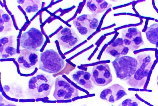
In bacteriology, gram-positive bacteria are bacteria that give a positive result in the Gram stain test, which is traditionally used to quickly classify bacteria into two broad categories according to their type of cell wall. Gram-positive bacteria take up the crystal violet stain used in the test, and then appear to be purple-coloured when seen through an optical microscope. This is because the thick peptidoglycan layer in the bacterial cell wall retains the stain after it is washed away from the rest of the sample, in the decolorization stage of the test. Conversely, gram-negative bacteria cannot retain the violet stain after the decolorization step; alcohol used in this stage degrades the outer membrane of gram-negative cells, making the cell wall more porous and incapable of retaining the crystal violet stain. Their peptidoglycan layer is much thinner and sandwiched between an inner cell membrane and a bacterial outer membrane, causing them to take up the counterstain ... [...More Info...] [...Related Items...] OR: [Wikipedia] [Google] [Baidu] |
Salmonella Enterica
''Salmonella enterica'' (formerly ''Salmonella choleraesuis'') is a rod-headed, flagellate, facultative anaerobic, Gram-negative bacterium and a species of the genus '' Salmonella''. A number of its serovars are serious human pathogens. Epidemiology Most cases of salmonellosis are caused by food infected with ''S. enterica'', which often infects cattle and poultry, though other animals such as domestic cats and hamsters have also been shown to be sources of infection in humans. Investigations of vacuum cleaner bags have shown that households can act as a reservoir of the bacterium; this is more likely if the household has contact with an infection source (i.e., members working with cattle or in a veterinary clinic). Raw chicken eggs and goose eggs can harbor ''S. enterica'', initially in the egg whites, although most eggs are not infected. As the egg ages at room temperature, the yolk membrane begins to break down and ''S. enterica'' can spread into the yolk. Refrigerati ... [...More Info...] [...Related Items...] OR: [Wikipedia] [Google] [Baidu] |
Ultrastructure
Ultrastructure (or ultra-structure) is the architecture of cells and biomaterials that is visible at higher magnifications than found on a standard optical light microscope. This traditionally meant the resolution and magnification range of a conventional transmission electron microscope (TEM) when viewing biological specimens such as cells, tissue, or organs. Ultrastructure can also be viewed with scanning electron microscopy and super-resolution microscopy, although TEM is a standard histology technique for viewing ultrastructure. Such cellular structures as organelles, which allow the cell to function properly within its specified environment, can be examined at the ultrastructural level. Ultrastructure, along with molecular phylogeny, is a reliable phylogenetic way of classifying organisms. Features of ultrastructure are used industrially to control material properties and promote biocompatibility. History In 1931, German engineers Max Knoll and Ernst Ruska invented ... [...More Info...] [...Related Items...] OR: [Wikipedia] [Google] [Baidu] |
Bacteria
Bacteria (; singular: bacterium) are ubiquitous, mostly free-living organisms often consisting of one biological cell. They constitute a large domain of prokaryotic microorganisms. Typically a few micrometres in length, bacteria were among the first life forms to appear on Earth, and are present in most of its habitats. Bacteria inhabit soil, water, acidic hot springs, radioactive waste, and the deep biosphere of Earth's crust. Bacteria are vital in many stages of the nutrient cycle by recycling nutrients such as the fixation of nitrogen from the atmosphere. The nutrient cycle includes the decomposition of dead bodies; bacteria are responsible for the putrefaction stage in this process. In the biological communities surrounding hydrothermal vents and cold seeps, extremophile bacteria provide the nutrients needed to sustain life by converting dissolved compounds, such as hydrogen sulphide and methane, to energy. Bacteria also live in symbiotic and parasitic re ... [...More Info...] [...Related Items...] OR: [Wikipedia] [Google] [Baidu] |
Electron Cryotomography
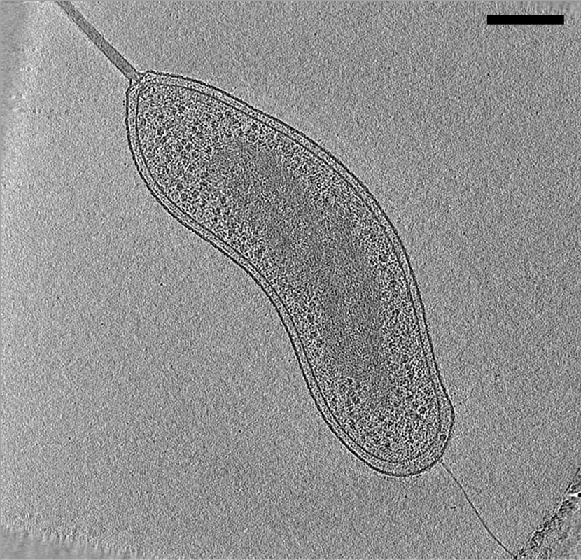
Electron cryotomography (CryoET) is an imaging technique used to produce high-resolution (~1–4 nm) three-dimensional views of samples, often (but not limited to) biological macromolecules and Cell (biology), cells. CryoET is a specialized application of transmission electron cryomicroscopy (CryoTEM) in which samples are imaged as they are tilted, resulting in a series of 2D images that can be combined to produce a 3D reconstruction, similar to a CT scan of the human body. In contrast to other electron tomography techniques, samples are imaged under Cryogenics, cryogenic conditions (< −150 °C). For cellular material, the structure is immobilized in non-crystalline, vitreous ice, allowing them to be imaged without dehydration or chemical fixation, which would otherwise disrupt or distort biological structures. Description of technique [...More Info...] [...Related Items...] OR: [Wikipedia] [Google] [Baidu] |
Transmission Electron Microscopy
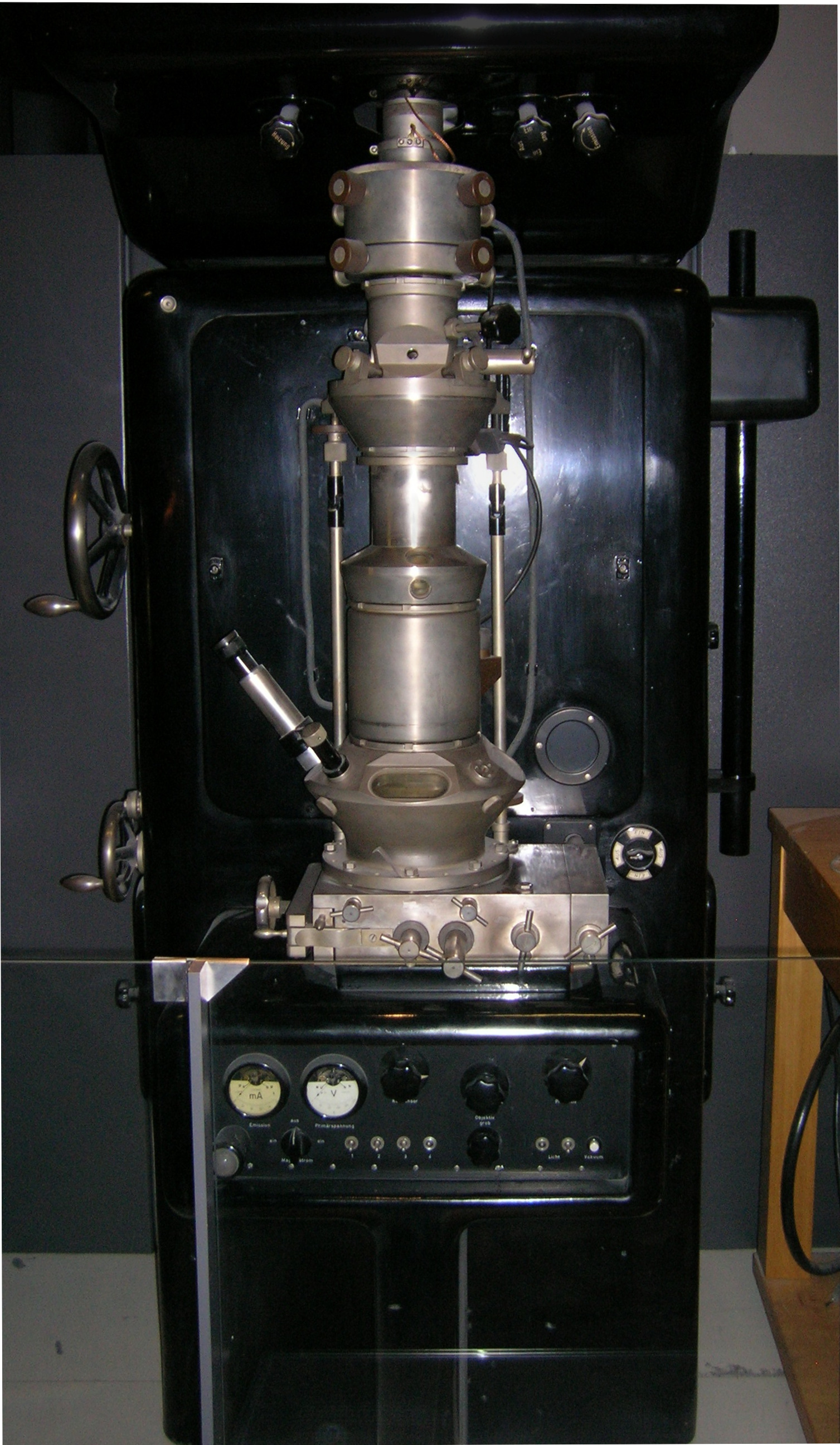
Transmission electron microscopy (TEM) is a microscopy technique in which a beam of electrons is transmitted through a specimen to form an image. The specimen is most often an ultrathin section less than 100 nm thick or a suspension on a grid. An image is formed from the interaction of the electrons with the sample as the beam is transmitted through the specimen. The image is then magnified and focused onto an imaging device, such as a fluorescent screen, a layer of photographic film, or a sensor such as a scintillator attached to a charge-coupled device. Transmission electron microscopes are capable of imaging at a significantly higher resolution than light microscopes, owing to the smaller de Broglie wavelength of electrons. This enables the instrument to capture fine detail—even as small as a single column of atoms, which is thousands of times smaller than a resolvable object seen in a light microscope. Transmission electron microscopy is a major analytical method ... [...More Info...] [...Related Items...] OR: [Wikipedia] [Google] [Baidu] |
.jpg)
