|
Miliary Tuberculosis
Miliary tuberculosis is a form of tuberculosis that is characterized by a wide dissemination into the human body and by the tiny size of the lesions (1–5 mm). Its name comes from a distinctive pattern seen on a chest radiograph of many tiny spots distributed throughout the lung fields with the appearance similar to millet seeds—thus the term "miliary" tuberculosis. Miliary TB may infect any number of organs, including the lungs, liver, and spleen. Signs and symptoms Patients with miliary tuberculosis often experience non-specific signs, such as coughing and enlarged lymph nodes. Miliary tuberculosis can also present with enlarged liver (40% of cases), enlarged spleen (15%), inflammation of the pancreas (<5%), and multiple organ dysfunction with |
Infectious Disease (medical Speciality)
Infectious diseases (ID), also known as infectiology, is a medical specialty dealing with the diagnosis and treatment of infections. An infectious diseases specialist's practice consists of managing nosocomial ( healthcare-acquired) infections or community-acquired infections. An ID specialist investigates and determines the cause of a disease (bacteria, virus, parasite, fungus or prions). Once the cause is known, an ID specialist can then run various tests to determine the best drug to treat the disease. While infectious diseases have always been around, the infectious disease specialty did not exist until the late 1900s after scientists and physicians in the 19th century paved the way with research on the sources of infectious disease and the development of vaccines. Scope Infectious diseases specialists typically serve as consultants to other physicians in cases of complex infections, and often manage patients with HIV/AIDS and other forms of immunodeficiency. Although many co ... [...More Info...] [...Related Items...] OR: [Wikipedia] [Google] [Baidu] |
Macrophage
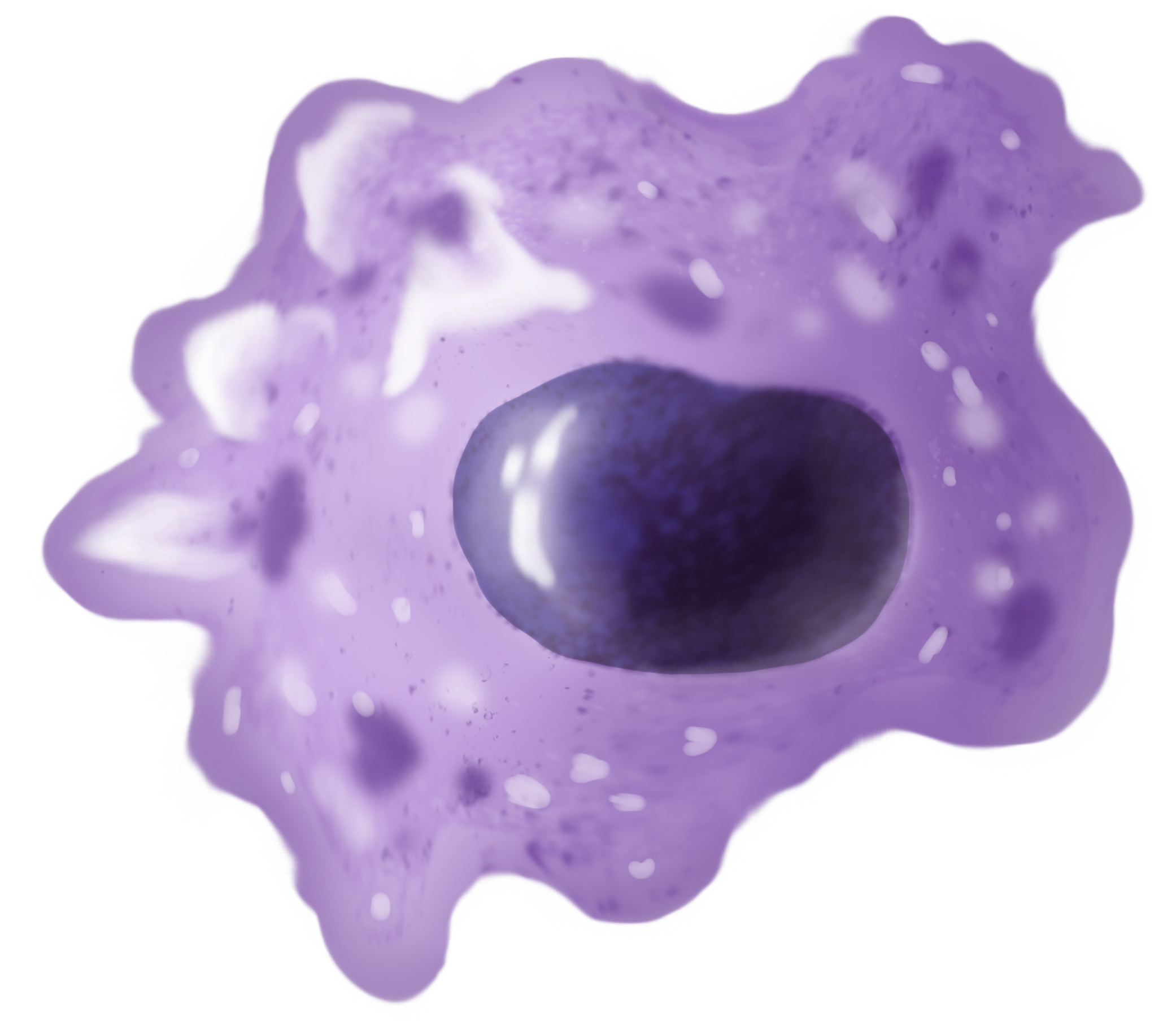
Macrophages (; abbreviated MPhi, φ, MΦ or MP) are a type of white blood cell of the innate immune system that engulf and digest pathogens, such as cancer cells, microbes, cellular debris and foreign substances, which do not have proteins that are specific to healthy body cells on their surface. This self-protection method can be contrasted with that employed by Natural killer cell, Natural Killer cells. This process of engulfment and digestion is called phagocytosis; it acts to defend the host against infection and injury. Macrophages are found in essentially all tissues, where they patrol for potential pathogens by amoeboid movement. They take various forms (with various names) throughout the body (e.g., histiocytes, Kupffer cells, alveolar macrophages, microglia, and others), but all are part of the mononuclear phagocyte system. Besides phagocytosis, they play a critical role in nonspecific defense (innate immunity) and also help initiate specific defense mechanisms (adapti ... [...More Info...] [...Related Items...] OR: [Wikipedia] [Google] [Baidu] |
Tuberculous Meningitis
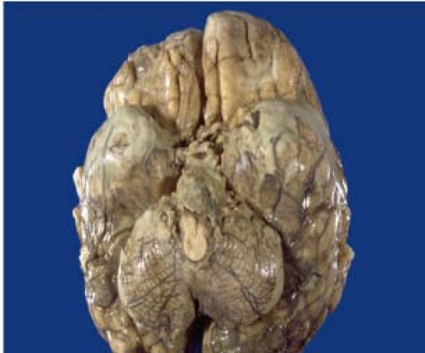
Tuberculous meningitis, also known as TB meningitis or tubercular meningitis, is a specific type of bacterial meningitis caused by the ''Mycobacterium tuberculosis'' infection of the meninges—the system of membranes which envelop the central nervous system. Signs and symptoms Fever and headache are the cardinal features; confusion is a late feature and coma bears a poor prognosis. Meningism is absent in a fifth of patients with TB meningitis. Patients may also have focal neurological deficits. Causes ''Mycobacterium tuberculosis'' of the meninges is the cardinal feature and the inflammation is concentrated towards the base of the brain. When the inflammation is in the brain stem subarachnoid area, cranial nerve roots may be affected. The symptoms will mimic those of space-occupying lesions. Blood-borne spread certainly occurs, presumably by crossing the blood–brain barrier, but a proportion of patients may get TB meningitis from rupture of a cortical focus in the brain; a ... [...More Info...] [...Related Items...] OR: [Wikipedia] [Google] [Baidu] |
Electrocardiography
Electrocardiography is the process of producing an electrocardiogram (ECG or EKG), a recording of the heart's electrical activity through repeated cardiac cycles. It is an electrogram of the heart which is a graph of voltage versus time of the electrical activity of the heart using electrodes placed on the skin. These electrodes detect the small electrical changes that are a consequence of cardiac muscle depolarization followed by repolarization during each cardiac cycle (heartbeat). Changes in the normal ECG pattern occur in numerous cardiac abnormalities, including: * Cardiac rhythmicity, Cardiac rhythm disturbances, such as atrial fibrillation and ventricular tachycardia; * Inadequate coronary artery blood flow, such as myocardial ischemia and myocardial infarction; * and electrolyte disturbances, such as hypokalemia. Traditionally, "ECG" usually means a 12-lead ECG taken while lying down as discussed below. However, other devices can record the electrical activity of ... [...More Info...] [...Related Items...] OR: [Wikipedia] [Google] [Baidu] |
Fundoscopy
Ophthalmoscopy, also called funduscopy, is a test that allows a health professional to see inside the fundus of the eye and other structures using an ophthalmoscope (or funduscope). It is done as part of an eye examination and may be done as part of a routine physical examination. It is crucial in determining the health of the retina, optic disc, and vitreous humor. The pupil is a hole through which the eye's interior can be viewed. For better viewing, the pupil can be opened wider (dilated; mydriasis) before ophthalmoscopy using medicated eye drops (dilated fundus examination). However, undilated examination is more convenient (albeit not as comprehensive), and is the most common type in primary care. An alternative or complement to ophthalmoscopy is to perform a fundus photography, where the image can be analysed later by a professional. Types There are two major types of ophthalmoscopy: * direct ophthalmoscopy, which produces an upright (unreversed) image of approximately ... [...More Info...] [...Related Items...] OR: [Wikipedia] [Google] [Baidu] |
Blood Cultures
A blood culture is a medical laboratory test used to detect bacteria or fungi in a person's blood. Under normal conditions, the blood does not contain microorganisms: their presence can indicate a bloodstream infection such as bacteremia or fungemia, which in severe cases may result in sepsis. By culturing the blood, microbes can be identified and tested for resistance to antimicrobial drugs, which allows clinicians to provide an effective treatment. To perform the test, blood is drawn into bottles containing a liquid formula that enhances microbial growth, called a culture medium. Usually, two containers are collected during one draw, one of which is designed for aerobic organisms that require oxygen, and one of which is for anaerobic organisms, that do not. These two containers are referred to as a ''set'' of blood cultures. Two sets of blood cultures are sometimes collected from two different blood draw sites. If an organism only appears in one of the two sets, it is mo ... [...More Info...] [...Related Items...] OR: [Wikipedia] [Google] [Baidu] |
Computed Tomography
A computed tomography scan (CT scan), formerly called computed axial tomography scan (CAT scan), is a medical imaging technique used to obtain detailed internal images of the body. The personnel that perform CT scans are called radiographers or radiology technologists. CT scanners use a rotating X-ray tube and a row of detectors placed in a gantry to measure X-ray attenuations by different tissues inside the body. The multiple X-ray measurements taken from different angles are then processed on a computer using tomographic reconstruction algorithms to produce tomographic (cross-sectional) images (virtual "slices") of a body. CT scans can be used in patients with metallic implants or pacemakers, for whom magnetic resonance imaging (MRI) is contraindicated. Since its development in the 1970s, CT scanning has proven to be a versatile imaging technique. While CT is most prominently used in medical diagnosis, it can also be used to form images of non-living objects. The 1979 N ... [...More Info...] [...Related Items...] OR: [Wikipedia] [Google] [Baidu] |
Biopsy
A biopsy is a medical test commonly performed by a surgeon, interventional radiologist, an interventional radiologist, or an interventional cardiology, interventional cardiologist. The process involves the extraction of sampling (medicine), sample Cell (biology), cells or Biological tissue, tissues for examination to determine the presence or extent of a disease. The tissue is then Histopathology, fixed, dehydrated, embedded, sectioned, stained and mounted before it is generally examined under a microscope by a pathologist; it may also be analyzed chemically. When an entire lump or suspicious area is removed, the procedure is called an excisional biopsy. An incisional biopsy or core biopsy samples a portion of the abnormal tissue without attempting to remove the entire lesion or tumor. When a sample of tissue or fluid is removed with a needle in such a way that cells are removed without preserving the histological architecture of the tissue cells, the procedure is called a needle as ... [...More Info...] [...Related Items...] OR: [Wikipedia] [Google] [Baidu] |
Bronchoscopy
Bronchoscopy is an endoscopic technique of visualizing the inside of the airways for diagnostic and therapeutic purposes. An instrument (bronchoscope) is inserted into the airways, usually through the nose or mouth, or occasionally through a tracheostomy. This allows the practitioner to examine the patient's airways for abnormalities such as foreign bodies, bleeding, tumors, or inflammation. Specimens may be taken from inside the lungs. The construction of bronchoscopes ranges from rigid metal tubes with attached lighting devices to flexible optical fiber instruments with realtime video equipment. History The German laryngologist Gustav Killian is attributed with performing the first bronchoscopy in 1897. Killian used a rigid bronchoscope to remove a pork bone. The procedure was done in an awake patient using topical cocaine as a local anesthetic. From this time until the 1970s, rigid bronchoscopes were used exclusively. Chevalier Jackson refined the rigid bronchoscope in th ... [...More Info...] [...Related Items...] OR: [Wikipedia] [Google] [Baidu] |
Sputum Culture
A sputum culture is a test to detect and identify bacteria or fungi that infect the lungs or breathing passages. Sputum is a thick fluid produced in the lungs and in the adjacent airways. Normally, fresh morning sample is preferred for the bacteriological examination of sputum. A sample of sputum is collected in a sterile, wide-mouthed, dry, leak-proof and break-resistant plastic-container and sent to the laboratory for testing. Sampling may be performed by sputum being expectorated (produced by coughing), induced (saline is sprayed in the lungs to induce sputum production), or taken via an endotracheal tube with a protected specimen brush (commonly used on patients on respirators) in an intensive care setting. For selected organisms such as Cytomegalovirus or " Pneumocystis jiroveci" in specific clinical settings (immunocompromised patients) a bronchoalveolar lavage might be taken by an experienced pneumologist. If no bacteria or fungi grow, the culture is negative. If organi ... [...More Info...] [...Related Items...] OR: [Wikipedia] [Google] [Baidu] |
Chest X-ray
A chest radiograph, chest X-ray (CXR), or chest film is a Projectional radiography, projection radiograph of the chest used to diagnose conditions affecting the chest, its contents, and nearby structures. Chest radiographs are the most common film taken in medicine. Like all methods of radiography, chest radiography employs ionizing radiation in the form of X-rays to generate images of the chest. The mean radiation dose to an adult from a chest radiograph is around 0.02 Sievert, mSv (2 Roentgen equivalent man, mrem) for a front view (PA, or posteroanterior) and 0.08 mSv (8 mrem) for a side view (LL, or latero-lateral). Together, this corresponds to a background radiation equivalent time of about 10 days. Medical uses Conditions commonly identified by chest radiography * Pneumonia * Pneumothorax * Interstitial lung disease * Heart failure * Fracture (bone), Bone fracture * Hiatal hernia * Pulmonary tuberculosis Chest radiographs are used to diagnose many conditions involving th ... [...More Info...] [...Related Items...] OR: [Wikipedia] [Google] [Baidu] |


