|
Middle Meatus
In anatomy, the term nasal meatus can refer to any of the three meatuses (passages) through the skulls nasal cavity: the superior meatus (''meatus nasi superior''), middle meatus (''meatus nasi medius''), and inferior meatus (''meatus nasi inferior''). The nasal meatuses are the spaces beneath each of the corresponding nasal conchae. In the case where a fourth, supreme nasal concha is present, there is a fourth supreme nasal meatus. Structure The superior meatus is the smallest of the three. It is a narrow cavity located obliquely below the superior concha. This meatus is short, lies above and extends from the middle part of the middle concha below. From behind, the sphenopalatine foramen opens into the cavity of the superior meatus and the meatus communicates with the posterior ethmoidal cells. Above and at the back of the superior concha is the sphenoethmoidal recess which the sphenoidal sinus opens into. The superior meatus occupies the middle third of the nasal cavity’s l ... [...More Info...] [...Related Items...] OR: [Wikipedia] [Google] [Baidu] |
Anatomy
Anatomy () is the branch of morphology concerned with the study of the internal structure of organisms and their parts. Anatomy is a branch of natural science that deals with the structural organization of living things. It is an old science, having its beginnings in prehistoric times. Anatomy is inherently tied to developmental biology, embryology, comparative anatomy, evolutionary biology, and phylogeny, as these are the processes by which anatomy is generated, both over immediate and long-term timescales. Anatomy and physiology, which study the structure and function of organisms and their parts respectively, make a natural pair of related disciplines, and are often studied together. Human anatomy is one of the essential basic sciences that are applied in medicine, and is often studied alongside physiology. Anatomy is a complex and dynamic field that is constantly evolving as discoveries are made. In recent years, there has been a significant increase in the use of ... [...More Info...] [...Related Items...] OR: [Wikipedia] [Google] [Baidu] |
Sphenoidal Sinus
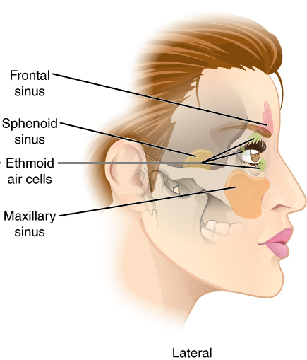
The sphenoid sinus is a paired paranasal sinus in the Body of sphenoid bone, body of the sphenoid bone. It is one pair of the four paired paranasal sinuses.Illustrated Anatomy of the Head and Neck, Fehrenbach and Herring, Elsevier, 2012, page 64 The two sphenoid sinuses are separated from each other by a septum. Each sphenoid sinus communicates with the nasal cavity via the opening of sphenoidal sinus. The two sphenoid sinuses vary in size and shape, and are usually asymmetrical. Structure On average, a sphenoid sinus measures 2.2 cm vertical height, 2 cm in transverse breadth; and 2.2 cm antero-posterior depth. Each spehoid sinus is in the body of sphenoid bone, just under the sella turcica. The sphenoid sinuses are separated from each other medially by the septum of sphenoidal sinuses, which is usually asymmetrical. An opening of sphenoidal sinus forms a passage between each sphenoidal sinus and the nasal cavity. Posteriorly, an opening of sphenoidal sinus opens ... [...More Info...] [...Related Items...] OR: [Wikipedia] [Google] [Baidu] |
Nasolacrimal Canal
The nasolacrimal duct (also called the tear duct) carries tears from the lacrimal sac of the eye into the nasal cavity. The duct begins in the eye socket between the maxillary and lacrimal bones, from where it passes downwards and backwards. The opening of the nasolacrimal duct into the inferior nasal meatus of the nasal cavity is partially covered by a mucosal fold ( valve of Hasner or ''plica lacrimalis''). Excess tears flow through the nasolacrimal duct which drains into the inferior nasal meatus. This is the reason the nose starts to run when a person is crying or has watery eyes from an allergy, and why one can sometimes taste eye drops. This is for the same reason when applying some eye drops it is often advised to close the nasolacrimal duct by pressing it with a finger to prevent the medicine from escaping the eye and having unwanted side effects elsewhere in the body as it will proceed through the canal to the nasal cavity. Like the lacrimal sac, the duct is lined by st ... [...More Info...] [...Related Items...] OR: [Wikipedia] [Google] [Baidu] |
Maxillary Sinus
The pyramid-shaped maxillary sinus (or antrum of Nathaniel Highmore (surgeon), Highmore) is the largest of the paranasal sinuses, located in the maxilla. It drains into the middle meatus of the noseHuman Anatomy, Jacobs, Elsevier, 2008, page 209-210 through the semilunar hiatus. It is located to the side of the nasal cavity, and below the orbit. Structure It is the largest air sinus in the body. It has a mean volume of about 10 ml. It is situated within the body of the maxilla, but may extend into its Maxilla, zygomatic and Maxilla, alveolar processes when large. It is pyramid-shaped, with the apex at the maxillary zygomatic process, and the base represented by the lateral nasal wall. It has three recesses: an alveolar recess pointed inferiorly, bounded by the alveolar process of the maxilla; a zygomatic recess pointed laterally, bounded by the zygomatic bone; and an infraorbital recess pointed superiorly, bounded by the inferior Orbital surface of the body of the maxilla, orbita ... [...More Info...] [...Related Items...] OR: [Wikipedia] [Google] [Baidu] |
Sinus (anatomy)
A sinus is a sac or cavity in any organ or tissue, or an abnormal cavity or passage. In common usage, "sinus" usually refers to the paranasal sinuses, which are air cavities in the cranial bones, especially those near the nose and connecting to it. Most individuals have four paired cavities located in the cranial bone or skull. Etymology ''Sinus'' is Latin for "bay", "pocket", "curve", or "bosom". In anatomy, the term is used in various contexts. The word "sinusitis" is used to indicate that one or more of the membrane linings found in the sinus cavities has become inflamed or infected. It is however distinct from a fistula, which is a tract connecting two epithelial surfaces. If left untreated, infections occurring in the sinus cavities can affect the chest and lungs. Sinuses in the body * Paranasal sinuses ** Maxillary ** Ethmoid ** Sphenoid ** Frontal * Dural venous sinuses ** Anterior midline *** Cavernous *** Superior petrosal *** Inferior petrosal ** Central sul ... [...More Info...] [...Related Items...] OR: [Wikipedia] [Google] [Baidu] |
Ethmoidal Cells
The ethmoid sinuses or ethmoid air cells of the ethmoid bone are one of the four paired paranasal sinuses. Unlike the other three pairs of paranasal sinuses which consist of one or two large cavities, the ethmoidal sinuses entail a number of small air-filled cavities ("air cells"). The cells are located within the lateral mass (labyrinth) of each ethmoid bone and are variable in both size and number.Illustrated Anatomy of the Head and Neck, Fehrenbach and Herring, Elsevier, 2012, page 64 The cells are grouped into anterior, middle, and posterior groups; the groups differ in their drainage modalities, though all ultimately drain into either the superior or the middle nasal meatus of the lateral wall of the nasal cavity. Structure The ethmoid air cells consist of numerous thin-walled cavities in the ethmoidal labyrinthOtorhinolaryngology, Head and Neck Surgery, Anniko, Springer, 2010, page 188 that represent invaginations of the mucous membrane of the nasal wall into the ethmoid b ... [...More Info...] [...Related Items...] OR: [Wikipedia] [Google] [Baidu] |
Bulla Ethmoidalis
The ethmoid bulla (or ethmoidal bulla) is a rounded elevation upon the lateral wall of the middle nasal meatus (nasal cavity inferior to the middle nasal concha) produced by one or more of the underlying middle ethmoidal air cells (which open into the nasal cavity upon or superior to the ethmoidal bulla). It varies significantly based on the size of the underlying air cells. Structure The ethmoid bulla is formed by is the largest and least variable of the middle ethmoidal air cells. The size of the bulla varies with that of its contained cells. The bulla may be a pneumatised cell or a bony prominence found in middle meatus. Relations The hiatus semilunaris is situated (sources differ) inferior/anterior to the ethmoid bulla. The maxillary sinus also opens below the bulla. Development The ethmoid bulla begins to develop between 8 weeks and 12 weeks of gestation Gestation is the period of development during the carrying of an embryo, and later fetus, inside viviparo ... [...More Info...] [...Related Items...] OR: [Wikipedia] [Google] [Baidu] |
Uncinate Process Of The Ethmoid
In the ethmoid bone, a sickle shaped projection, the uncinate process, projects posteroinferiorly from the ethmoid labyrinth. Between the posterior edge of this process and the anterior surface of the ethmoid bulla, there is a two-dimensional space, resembling a crescent shape. This space continues laterally as a three-dimensional slit-like space - the ethmoidal infundibulum. This is bounded by the uncinate process, medially, the orbital lamina of ethmoid bone The orbital lamina of ethmoid bone (or lamina papyracea or orbital lamina) is a smooth, oblong, paper-thin bone plate which forms the lateral wall of the labyrinth of the ethmoid bone. It covers the middle and posterior ethmoidal cells, and forms ... (lamina papyracea), laterally, and the ethmoidal bulla, posterosuperiorly. This concept is easier to understand if one imagine the infundibulum as a prism so that its medial face is the hiatus semilunaris. The "lateral face" of this infundibulum contains the ostium of the ma ... [...More Info...] [...Related Items...] OR: [Wikipedia] [Google] [Baidu] |
Hiatus Semilunaris
The semilunar hiatus (eg, hiatus semilunaris) is a crescent-shaped/semicircular/ curved slit/groove upon the lateral wall of the nasal cavity at the middle nasal meatus just inferior to the ethmoidal bulla. It is the location of the openings for the frontal sinus, maxillary sinus, and anterior ethmoidal sinus. It is bounded inferiorly and anteriorly by the sharp concave margin of the uncinate process of the ethmoid bone, superiorly by the ethmoidal bulla, and posteriorly by the ethmoidal process of the inferior nasal concha The inferior nasal concha (inferior turbinated bone or inferior turbinal/turbinate) is one of the three paired nasal conchae in the human nose, nose. It extends horizontally along the lateral wall of the nasal cavity and consists of a wikt:lam .... It leads into the ethmoidal infundibulum; it marks the medial limit of the ethmoidal infundibulum. References External links * {{Authority control Nose Rhinology ... [...More Info...] [...Related Items...] OR: [Wikipedia] [Google] [Baidu] |
Inferior Concha
The inferior nasal concha (inferior turbinated bone or inferior turbinal/turbinate) is one of the three paired nasal conchae in the nose. It extends horizontally along the lateral wall of the nasal cavity and consists of a lamina of spongy bone, curled upon itself like a scroll, (''turbinate'' meaning inverted cone). The inferior nasal conchae are considered a pair of facial bones. As the air passes through the turbinates, the air is churned against these mucosa-lined bones in order to receive warmth, moisture and cleansing. Superior to inferior nasal concha are the middle nasal concha and superior nasal concha which both arise from the ethmoid bone, of the cranial portion of the skull. Hence, these two are considered as a part of the cranial bones. It has two surfaces, two borders, and two extremities. Structure Surfaces The medial surface is convex, perforated by numerous apertures, and traversed by longitudinal grooves for the lodgement of vessels. The lateral surface i ... [...More Info...] [...Related Items...] OR: [Wikipedia] [Google] [Baidu] |
Sphenoethmoidal Recess
The sphenoethmoidal recess is a small triangular space superior to the superior nasal meatus of the nasal cavity into which the sphenoidal sinus opens. The sphenoethmoidal recess is situated supero posterior to the superior nasal concha The superior nasal concha is a small, curved plate of bone representing a medial bony process of the labyrinth of the ethmoid bone. The superior nasal concha forms the roof of the superior nasal meatus. Anatomy Anatomical relations The super ..., between the superior nasal concha and the anterior aspect of the body of the sphenoid bone. Anatomy Variation When present, the supreme nasal concha is situated upon the lateral wall of the sphenoethmoid recess, with the supreme nasal meatus created below it occasionally presenting an opening of the posterior ethmoidal sinus. References External links * - "The turbinates have been cut and removed to illustrate the meatus and openings into them." Nose {{respiratory-stub ... [...More Info...] [...Related Items...] OR: [Wikipedia] [Google] [Baidu] |
Meatus
In anatomy, a meatus (, , : meatus or meatuses) is a natural body opening or canal. Meatus may refer to: * the , the opening of the * the internal auditory meatus, a canal in the of the |


