|
Microtia
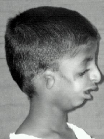
Microtia is a congenital deformity where the auricle (external ear) is underdeveloped. A completely undeveloped auricle is referred to as anotia. Because microtia and anotia have the same origin, it can be referred to as microtia-anotia. Microtia can be unilateral (one side only) or bilateral (affecting both sides). Microtia occurs in 1 out of about 8,000–10,000 births. In unilateral microtia, the right ear is most commonly affected. It may occur as a complication of taking Accutane (isotretinoin) during pregnancy. Classification According to the Altman-classification, there are four grades of microtia: *Grade I: A less than complete development of the external ear with identifiable structures and a small but present external ear canal *Grade II: A partially developed ear (usually the top portion is underdeveloped) with a closed stenotic external ear canal producing a conductive hearing loss. *Grade III: Absence of the external ear with a small peanut-like vestige structu ... [...More Info...] [...Related Items...] OR: [Wikipedia] [Google] [Baidu] |
Anotia
Anotia ("no ear") describes a rare congenital deformity that involves the complete absence of the auricle, the outer projected portion of the ear, and narrowing or absence of the ear canal. This contrasts with microtia, in which a small part of the auricle is present. Anotia and microtia may occur unilaterally (only one ear affected) or bilaterally (both ears affected). This deformity results in conductive hearing loss, deafness. Ear development Ear development begins in about the third week of human embryonic development, beginning with the formation of the otic placodes, an extension of the early hind brain. By the fourth week of development, the otic placodes invaginate, or sink inward forming pits which close themselves off for the outer surface ectoderm and begin forming the inner ear labyrinthe on the inside. Outer ear development begins in about the fifth week of human embryonic development. Upon the pharyngeal arches, auricle hillocks begin to form. By the seventh w ... [...More Info...] [...Related Items...] OR: [Wikipedia] [Google] [Baidu] |
Auricle (anatomy)
The auricle or auricula is the visible part of the ear that is outside the head. It is also called the pinna (Latin for 'wing' or ' fin', : pinnae), a term that is used more in zoology. Structure The diagram shows the shape and location of most of these components: * ''antihelix'' forms a 'Y' shape where the upper parts are: ** ''Superior crus'' (to the left of the ''fossa triangularis'' in the diagram) ** ''Inferior crus'' (to the right of the ''fossa triangularis'' in the diagram) * ''Antitragus'' is below the ''tragus'' * ''Aperture'' is the entrance to the ear canal * ''Auricular sulcus'' is the depression behind the ear next to the head * ''Concha'' is the hollow next to the ear canal * Conchal angle is the angle that the back of the ''concha'' makes with the side of the head * ''Crus'' of the helix is just above the ''tragus'' * ''Cymba conchae'' is the narrowest end of the ''concha'' * External auditory meatus is the ear canal * ''Fossa triangularis'' is the depression ... [...More Info...] [...Related Items...] OR: [Wikipedia] [Google] [Baidu] |
Franceschetti–Klein Syndrome
Treacher Collins syndrome (TCS) is a genetic disorder characterized by deformities of the ears, eyes, cheekbones, and chin. The degree to which a person is affected, however, may vary from mild to severe. Complications may include breathing problems, problems seeing, cleft palate, and hearing loss. Those affected generally have normal intelligence. TCS is usually autosomal dominant. More than half the time it occurs as a result of a new mutation rather than being inherited. The involved genes may include ''TCOF1'', ''POLR1C'', or ''POLR1D''. Diagnosis is generally suspected based on symptoms and radiographs, X-rays, and potentially confirmation by genetic testing. Treacher Collins syndrome is not curable. Symptoms may be managed with reconstructive surgery, hearing aids, speech therapy, and other assistive devices. Life expectancy is generally normal. TCS occurs in about one in 50,000 people. The syndrome is named after Edward Treacher Collins, an England, English Surgery, s ... [...More Info...] [...Related Items...] OR: [Wikipedia] [Google] [Baidu] |
Congenital Disorder
A birth defect is an abnormal condition that is present at childbirth, birth, regardless of its cause. Birth defects may result in disability, disabilities that may be physical disability, physical, intellectual disability, intellectual, or developmental disability, developmental. The disabilities can range from mild to severe. Birth defects are divided into two main types: structural disorders in which problems are seen with the shape of a body part and functional disorders in which problems exist with how a body part works. Functional disorders include metabolic disorder, metabolic and degenerative disease, degenerative disorders. Some birth defects include both structural and functional disorders. Birth defects may result from genetic disorder, genetic or chromosome abnormality, chromosomal disorders, exposure to certain medications or chemicals, or certain vertically transmitted infection, infections during pregnancy. Risk factors include folate deficiency, alcohol drink, d ... [...More Info...] [...Related Items...] OR: [Wikipedia] [Google] [Baidu] |
Dilated Cardiomyopathy
Dilated cardiomyopathy (DCM) is a condition in which the heart becomes enlarged and cannot pump blood effectively. Symptoms vary from none to feeling tired, leg swelling, and shortness of breath. It may also result in chest pain or fainting. Complications can include heart failure, heart valve disease, or an irregular heartbeat. Causes include genetics, alcohol, cocaine, certain toxins, complications of pregnancy, and certain infections. Coronary artery disease and high blood pressure may play a role, but are not the primary cause. In many cases the cause remains unclear. It is a type of cardiomyopathy, a group of diseases that primarily affects the heart muscle. The diagnosis may be supported by an electrocardiogram, chest X-ray, or echocardiogram. In those with heart failure, treatment may include medications in the ACE inhibitor, beta blocker, and diuretic families. A low salt diet may also be helpful. In those with certain types of irregular heartbeat, blood thinner ... [...More Info...] [...Related Items...] OR: [Wikipedia] [Google] [Baidu] |
Diamond–Blackfan Anemia
Diamond–Blackfan anemia (DBA) is a congenital pure red blood cell aplasia that usually presents in infancy. DBA causes anemia, but has no effect on the other blood components (platelets, white blood cells). This is in contrast to Shwachman–Bodian–Diamond syndrome, in which the bone marrow defect results primarily in neutropenia, and Fanconi anemia, where all cell lines are affected resulting in pancytopenia. There is a risk to develop acute myelogenous leukemia (AML) and certain other cancers. A variety of other congenital abnormalities may also occur in DBA, such as triphalangeal thumbs, craniofacial abnormalities, and short stature. Signs and symptoms Diamond–Blackfan anemia is characterized by normocytic or macrocytic anemia (low red blood cell counts) with decreased erythroid progenitor cells in the bone marrow. This usually develops during the neonatal period. About 47% of affected individuals also have a variety of congenital abnormalities, including craniofa ... [...More Info...] [...Related Items...] OR: [Wikipedia] [Google] [Baidu] |
Congenital Disorder Of Glycosylation
A congenital disorder of glycosylation (previously called carbohydrate-deficient glycoprotein syndrome) is one of several rare inborn errors of metabolism in which glycosylation of a variety of tissue proteins and/or lipids is deficient or defective. Congenital disorders of glycosylation are sometimes known as CDG syndromes. They often cause serious, sometimes fatal, malfunction of several different organ systems (especially the nervous system, muscles, and intestines) in affected infants. The most common sub-type is PMM2-CDG (formerly known as CDG-Ia) where the genetic defect leads to the loss of phosphomannomutase 2 ( PMM2), the enzyme responsible for the conversion of mannose-6-phosphate into mannose-1-phosphate. Presentation Clinical features depend on the molecular pathology of the particular CDG subtype. Common manifestations include |
COG1
Conserved oligomeric Golgi complex subunit 1 is a protein that in humans is encoded by the ''COG1'' gene In biology, the word gene has two meanings. The Mendelian gene is a basic unit of heredity. The molecular gene is a sequence of nucleotides in DNA that is transcribed to produce a functional RNA. There are two types of molecular genes: protei .... The protein encoded by this gene is one of eight proteins (Cog1-8) which form a Golgi-localized complex (COG) required for normal Golgi morphology and function. It is thought that this protein is required for steps in the normal medial and trans-Golgi-associated processing of glycoconjugates and plays a role in the organization of the Golgi-localized complex. Interactions COG1 has been shown to interact with COG4 and COG3. References Further reading * * * * * * * * * External links GeneReviews/NCBI/NIH/UW entry on Congenital Disorders of Glycosylation Overview * {{gene-17-stub ... [...More Info...] [...Related Items...] OR: [Wikipedia] [Google] [Baidu] |
1p36 Deletion Syndrome
1p36 deletion syndrome is a congenital genetic disorder characterized by moderate to severe intellectual disability, delayed growth, hypotonia, seizures, limited speech ability, malformations, hearing and vision impairment, and distinct facial features. The symptoms may vary, depending on the exact location of the chromosomal deletion. The condition is caused by a genetic deletion (loss of a segment of DNA) on the outermost band on the short arm (p) of chromosome 1. It is one of the most common deletion syndromes. The syndrome is thought to affect one in every 5,000 to 10,000 births. Signs and symptoms There are a number of signs and symptoms characteristic of monosomy 1p36, but no one individual will display all of the possible features. In general, children will exhibit failure to thrive and global delays. Developmental and behavioral Most young children with 1p36 deletion syndrome have delayed development of speech and motor skills. Speech is severely affected, with many ... [...More Info...] [...Related Items...] OR: [Wikipedia] [Google] [Baidu] |
CHARGE Syndrome
CHARGE syndrome (formerly known as CHARGE association) is a rare syndrome caused by a genetic disorder. First described in 1979, the acronym "CHARGE" came into use for newborn children with the congenital features of coloboma of the eye, heart defects, atresia of the nasal choanae, restricted growth or development, genital or urinary abnormalities, and ear abnormalities and deafness. These features are no longer used in making a diagnosis of CHARGE syndrome, but the name remains. About two thirds of cases are due to a CHD7 mutation. CHARGE syndrome occurs only in 0.1–1.2 per 10,000 live births; as of 2009, it was the leading cause of congenital deafblindness in the US. Genetics CHARGE syndrome was formerly referred to as CHARGE association, which indicates a non-random pattern of congenital anomalies that occurs together more frequently than one would expect on the basis of chance, but for which a common cause has not been identified. Very few people with CHARGE will have ... [...More Info...] [...Related Items...] OR: [Wikipedia] [Google] [Baidu] |
Branchio-oto-renal Syndrome
Branchio-oto-renal syndrome (BOR) is an autosomal dominant genetic disorder involving the kidneys, ears, and neck. It is also known as Melnick-Fraser syndrome. Signs and symptoms The signs and symptoms of branchio-oto-renal syndrome are consistent with underdeveloped (hypoplastic) or absent kidneys with resultant chronic kidney disease or kidney failure. Ear anomalies include extra openings in front of the ears, extra pieces of skin in front of the ears (preauricular tags), or further malformation or absence of the outer ear ( pinna). Malformation or absence of the middle ear is also possible, individuals can have mild to profound hearing loss. People with BOR may also have cysts or fistulae along the sides of their neck. In some individuals and families, renal features are completely absent. The disease may then be termed "branchio-oto syndrome" (BO syndrome)., updated, 2015, Cause The cause of branchio-oto-renal syndrome are mutations in genes, EYA1, SIX1, and SIX5 (approxi ... [...More Info...] [...Related Items...] OR: [Wikipedia] [Google] [Baidu] |


