|
Methiodal
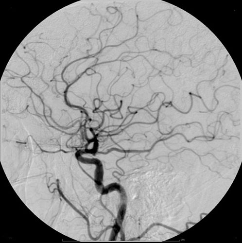
Methiodal is a pharmaceutical drug that was used as an iodinated contrast medium for X-ray imaging. Its uses included myelography (imaging of the spinal cord); for this use, cases of adhesive arachnoiditis have been reported, similar to those seen under the contrast medium iofendylate Iofendylate is a molecule that was used as a radiocontrast agent, typically for performing myelography studies. It was marketed under the trade names Pantopaque (in North America) and Myodil (rest of the world). Iofendylate is a highly lipophilic .... It is not known to be marketed anywhere in the world in 2021. References Radiocontrast agents Organoiodides Sulfonates {{pharmacology-stub ... [...More Info...] [...Related Items...] OR: [Wikipedia] [Google] [Baidu] |
Iodinated Contrast
Iodinated contrast is a form of water-soluble, intravenous radiocontrast agent containing iodine, which enhances the visibility of vascular structures and organs during radiography, radiographic procedures. Some pathologies, such as cancer, have particularly improved visibility with iodinated contrast. The radiodensity of iodinated contrast is 25–30 Hounsfield units (HU) per milligram of iodine per milliliter at a tube voltage of 100–120 kVp. Types Iodine-based contrast media are usually classified as ionic or nonionic. Both types are used most commonly in radiology due to their relatively harmless interaction with the body and their solubility. Contrast media are primarily used to visualize vessels and changes in tissues on radiography and CT Scan, CT (computerized tomography). Contrast media can also be used for tests of the urinary tract, uterus and fallopian tubes. It may cause the patient to feel as if they have had urinary incontinence. It also puts a metallic taste in t ... [...More Info...] [...Related Items...] OR: [Wikipedia] [Google] [Baidu] |
X-ray Imaging
Radiography is an imaging technology, imaging technique using X-rays, gamma rays, or similar ionizing radiation and non-ionizing radiation to view the internal form of an object. Applications of radiography include medical ("diagnostic" radiography and "therapeutic radiography") and industrial radiography. Similar techniques are used in airport security, (where "body scanners" generally use backscatter X-ray). To create an image in conventional radiography, a beam of X-rays is produced by an X-ray generator and it is projected towards the object. A certain amount of the X-rays or other radiation are absorbed by the object, dependent on the object's density and structural composition. The X-rays that pass through the object are captured behind the object by a X-ray detector, detector (either photographic film or a digital detector). The generation of flat two-dimensional images by this technique is called Projection radiography, projectional radiography. In computed tomography (C ... [...More Info...] [...Related Items...] OR: [Wikipedia] [Google] [Baidu] |
Myelography
Myelography is a type of radiographic examination that uses a contrast medium (e.g. iodised oil) to detect pathology of the spinal cord, including the location of a spinal cord injury, cysts, and tumors. Historically the procedure involved the injection of a radiocontrast agent into the cervical or lumbar spine, followed by several X-ray projections. Today, myelography has largely been replaced by the use of MRI scans, although the technique is still sometimes used under certain circumstances – though now usually in conjunction with CT rather than X-ray projections. Types Cervical myelography This procedure is used to look for the level of where spinal cord disease occurs or compression of the spinal cord at the neck region for those who are unable or unwilling to undergone MRI scan of the spine. Lumbar myelography This procedure is to look for the level of spinal cord disease such as lumbar nerve root compression, cauda equina syndrome, conus medullaris lesions, and spinal ... [...More Info...] [...Related Items...] OR: [Wikipedia] [Google] [Baidu] |
Spinal Cord
The spinal cord is a long, thin, tubular structure made up of nervous tissue that extends from the medulla oblongata in the lower brainstem to the lumbar region of the vertebral column (backbone) of vertebrate animals. The center of the spinal cord is hollow and contains a structure called the central canal, which contains cerebrospinal fluid. The spinal cord is also covered by meninges and enclosed by the neural arches. Together, the brain and spinal cord make up the central nervous system. In humans, the spinal cord is a continuation of the brainstem and anatomically begins at the occipital bone, passing out of the foramen magnum and then enters the spinal canal at the beginning of the cervical vertebrae. The spinal cord extends down to between the first and second lumbar vertebrae, where it tapers to become the cauda equina. The enclosing bony vertebral column protects the relatively shorter spinal cord. It is around long in adult men and around long in adult women. The diam ... [...More Info...] [...Related Items...] OR: [Wikipedia] [Google] [Baidu] |
Arachnoiditis
Arachnoiditis is an inflammatory condition of the arachnoid mater or 'arachnoid', one of the membranes known as meninges that surround and protect the central nervous system. The outermost layer of the meninges is the dura mater (Latin for hard) and adheres to inner surface of the skull and vertebrae. The arachnoid is under or "deep" to the dura and is a thin membrane that adheres directly to the surface of the brain and spinal cord. Signs and symptoms Arachnoid inflammation can lead to many painful and debilitating symptoms which can vary greatly in each case, and not all people experience all symptoms. Chronic pain is common, including neuralgia, while numbness and tingling of the extremities can occur with spinal cord involvement, and bowel, bladder, and sexual functioning can be affected if the lower part of the spinal cord is involved. While arachnoiditis has no consistent pattern of symptoms, it frequently affects the nerves that supply the legs and lower back. Many pat ... [...More Info...] [...Related Items...] OR: [Wikipedia] [Google] [Baidu] |
Iofendylate
Iofendylate is a molecule that was used as a radiocontrast agent, typically for performing myelography studies. It was marketed under the trade names Pantopaque (in North America) and Myodil (rest of the world). Iofendylate is a highly lipophilic (oily) substance and as such it was recommended that the physician remove it from the patient at the end of the myelography study, which was a difficult and painful part of the procedure. Moreover, because complete removal could not always be achieved (or even attempted by some physicians), iofendylate's persistence in the body might sometimes lead to arachnoiditis, a potentially painful and debilitating lifelong disorder of the spine. As a result, the substance, which was used extensively for over three decades, became the subject of multiple lawsuits filed around the world. Iofendylate's use ceased when water-soluble agents suitable for spinal imaging (such as metrizamide) became available in the late 1970s. With those substances it wa ... [...More Info...] [...Related Items...] OR: [Wikipedia] [Google] [Baidu] |
Radiocontrast Agents
Radiocontrast agents are substances used to enhance the visibility of internal structures in X-ray-based imaging techniques such as computed tomography (contrast CT), projectional radiography, and fluoroscopy. Radiocontrast agents are typically iodine, or more rarely barium sulfate. The contrast agents absorb external X-rays, resulting in decreased exposure on the X-ray detector. This is different from radiopharmaceuticals used in nuclear medicine which emit radiation. Magnetic resonance imaging (MRI) functions through different principles and thus MRI contrast agents have a different mode of action. These compounds work by altering the magnetic properties of nearby hydrogen nuclei. Types and uses Radiocontrast agents used in X-ray examinations can be grouped in positive (iodinated agents, barium sulfate), and negative agents (air, carbon dioxide, methylcellulose). Iodine (circulatory system) Iodinated contrast contains iodine. It is the main type of radiocontrast used for intra ... [...More Info...] [...Related Items...] OR: [Wikipedia] [Google] [Baidu] |
Organoiodides
Organoiodine chemistry is the study of the synthesis and properties of organoiodine compounds, or organoiodides, organic compounds that contain one or more carbon–iodine bonds. They occur widely in organic chemistry, but are relatively rare in nature. The thyroxine hormones are organoiodine compounds that are required for health and the reason for government-mandated iodization of salt. Structure, bonding, general properties Almost all organoiodine compounds feature iodide connected to one carbon center. These are usually classified as derivatives of I−. Some organoiodine compounds feature iodine in higher oxidation states. The C–I bond is the weakest of the carbon–halogen bonds. These bond strengths correlate with the electronegativity of the halogen, decreasing in the order F > Cl > Br > I. This periodic order also follows the atomic radius of halogens and the length of the carbon-halogen bond. For example, in the molecules represented by CH3X, where X ... [...More Info...] [...Related Items...] OR: [Wikipedia] [Google] [Baidu] |


