|
Mediastinal Cyst
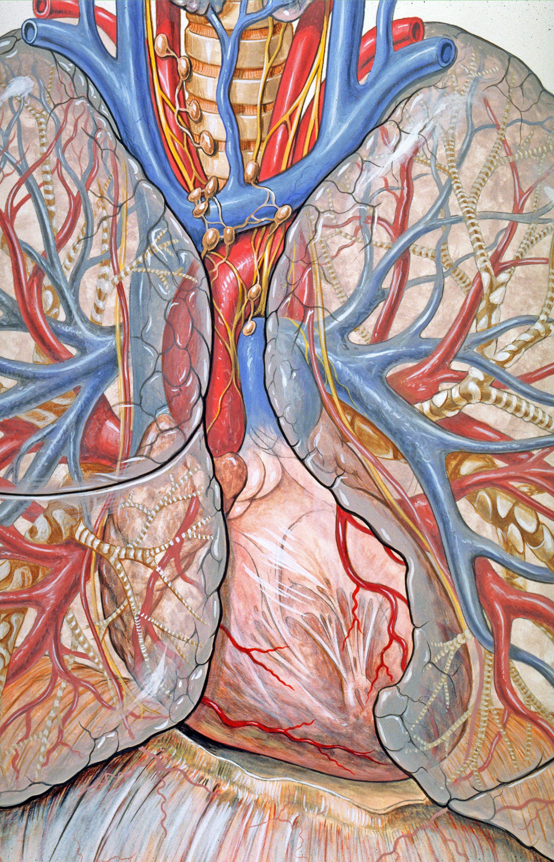
The mediastinum (from ;: mediastina) is the central compartment of the thoracic cavity. Surrounded by loose connective tissue, it is a region that contains vital organs and structures within the thorax, mainly the heart and its vessels, the esophagus, the trachea, the vagus, phrenic and cardiac nerves, the thoracic duct, the thymus and the lymph nodes of the central chest. Anatomy The mediastinum lies within the thorax and is enclosed on the right and left by pleurae. It is surrounded by the chest wall in front, the lungs to the sides and the spine at the back. It extends from the sternum in front to the vertebral column behind. It contains all the organs of the thorax except the lungs. It is continuous with the loose connective tissue of the neck. The mediastinum can be divided into an upper (or superior) and lower (or inferior) part: * The superior mediastinum starts at the superior thoracic aperture and ends at the thoracic plane. * The inferior mediastinum from this l ... [...More Info...] [...Related Items...] OR: [Wikipedia] [Google] [Baidu] |
Pericardial Cavity
The pericardium (: pericardia), also called pericardial sac, is a double-walled sac containing the heart and the roots of the great vessels. It has two layers, an outer layer made of strong inelastic connective tissue (fibrous pericardium), and an inner layer made of serous membrane (serous pericardium). It encloses the pericardial cavity, which contains pericardial fluid, and defines the middle mediastinum. It separates the heart from interference of other structures, protects it against infection and blunt trauma, and lubricates the heart's movements. The English name originates from the Ancient Greek prefix ''peri-'' (περί) 'around' and the suffix ''-cardion'' (κάρδιον) 'heart'. Anatomy The pericardium is a tough fibroelastic sac which covers the heart from all sides except at the cardiac root (where the great vessels join the heart) and the bottom (where only the serous pericardium exists to cover the upper surface of the central tendon of diaphragm). The ... [...More Info...] [...Related Items...] OR: [Wikipedia] [Google] [Baidu] |
Spine (anatomy)
The spinal column, also known as the vertebral column, spine or backbone, is the core part of the axial skeleton in vertebrates. The vertebral column is the defining and eponymous characteristic of the vertebrate. The spinal column is a segmented column of vertebrae that surrounds and protects the spinal cord. The vertebrae are separated by intervertebral discs in a series of cartilaginous joints. The dorsal portion of the spinal column houses the spinal canal, an elongated body cavity, cavity formed by the alignment of the vertebral neural arches that encloses and protects the spinal cord, with spinal nerves exiting via the intervertebral foramina to innervate each body segment. There are around 50,000 species of animals that have a vertebral column. The human spine is one of the most-studied examples, as the general structure of human vertebrae is fairly homology (biology), typical of that found in other mammals, reptiles, and birds. The shape of the vertebral body does, howev ... [...More Info...] [...Related Items...] OR: [Wikipedia] [Google] [Baidu] |
Intervertebral Disc
An intervertebral disc (British English), also spelled intervertebral disk (American English), lies between adjacent vertebrae in the vertebral column. Each disc forms a fibrocartilaginous joint (a symphysis), to allow slight movement of the vertebrae, to act as a ligament to hold the vertebrae together, and to function as a shock absorber for the spine. Structure Intervertebral discs consist of an outer fibrous ring, the ''anulus (or annulus) fibrosus disci intervertebralis'', which surrounds an inner gel-like center, the ''nucleus pulposus''. The ''anulus fibrosus'' consists of several layers (laminae) of fibrocartilage made up of both type I and type II collagen. Type I is concentrated toward the edge of the ring, where it provides greater strength. The stiff laminae can withstand compressive forces. The fibrous intervertebral disc contains the ''nucleus pulposus'' and this helps to distribute pressure evenly across the disc. This prevents the development of stress conc ... [...More Info...] [...Related Items...] OR: [Wikipedia] [Google] [Baidu] |
Thoracic Vertebrae
In vertebrates, thoracic vertebrae compose the middle segment of the vertebral column, between the cervical vertebrae and the lumbar vertebrae. In humans, there are twelve thoracic vertebra (anatomy), vertebrae of intermediate size between the cervical and lumbar vertebrae; they increase in size going towards the lumbar vertebrae. They are distinguished by the presence of Zygapophysial joint, facets on the sides of the bodies for Articulation (anatomy), articulation with the head of rib, heads of the ribs, as well as facets on the transverse processes of all, except the eleventh and twelfth, for articulation with the tubercle (rib), tubercles of the ribs. By convention, the human thoracic vertebrae are numbered T1–T12, with the first one (T1) located closest to the skull and the others going down the spine toward the lumbar region. General characteristics These are the general characteristics of the second through eighth thoracic vertebrae. The first and ninth through twelfth v ... [...More Info...] [...Related Items...] OR: [Wikipedia] [Google] [Baidu] |
Anatomical Plane
An anatomical plane is a hypothetical plane used to transect the body, in order to describe the location of structures or the direction of movements. In human anatomy and non-human anatomy, four principal planes are used: the median plane, sagittal plane, coronal plane, and transverse plane. * The median plane or midsagittal plane passes through the middle of the body, dividing it into left and right halves. * A para sagittal plane is any plane that runs parallel to the median plane, also dividing the body into left and right sections. * The dorsal plane divides the body into dorsal (towards the backbone) and ventral (towards the belly) parts. In human anatomy coronal plane is preferred, or sometimes the frontal plane, and the description may reference splitting the body into front and back parts, but this phrasing is not as clear for animals with a horizontal spine like quadrupeds or fish. * The transverse plane, also called the axial plane or horizontal plane, is perpendi ... [...More Info...] [...Related Items...] OR: [Wikipedia] [Google] [Baidu] |
Radiologists
Radiology ( ) is the medical specialty that uses medical imaging to diagnose diseases and guide treatment within the bodies of humans and other animals. It began with radiography (which is why its name has a root referring to radiation), but today it includes all imaging modalities. This includes technologies that use no ionizing electromagnetic radiation, such as ultrasonography and magnetic resonance imaging (MRI), as well as others that do use radiation, such as computed tomography (CT), fluoroscopy, and nuclear medicine including positron emission tomography (PET). Interventional radiology is the performance of usually minimally invasive medical procedures with the guidance of imaging technologies such as those mentioned above. The modern practice of radiology involves a team of several different healthcare professionals. A radiologist, who is a medical doctor with specialized post-graduate training, interprets medical images, communicates these findings to other physicians ... [...More Info...] [...Related Items...] OR: [Wikipedia] [Google] [Baidu] |
Surgeon
In medicine, a surgeon is a medical doctor who performs surgery. Even though there are different traditions in different times and places, a modern surgeon is a licensed physician and received the same medical training as physicians before specializing in surgery. In some countries and jurisdictions, the title of 'surgeon' is restricted to maintain the integrity of the craft group in the medical profession. A specialist regarded as a legally recognized surgeon includes podiatry, dentistry, and veterinary medicine. It is estimated that surgeons perform over 300 million surgical procedures globally each year. History The first person to document a surgery was the 6th century BC Indian physician-surgeon, Sushruta. He specialized in cosmetic plastic surgery and even documented an open rhinoplasty procedure.Papel, Ira D. and Frodel, John (2008) ''Facial Plastic and Reconstructive Surgery''. Thieme Medical Pub. His Masterpiece, magnum opus ''Suśruta-saṃhitā'' is one of the m ... [...More Info...] [...Related Items...] OR: [Wikipedia] [Google] [Baidu] |
Anatomist
Anatomy () is the branch of morphology concerned with the study of the internal structure of organisms and their parts. Anatomy is a branch of natural science that deals with the structural organization of living things. It is an old science, having its beginnings in prehistoric times. Anatomy is inherently tied to developmental biology, embryology, comparative anatomy, evolutionary biology, and phylogeny, as these are the processes by which anatomy is generated, both over immediate and long-term timescales. Anatomy and physiology, which study the structure and function of organisms and their parts respectively, make a natural pair of related disciplines, and are often studied together. Human anatomy is one of the essential basic sciences that are applied in medicine, and is often studied alongside physiology. Anatomy is a complex and dynamic field that is constantly evolving as discoveries are made. In recent years, there has been a significant increase in the use of ... [...More Info...] [...Related Items...] OR: [Wikipedia] [Google] [Baidu] |
Pericardium
The pericardium (: pericardia), also called pericardial sac, is a double-walled sac containing the heart and the roots of the great vessels. It has two layers, an outer layer made of strong inelastic connective tissue (fibrous pericardium), and an inner layer made of serous membrane (serous pericardium). It encloses the pericardial cavity, which contains pericardial fluid, and defines the middle mediastinum. It separates the heart from interference of other structures, protects it against infection and blunt trauma, and lubricates the heart's movements. The English name originates from the Ancient Greek prefix ''peri-'' (περί) 'around' and the suffix ''-cardion'' (κάρδιον) 'heart'. Anatomy The pericardium is a tough fibroelastic sac which covers the heart from all sides except at the cardiac root (where the great vessels join the heart) and the bottom (where only the serous pericardium exists to cover the upper surface of the central tendon of diaphragm). ... [...More Info...] [...Related Items...] OR: [Wikipedia] [Google] [Baidu] |
Thoracic Diaphragm
The thoracic diaphragm, or simply the diaphragm (; ), is a sheet of internal Skeletal striated muscle, skeletal muscle in humans and other mammals that extends across the bottom of the thoracic cavity. The diaphragm is the most important Muscles of respiration, muscle of respiration, and separates the thoracic cavity, containing the heart and lungs, from the abdominal cavity: as the diaphragm contracts, the volume of the thoracic cavity increases, creating a negative pressure there, which draws air into the lungs. Its high oxygen consumption is noted by the many mitochondria and capillaries present; more than in any other skeletal muscle. The term ''diaphragm'' in anatomy, created by Gerard of Cremona, can refer to other flat structures such as the urogenital diaphragm or Pelvic floor, pelvic diaphragm, but "the diaphragm" generally refers to the thoracic diaphragm. In humans, the diaphragm is slightly asymmetric—its right half is higher up (superior) to the left half, since th ... [...More Info...] [...Related Items...] OR: [Wikipedia] [Google] [Baidu] |
Thoracic Plane
The mediastinum (from ;: mediastina) is the central compartment of the thoracic cavity. Surrounded by loose connective tissue, it is a region that contains vital organs and structures within the thorax, mainly the heart and its vessels, the esophagus, the trachea, the vagus nerve, vagus, phrenic nerve, phrenic and cardiac nerves, the thoracic duct, the thymus and the lymph nodes of the central chest. Anatomy The mediastinum lies within the thorax and is enclosed on the right and left by pulmonary pleurae, pleurae. It is surrounded by the chest wall in front, the lungs to the sides and the Spine (anatomy), spine at the back. It extends from the sternum in front to the vertebral column behind. It contains all the organs of the thorax except the lungs. It is continuous with the loose connective tissue of the neck. The mediastinum can be divided into an upper (or superior) and lower (or inferior) part: * The superior mediastinum starts at the superior thoracic aperture and end ... [...More Info...] [...Related Items...] OR: [Wikipedia] [Google] [Baidu] |
Superior Thoracic Aperture
The superior thoracic aperture, also known as the thoracic outlet, or thoracic inlet refers to the opening at the top of the thoracic cavity. It is also clinically referred to as the thoracic outlet, in the case of thoracic outlet syndrome. A lower thoracic opening is the ''inferior thoracic aperture''. Structure The superior thoracic aperture is essentially a hole surrounded by a bony ring, through which several vital structures pass. It is bounded by: the first thoracic vertebra (T1) ''posteriorly''; the first pair of ribs ''laterally'', forming lateral C-shaped curves posterior to anterior; and the costal cartilage of the first rib and the superior border of the manubrium ''anteriorly''. Dimensions The adult thoracic outlet is around 6.5 cm antero-posteriorly and 11 cm transversely. Because of the obliquity of the first pair of ribs, the aperture slopes antero-inferiorly. Relations The clavicle articulates with the manubrium to form the anterior border of the thoracic outle ... [...More Info...] [...Related Items...] OR: [Wikipedia] [Google] [Baidu] |


