|
Mechanism Of Autism
The mechanisms of autism are the molecular and cellular Biological process, processes believed to cause or contribute to the symptoms of autism. Multiple processes are hypothesized to explain different autism spectrum features. These hypotheses include defects in synapse structure and function, reduced synaptic plasticity, disrupted neural circuit function, gut–brain axis dyshomeostasis, neuroinflammation, and altered brain structure or connectivity. Autism symptoms stem from maturation-related changes in brain systems. The mechanisms of autism are divided into two main areas: pathophysiology of brain structures and processes, and neuropsychological linkages between brain structures and behaviors, with multiple pathophysiologies linked to various autism behaviors. Evidence suggests gut–brain axis abnormalities may contribute to autism. Studies propose that immune, Gastrointestinal tract, gastrointestinal inflammation, autonomic nervous system dysfunction, gut microbiota alter ... [...More Info...] [...Related Items...] OR: [Wikipedia] [Google] [Baidu] |
Biological Process
Biological processes are those processes that are necessary for an organism to live and that shape its capacities for interacting with its environment. Biological processes are made of many chemical reactions or other events that are involved in the persistence and transformation of life forms. Regulation of biological processes occurs when any process is modulated in its frequency, rate or extent. Biological processes are regulated by many means; examples include the control of gene expression, protein modification or interaction with a protein or substrate molecule. * Homeostasis: regulation of the internal environment to maintain a constant state; for example, sweating to reduce temperature * Biological organization, Organization: being structurally composed of one or more cell (biology), cells – the basic units of life * Metabolism: transformation of energy by converting chemicals and energy into cellular components (anabolism) and decomposing organic matter (catabolis ... [...More Info...] [...Related Items...] OR: [Wikipedia] [Google] [Baidu] |
Alzheimer's Disease
Alzheimer's disease (AD) is a neurodegenerative disease and the cause of 60–70% of cases of dementia. The most common early symptom is difficulty in remembering recent events. As the disease advances, symptoms can include problems with language, disorientation (including easily getting lost), mood swings, loss of motivation, self-neglect, and behavioral issues. As a person's condition declines, they often withdraw from family and society. Gradually, bodily functions are lost, ultimately leading to death. Although the speed of progression can vary, the average life expectancy following diagnosis is three to twelve years. The causes of Alzheimer's disease remain poorly understood. There are many environmental and genetic risk factors associated with its development. The strongest genetic risk factor is from an allele of apolipoprotein E. Other risk factors include a history of head injury, clinical depression, and high blood pressure. The progression of the di ... [...More Info...] [...Related Items...] OR: [Wikipedia] [Google] [Baidu] |
SHANK3
SH3 and multiple ankyrin repeat domains 3 (Shank3), also known as proline-rich synapse-associated protein 2 (ProSAP2), is a protein that in humans is encoded by the ''SHANK3'' gene on chromosome 22. Additional isoforms have been described for this gene but they have not yet been experimentally verified. Function This gene is one of the 3-member Shank gene family (Shank1-3). The gene encodes a protein that contains 5 interaction domains or motifs including the ankyrin repeats domain (ANK), a src 3 domain ( SH3), a proline-rich domain, a PDZ domain and a sterile α motif domain (SAM). Shank proteins are multidomain scaffold proteins of the postsynaptic density that connect neurotransmitter receptors, ion channels, and other membrane proteins to the actin cytoskeleton and G-protein-coupled signaling pathways. Shank proteins also play a role in synapse formation and dendritic spine maturation. Clinical significance Mutations in this gene are associated with autism spectrum d ... [...More Info...] [...Related Items...] OR: [Wikipedia] [Google] [Baidu] |
Presynaptic
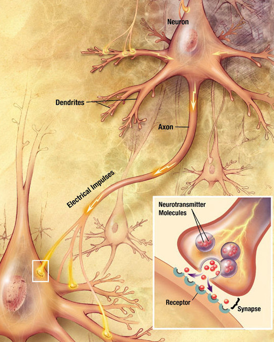
In the nervous system, a synapse is a structure that allows a neuron (or nerve cell) to pass an electrical or chemical signal to another neuron or a target effector cell. Synapses can be classified as either chemical or electrical, depending on the mechanism of signal transmission between neurons. In the case of electrical synapses, neurons are coupled bidirectionally with each other through gap junctions and have a connected cytoplasmic milieu. These types of synapses are known to produce synchronous network activity in the brain, but can also result in complicated, chaotic network level dynamics. Therefore, signal directionality cannot always be defined across electrical synapses. Chemical synapses, on the other hand, communicate through neurotransmitters released from the presynaptic neuron into the synaptic cleft. Upon release, these neurotransmitters bind to specific receptors on the postsynaptic membrane, inducing an electrical or chemical response in the target neuron. T ... [...More Info...] [...Related Items...] OR: [Wikipedia] [Google] [Baidu] |
Postsynaptic
Chemical synapses are biological junctions through which neurons' signals can be sent to each other and to non-neuronal cells such as those in muscles or glands. Chemical synapses allow neurons to form circuits within the central nervous system. They are crucial to the biological computations that underlie perception and thought. They allow the nervous system to connect to and control other systems of the body. At a chemical synapse, one neuron releases neurotransmitter molecules into a small space (the synaptic cleft) that is adjacent to another neuron. The neurotransmitters are contained within small sacs called synaptic vesicles, and are released into the synaptic cleft by exocytosis. These molecules then bind to neurotransmitter receptors on the postsynaptic cell. Finally, the neurotransmitters are cleared from the synapse through one of several potential mechanisms including enzymatic degradation or re-uptake by specific transporters either on the presynaptic cell or o ... [...More Info...] [...Related Items...] OR: [Wikipedia] [Google] [Baidu] |
Neuroligin
Neuroligin (NLGN), a Transmembrane protein, type I membrane protein, is a Cell adhesion molecule, cell adhesion protein on the Chemical synapse#Structure, postsynaptic membrane that mediates the formation and maintenance of synapses between neurons. Neuroligins act as ligands for Neurexin, β-neurexins, which are cell adhesion proteins located presynaptically. Neuroligin and β-neurexin "shake hands", resulting in the connection between two neurons and the production of a synapse. Neuroligins also affect the properties of neural networks by specifying synaptic functions, and they mediate signalling by recruiting and stabilizing key synaptic components. Neuroligins interact with other postsynaptic proteins to localize neurotransmitter receptors and channels in the postsynaptic density as the cell matures. Additionally, neuroligins are expressed in human peripheral tissues and have been found to play a role in angiogenesis. In humans, alterations in genes encoding neuroligins ... [...More Info...] [...Related Items...] OR: [Wikipedia] [Google] [Baidu] |
Cell Adhesion Molecule
Cell adhesion molecules (CAMs) are a subset of cell surface proteins that are involved in the binding of cells with other cells or with the extracellular matrix (ECM), in a process called cell adhesion. In essence, CAMs help cells stick to each other and to their surroundings. CAMs are crucial components in maintaining tissue structure and function. In fully developed animals, these molecules play an integral role in generating force and movement and consequently ensuring that organs are able to execute their functions normally. In addition to serving as "molecular glue", CAMs play important roles in the cellular mechanisms of growth, contact inhibition, and apoptosis. Aberrant expression of CAMs may result in a wide range of pathologies, ranging from frostbite to cancer. Structure CAMs are typically single-pass transmembrane receptors and are composed of three conserved domains: an intracellular domain that interacts with the cytoskeleton, a transmembrane domain, and an extrace ... [...More Info...] [...Related Items...] OR: [Wikipedia] [Google] [Baidu] |
Neurotypical
The neurodiversity paradigm is a framework for understanding human brain function that considers the diversity within sensory processing, motor abilities, social comfort, cognition, and focus as neurobiological differences. This diversity falls on a spectrum of neurocognitive differences. The neurodiversity paradigm argues that diversity in neurocognition is part of humanity and that some neurodivergences generally classified as disorders, such as autism, are differences with strengths and weaknesses as well as disabilities that are not necessarily pathological. The neurodiversity movement started in the late 1980s and early 1990s with the start of Autism Network International. Much of the correspondence that led to the formation of the movement happened over autism conferences, namely the autistic-led Autreat, penpal lists, and Usenet. The framework grew out of the disability rights movement and builds on the social model of disability, arguing that disability partly arises ... [...More Info...] [...Related Items...] OR: [Wikipedia] [Google] [Baidu] |
Cerebellar Vermis
The cerebellar vermis (from Latin ''vermis,'' "worm") is located in the medial, cortico-nuclear zone of the cerebellum, which is in the posterior cranial fossa, posterior fossa of the cranium. The primary fissure in the vermis curves ventrolaterally to the anatomical terms of location, superior surface of the cerebellum, dividing it into anterior and posterior (anatomy), posterior lobe (anatomy), lobes. Functionally, the vermis is associated with bodily Neutral spine, posture and Motion (physics), locomotion. The vermis is included within the Anatomy of the cerebellum#Phylogenetic and functional divisions, spinocerebellum and receives somatic sensory input from the head and proximal body parts via spinal cord, ascending spinal pathways. The cerebellum develops in a rostro-caudal manner, with Anatomical terms of location#Directional terms, rostral regions in the midline giving rise to the vermis, and Caudal (anatomical term), caudal regions developing into the cerebellar hemisphere ... [...More Info...] [...Related Items...] OR: [Wikipedia] [Google] [Baidu] |
Macrocephaly
Macrocephaly is a condition in which circumference of the human head is abnormally large. It may be pathological or harmless, and can be a Heredity, familial genetic characteristic. People diagnosed with macrocephaly will receive further medical tests to determine whether the syndrome is accompanied by particular Disorder (medicine), disorders. Those with benign or familial macrocephaly are considered to have megalencephaly. Causes Many people with abnormally large heads or large skulls are healthy, but macrocephaly may be pathological. Pathologic macrocephaly may be due to megalencephaly (enlarged brain), hydrocephalus (abnormally increased cerebrospinal fluid), cranial hyperostosis (bone overgrowth), and other conditions. Pathologic macrocephaly is called "syndromic", when it is associated with any other noteworthy condition, and "nonsyndromic" otherwise. Pathologic macrocephaly may be caused by congenital anatomic abnormalities, genetic conditions, or by environmental events. ... [...More Info...] [...Related Items...] OR: [Wikipedia] [Google] [Baidu] |
Microglia
Microglia are a type of glia, glial cell located throughout the brain and spinal cord of the central nervous system (CNS). Microglia account for about around 5–10% of cells found within the brain. As the resident macrophage cells, they act as the first and main form of active immune defense in the CNS. Microglia originate in the yolk sac under tightly regulated molecular conditions. These cells (and other neuroglia including astrocytes) are distributed in large non-overlapping regions throughout the CNS. Microglia are key cells in overall brain maintenancethey are constantly scavenging the CNS for senile plaques, plaques, damaged or unnecessary neurons and synapses, and infectious agents. Since these processes must be efficient to prevent potentially fatal damage, microglia are extremely sensitive to even small pathological changes in the CNS. This sensitivity is achieved in part by the presence of unique potassium channels that respond to even small changes in extracellular pota ... [...More Info...] [...Related Items...] OR: [Wikipedia] [Google] [Baidu] |
Astrocyte
Astrocytes (from Ancient Greek , , "star" and , , "cavity", "cell"), also known collectively as astroglia, are characteristic star-shaped glial cells in the brain and spinal cord. They perform many functions, including biochemical control of endothelial cells that form the blood–brain barrier, provision of nutrients to the nervous tissue, maintenance of extracellular ion balance, regulation of cerebral blood flow, and a role in the repair and scarring process of the brain and spinal cord following infection and traumatic injuries. The proportion of astrocytes in the brain is not well defined; depending on the counting technique used, studies have found that the astrocyte proportion varies by region and ranges from 20% to around 40% of all glia. Another study reports that astrocytes are the most numerous cell type in the brain. Astrocytes are the major source of cholesterol in the central nervous system. Apolipoprotein E transports cholesterol from astrocytes to neurons and ot ... [...More Info...] [...Related Items...] OR: [Wikipedia] [Google] [Baidu] |


