|
Mastigoneme
Mastigonemes are lateral "hairs" that attach to protistan flagella. Flimsy hairs attach to the flagella of euglenid flagellates, while stiff hairs occur in stramenopile and cryptophyte protists.Hoek, C. van den, Mann, D. G. and Jahns, H. M. (1995). Algae : An introduction to phycology', Cambridge University Press, UK. Stramenopile hairs are approximately 15 nm in diameter, and usually consist of flexible basal part that inserts into the cell membrane, a tubular shaft that itself terminates in smaller "hairs". They reverse the thrust caused when a flagellum beats. The consequence is that the cell is drawn into the water and particles of food are drawn to the surface of heterotrophic species. Typology of flagella with hairs: *whiplash flagella (= smooth, acronematic flagella): without hairs but may have extensions, e.g., in Opisthokonta *hairy flagella (= tinsel, flimmer, pleuronematic flagella): with hairs (= mastigonemes ''sensu lato''), divided in: **with fine hairs (= ... [...More Info...] [...Related Items...] OR: [Wikipedia] [Google] [Baidu] |
Heterokonts
The stramenopiles, also called heterokonts, are protists distinguished by the presence of stiff tripartite external hairs. In most species, the hairs are attached to flagella, in some they are attached to other areas of the cellular surface, and in some they have been secondarily lost (in which case relatedness to stramenopile ancestors is evident from other shared cytological features or from genetic similarity). Stramenopiles represent one of the three major clades in the SAR supergroup, along with Alveolata and Rhizaria. Stramenopiles are eukaryotes; most are single-celled, but some are multicellular including some large seaweeds, the brown algae. The group includes a variety of algal protists, heterotrophic flagellates, opalines and closely related proteromonad flagellates (all endobionts in other organisms); the actinophryid Heliozoa, and oomycetes. The tripartite hairs characteristic of the group have been lost in some of the included taxa – for example in most dia ... [...More Info...] [...Related Items...] OR: [Wikipedia] [Google] [Baidu] |
Euglenid
Euglenids or euglenoids are one of the best-known groups of eukaryotic flagellates: single-celled organisms with flagella, or whip-like tails. They are classified in the phylum Euglenophyta, class Euglenida or Euglenoidea. Euglenids are commonly found in fresh water, especially when it is rich in organic materials, but they have a few marine and endosymbiotic members. Many euglenids feed by phagocytosis, or strictly by diffusion. A monophyletic subgroup known as Euglenophyceae have chloroplasts and produce their own food through photosynthesis. This group contains the carbohydrate paramylon. Euglenids split from other Euglenozoa (a larger group of flagellates) more than a billion years ago. The plastids (membranous organelles) in all extant photosynthetic species result from secondary endosymbiosis between a euglenid and a green alga. Structure Euglenoids are distinguished mainly by the presence of a type of cell covering called a pellicle. Within its taxon, the pellic ... [...More Info...] [...Related Items...] OR: [Wikipedia] [Google] [Baidu] |
Stramenopile
The stramenopiles, also called heterokonts, are protists distinguished by the presence of stiff tripartite external hairs. In most species, the hairs are attached to flagella, in some they are attached to other areas of the cellular surface, and in some they have been secondarily lost (in which case relatedness to stramenopile ancestors is evident from other shared cytological features or from genetic similarity). Stramenopiles represent one of the three major clades in the SAR supergroup, along with Alveolata and Rhizaria. Stramenopiles are eukaryotes; most are single-celled, but some are multicellular including some large seaweeds, the brown algae. The group includes a variety of algal protists, heterotrophic flagellates, opalines and closely related proteromonad flagellates (all endobionts in other organisms); the actinophryid Heliozoa, and oomycetes. The tripartite hairs characteristic of the group have been lost in some of the included taxa – for example in most ... [...More Info...] [...Related Items...] OR: [Wikipedia] [Google] [Baidu] |
Heterokonta
The stramenopiles, also called heterokonts, are protists distinguished by the presence of stiff tripartite external hairs. In most species, the hairs are attached to flagella, in some they are attached to other areas of the cellular surface, and in some they have been secondarily lost (in which case relatedness to stramenopile ancestors is evident from other shared cytological features or from genetic similarity). Stramenopiles represent one of the three major clades in the SAR supergroup, along with Alveolata and Rhizaria. Stramenopiles are eukaryotes; most are single-celled, but some are multicellular including some large seaweeds, the brown algae. The group includes a variety of algal protists, heterotrophic flagellates, opalines and closely related proteromonad flagellates (all endobionts in other organisms); the actinophryid Heliozoa, and oomycetes. The tripartite hairs characteristic of the group have been lost in some of the included taxa – for example in most di ... [...More Info...] [...Related Items...] OR: [Wikipedia] [Google] [Baidu] |
Cryptomonad
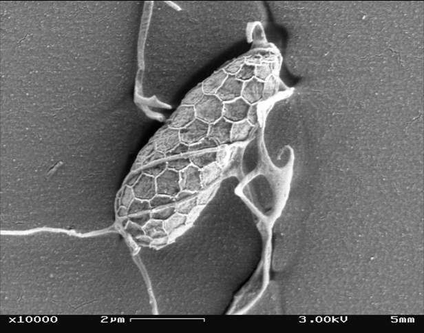
The cryptomonads (or cryptophytes) are a superclass of algae, most of which have plastids. They are traditionally considered a division of algae among phycologists, under the name of Cryptophyta. They are common in freshwater, and also occur in marine and brackish habitats. Each cell is around 10–50 μm in size and flattened in shape, with an anterior groove or pocket. At the edge of the pocket there are typically two slightly unequal flagella. Some may exhibit mixotrophy. They are classified as superclass Cryptomonada, which is divided into two classes: heterotrophic Goniomonadea and phototrophic Cryptophyceae. The two groups are united under three shared morphological characteristics: presence of a periplast, ejectisomes with secondary scroll, and mitochondrial cristae with flat tubules. Genetic studies as early as 1994 also supported the hypothesis that ''Goniomonas'' was sister to Cryptophyceae. A study in 2018 found strong evidence that the common ancestor of Crypto ... [...More Info...] [...Related Items...] OR: [Wikipedia] [Google] [Baidu] |
Cryptophyceae
The cryptophyceae are a class of algae, most of which have plastids. About 230 species are known, and they are common in freshwater, and also occur in marine and brackish habitats. Each cell is around 10–50 μm in size and flattened in shape, with an anterior groove or pocket. At the edge of the pocket there are typically two slightly unequal flagella. Some exhibit mixotrophy. Characteristics Cryptophytes are distinguished by the presence of characteristic extrusomes called ejectosomes or ejectisomes, which consist of two connected spiral ribbons held under tension. If the cells are irritated either by mechanical, chemical or light stress, they discharge, propelling the cell in a zig-zag course away from the disturbance. Large ejectosomes, visible under the light microscope, are associated with the pocket; smaller ones occur underneath the periplast, the cryptophyte-specific cell surrounding. Except for '' Chilomonas'', which has leucoplasts, cryptophytes have one or ... [...More Info...] [...Related Items...] OR: [Wikipedia] [Google] [Baidu] |
Eukaryotic Cell Anatomy
The eukaryotes ( ) constitute the domain of Eukaryota or Eukarya, organisms whose cells have a membrane-bound nucleus. All animals, plants, fungi, seaweeds, and many unicellular organisms are eukaryotes. They constitute a major group of life forms alongside the two groups of prokaryotes: the Bacteria and the Archaea. Eukaryotes represent a small minority of the number of organisms, but given their generally much larger size, their collective global biomass is much larger than that of prokaryotes. The eukaryotes emerged within the archaeal kingdom Promethearchaeati and its sole phylum Promethearchaeota. This implies that there are only two domains of life, Bacteria and Archaea, with eukaryotes incorporated among the Archaea. Eukaryotes first emerged during the Paleoproterozoic, likely as flagellated cells. The leading evolutionary theory is they were created by symbiogenesis between an anaerobic Promethearchaeati archaean and an aerobic proteobacterium, which formed the ... [...More Info...] [...Related Items...] OR: [Wikipedia] [Google] [Baidu] |
Algal Anatomy
Algae ( , ; : alga ) is an informal term for any organisms of a large and diverse group of photosynthetic organisms that are not plants, and includes species from multiple distinct clades. Such organisms range from unicellular microalgae, such as cyanobacteria, ''Chlorella'', and diatoms, to multicellular macroalgae such as kelp or brown algae which may grow up to in length. Most algae are aquatic organisms and lack many of the distinct cell and tissue types, such as stomata, xylem, and phloem that are found in land plants. The largest and most complex marine algae are called seaweeds. In contrast, the most complex freshwater forms are the Charophyta, a division of green algae which includes, for example, ''Spirogyra'' and stoneworts. Algae that are carried passively by water are plankton, specifically phytoplankton. Algae constitute a polyphyletic group because they do not include a common ancestor, and although eukaryotic algae with chlorophyll-bearing plastids seem to ... [...More Info...] [...Related Items...] OR: [Wikipedia] [Google] [Baidu] |
Electron Microscopy
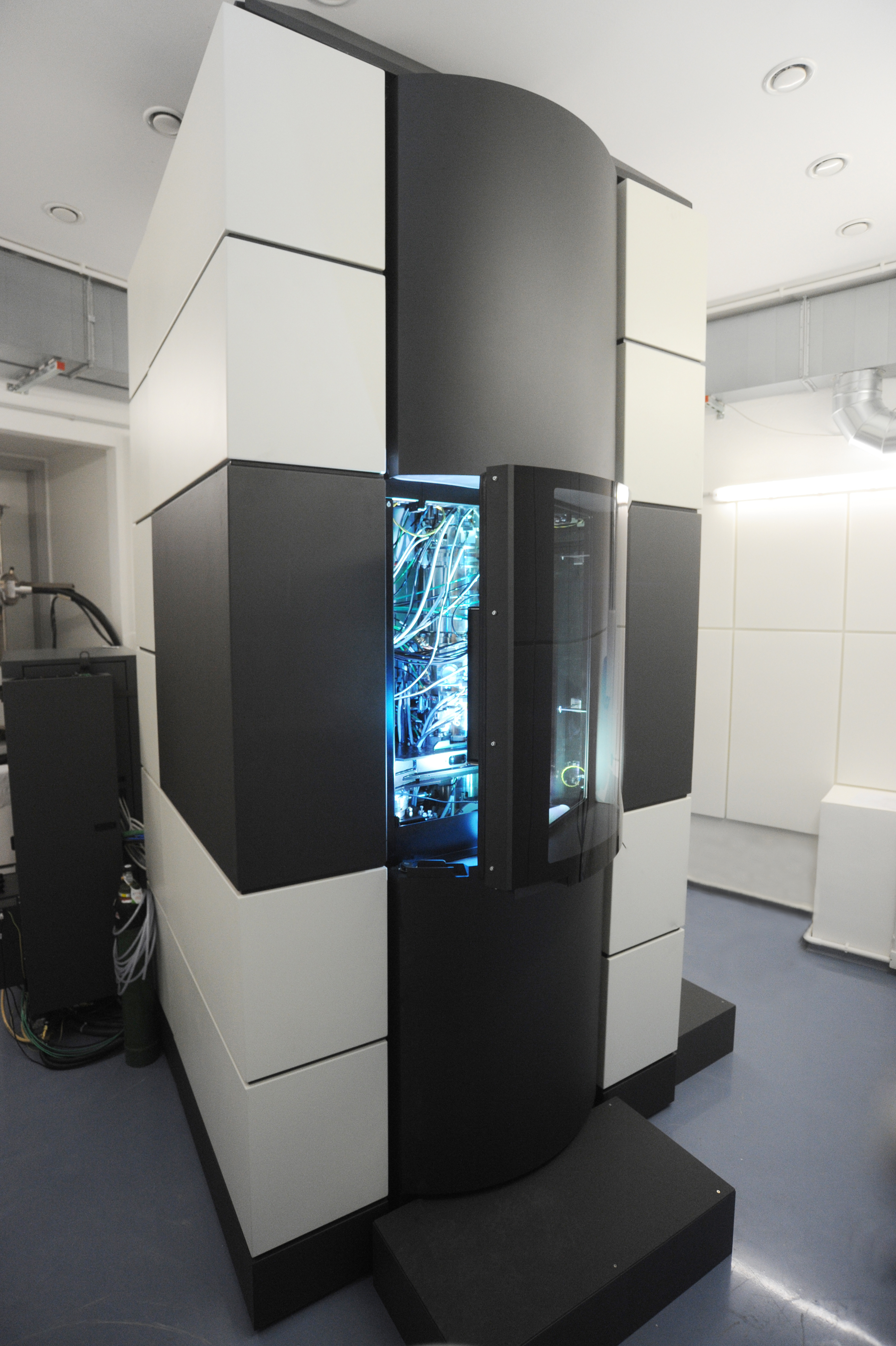
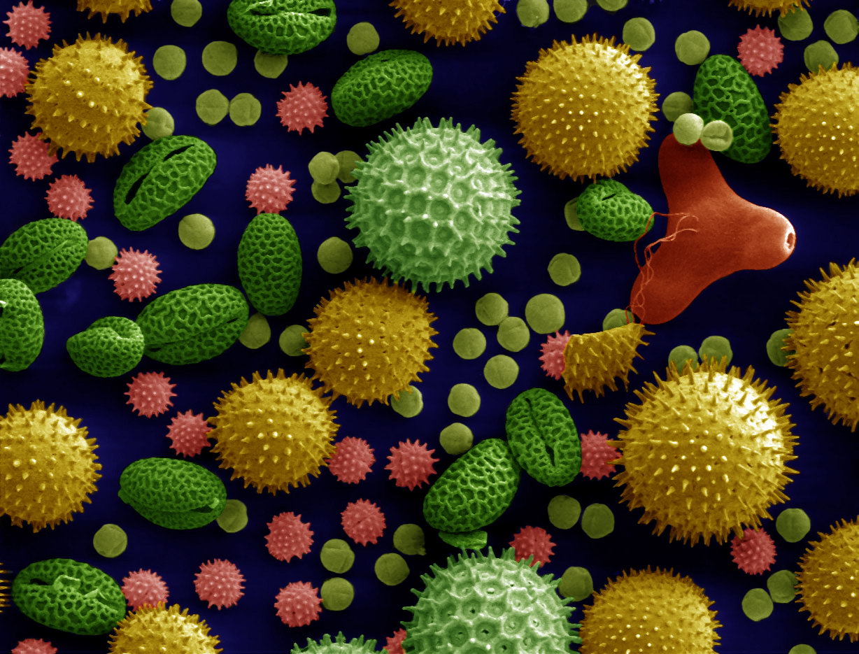
An electron microscope is a microscope that uses a beam of electrons as a source of illumination. It uses electron optics that are analogous to the glass lenses of an optical light microscope to control the electron beam, for instance focusing it to produce magnified images or electron diffraction patterns. As the wavelength of an electron can be up to 100,000 times smaller than that of visible light, electron microscopes have a much higher resolution of about 0.1 nm, which compares to about 200 nm for light microscopes. ''Electron microscope'' may refer to: * Transmission electron microscope (TEM) where swift electrons go through a thin sample * Scanning transmission electron microscope (STEM) which is similar to TEM with a scanned electron probe * Scanning electron microscope (SEM) which is similar to STEM, but with thick samples * Electron microprobe similar to a SEM, but more for chemical analysis * Low-energy electron microscope (LEEM), used to image surfaces * ... [...More Info...] [...Related Items...] OR: [Wikipedia] [Google] [Baidu] |
Visual Artifact
Visual artifacts (also artefacts) are anomalies apparent during visual representation as in digital graphics and other forms of imagery, especially photography and microscopy. In digital graphics * Image quality factors, different types of visual artifacts * Compression artifacts * Digital artifacts, visual artifacts resulting from digital image processing * Noise * Screen-door effect, also known as fixed-pattern noise (FPN), a visual artifact of digital projection technology * Ghosting (television) *Screen burn-in * Distortion * Silk screen effect * Rainbow effect * Screen tearing * Moiré pattern * Color banding In video entertainment Many people who use their computers as a hobby experience artifacting due to a hardware or software malfunction. The cases can differ but the usual causes are: * Temperature issues, such as failure of cooling fan. * Unsuited video card (graphics card) drivers. * Drivers that have values that the graphics card is not suited with. * Overcl ... [...More Info...] [...Related Items...] OR: [Wikipedia] [Google] [Baidu] |
Light Microscopy
Microscopy is the technical field of using microscopes to view subjects too small to be seen with the naked eye (objects that are not within the resolution range of the normal eye). There are three well-known branches of microscopy: optical, electron, and scanning probe microscopy, along with the emerging field of X-ray microscopy. Optical microscopy and electron microscopy involve the diffraction, reflection, or refraction of electromagnetic radiation/electron beams interacting with the specimen, and the collection of the scattered radiation or another signal in order to create an image. This process may be carried out by wide-field irradiation of the sample (for example standard light microscopy and transmission electron microscopy) or by scanning a fine beam over the sample (for example confocal laser scanning microscopy and scanning electron microscopy). Scanning probe microscopy involves the interaction of a scanning probe with the surface of the object of interest. The de ... [...More Info...] [...Related Items...] OR: [Wikipedia] [Google] [Baidu] |


