|
Macular Sparing
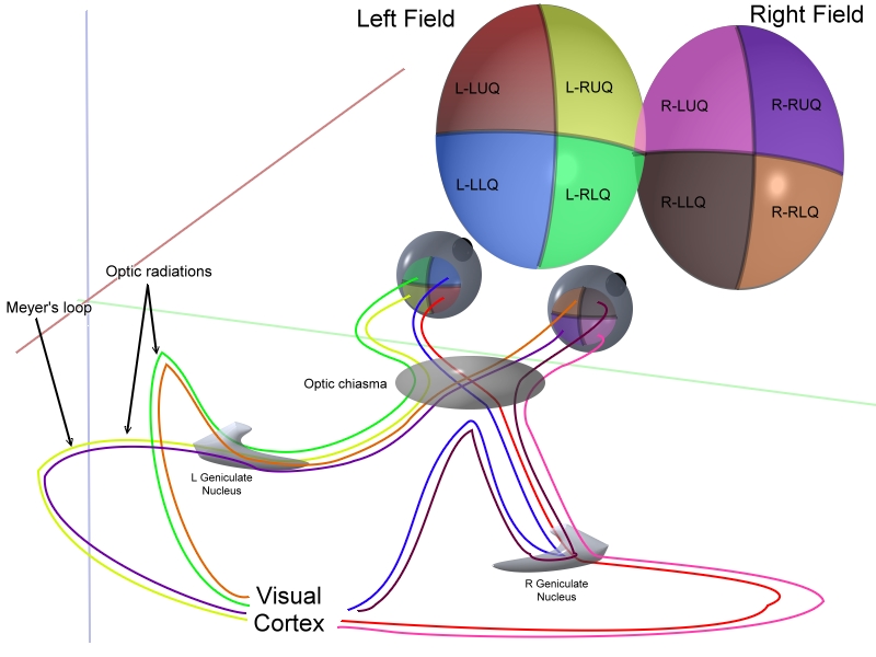
Macular sparing is visual field loss that preserves vision in the center of the visual field, otherwise known as the macula. It appears in people with damage to one hemisphere of their visual cortex, and occurs simultaneously with bilateral homonymous hemianopia or homonymous quadrantanopia. The exact mechanism behind this phenomenon is still uncertain.Whishaw, I. Q., & Kolb, B. (2015). Fundamentals of Human Neuropsychology (7th ed.). New York, NY: Worth Custom Publishing. The opposing effect, where vision in half of the center of the visual field is lost, is known as macular splitting.Windsor, R. L. (n.d.). Visual Fields in Brain Injury - Hemianopsia.net Everything you need to know about Hemianopsia. Retrieved from http://www.hemianopsia.net/visual-fields-in-brain-injury/ Causes The favored explanation for why the center visual field is preserved after large hemispheric lesions is that the macular regions of the cortex have a double vascular supply from the middle cerebral artery ... [...More Info...] [...Related Items...] OR: [Wikipedia] [Google] [Baidu] |
Visual Field Loss
The visual system is the physiological basis of visual perception (the ability to detect and process light). The system detects, transduces and interprets information concerning light within the visible range to construct an image and build a mental model of the surrounding environment. The visual system is associated with the eye and functionally divided into the optical system (including cornea and lens) and the neural system (including the retina and visual cortex). The visual system performs a number of complex tasks based on the ''image forming'' functionality of the eye, including the formation of monocular images, the neural mechanisms underlying stereopsis and assessment of distances to (depth perception) and between objects, motion perception, pattern recognition, accurate motor coordination under visual guidance, and colour vision. Together, these facilitate higher order tasks, such as object identification. The neuropsychological side of visual information proce ... [...More Info...] [...Related Items...] OR: [Wikipedia] [Google] [Baidu] |
Lateral Geniculate Nucleus
In neuroanatomy, the lateral geniculate nucleus (LGN; also called the lateral geniculate body or lateral geniculate complex) is a structure in the thalamus and a key component of the mammalian visual pathway. It is a small, ovoid, Anatomical terms of location#Dorsal_and_ventral, ventral projection of the thalamus where the thalamus connects with the optic nerve. There are two LGNs, one on the left and another on the right side of the thalamus. In humans, both LGNs have six layers of neurons (grey matter) alternating with optic fibers (white matter). The LGN receives information directly from the ascending retinal ganglion cells via the optic tract and from the reticular activating system. Neurons of the LGN send their axons through the optic radiation, a direct pathway to the primary visual cortex. In addition, the LGN receives many strong feedback connections from the primary visual cortex. In humans as well as other mammals, the two strongest pathways linking the eye to the bra ... [...More Info...] [...Related Items...] OR: [Wikipedia] [Google] [Baidu] |
Ophthalmology
Ophthalmology (, ) is the branch of medicine that deals with the diagnosis, treatment, and surgery of eye diseases and disorders. An ophthalmologist is a physician who undergoes subspecialty training in medical and surgical eye care. Following a medical degree, a doctor specialising in ophthalmology must pursue additional postgraduate residency training specific to that field. In the United States, following graduation from medical school, one must complete a four-year residency in ophthalmology to become an ophthalmologist. Following residency, additional specialty training (or fellowship) may be sought in a particular aspect of eye pathology. Ophthalmologists prescribe medications to treat ailments, such as eye diseases, implement laser therapy, and perform surgery when needed. Ophthalmologists provide both primary and specialty eye care—medical and surgical. Most ophthalmologists participate in academic research on eye diseases at some point in their training and many inc ... [...More Info...] [...Related Items...] OR: [Wikipedia] [Google] [Baidu] |
Visual Disturbances And Blindness
The visual system is the physiological basis of visual perception (the ability to detect and process light). The system detects, transduces and interprets information concerning light within the visible range to construct an image and build a mental model of the surrounding environment. The visual system is associated with the eye and functionally divided into the optical system (including cornea and lens) and the neural system (including the retina and visual cortex). The visual system performs a number of complex tasks based on the ''image forming'' functionality of the eye, including the formation of monocular images, the neural mechanisms underlying stereopsis and assessment of distances to (depth perception) and between objects, motion perception, pattern recognition, accurate motor coordination under visual guidance, and colour vision. Together, these facilitate higher order tasks, such as object identification. The neuropsychological side of visual information proces ... [...More Info...] [...Related Items...] OR: [Wikipedia] [Google] [Baidu] |
Bitemporal Hemianopsia
Bitemporal hemianopsia is the medical description of a type of partial blindness where vision is missing in the outer half of both the right and left visual field. It is usually associated with lesions of the optic chiasm, the area where the optic nerves from the right and left eyes cross near the pituitary gland. Causes In bitemporal hemianopsia, vision is missing in the outer (temporal or lateral) half of both the right and left visual fields. Information from the temporal visual field falls on the nasal (medial) retina. The nasal retina is responsible for carrying the information along the optic nerve, and crosses to the other side at the optic chiasm. When there is compression at the optic chiasm, the visual impulse from both nasal retina are affected, leading to inability to see the temporal, or peripheral, field of vision. This phenomenon is known as bitemporal hemianopsia. Knowing the neurocircuitry of visual signal flow through the optic tract is very important in unde ... [...More Info...] [...Related Items...] OR: [Wikipedia] [Google] [Baidu] |
Binasal Hemianopsia
Binasal hemianopsia is the medical description of a type of partial blindness where vision is missing in the inner half of both the right and left visual field. It is associated with certain lesions of the eye and of the central nervous system, such as congenital hydrocephalus. Causes In binasal hemianopsia, vision is missing in the inner (nasal or medial) half of both the right and left visual fields. Information from the nasal visual field falls on the temporal (lateral) retina. Those lateral retinal nerve fibers do not cross in the optic chiasm. Calcification of the internal carotid arteries can impinge the uncrossed, lateral retinal fibers, leading to loss of vision in the nasal field. Clinical testing of visual fields (by confrontation) can produce a false positive result, particularly in inferior nasal quadrants. Management Etymology The absence of Visual perception, vision in half of a visual field is described as ''hemianopsia''. The absence of visual perception in o ... [...More Info...] [...Related Items...] OR: [Wikipedia] [Google] [Baidu] |
Hemianopsia
Hemianopsia, or hemianopia, is a loss of vision or blindness ( anopsia) in half the visual field, usually on one side of the vertical midline. The most common causes of this damage are stroke, brain tumor, and trauma. This article deals only with permanent hemianopsia, and not with transitory or temporary hemianopsia, as identified by William Wollaston PRS in 1824. Temporary hemianopsia can occur in the aura phase of migraine. Etymology The word ''hemianopsia'' is from Greek origins, where: * ''hemi'' means "half", * ''an'' means "without", and * ''opsia'' means "seeing". Types When the pathology involves both eyes, it is either homonymous or heteronymous. Homonymous hemianopsia Paris as seen with left homonymous hemianopsia A homonymous hemianopsia is the loss of half of the visual field on the same side in both eyes. The visual images that we see to the right side travel from both eyes to the left side of the brain, while the visual images we see to the left side i ... [...More Info...] [...Related Items...] OR: [Wikipedia] [Google] [Baidu] |
Visual Acuity
Visual acuity (VA) commonly refers to the clarity of visual perception, vision, but technically rates an animal's ability to recognize small details with precision. Visual acuity depends on optical and neural factors. Optical factors of the eye influence the sharpness of an image on its retina. Neural factors include the health and functioning of the retina, of the neural pathways to the brain, and of the interpretative faculty of the brain. The most commonly referred-to visual acuity is ''distance acuity'' or ''far acuity'' (e.g., "20/20 vision"), which describes someone's ability to recognize small details at a far distance. This ability is compromised in people with myopia, also known as short-sightedness or near-sightedness. Another visual acuity is ''Near visual acuity, near acuity'', which describes someone's ability to recognize small details at a near distance. This ability is compromised in people with hyperopia, also known as long-sightedness or far-sightedness. A com ... [...More Info...] [...Related Items...] OR: [Wikipedia] [Google] [Baidu] |
Optic Tract
In neuroanatomy, the optic tract () is a part of the visual system in the brain. It is a continuation of the optic nerve that relays information from the optic chiasm to the ipsilateral lateral geniculate nucleus (LGN), pretectal nuclei, and superior colliculus. It is composed of two individual tracts, the left optic tract and the right optic tract, each of which conveys visual information exclusive to its respective contralateral half of the visual field. Each of these tracts is derived from a combination of temporal and nasal retinal fibers from each eye that corresponds to one half of the visual field. In more specific terms, the optic tract contains fibers from the ipsilateral temporal hemiretina and contralateral nasal hemiretina. Anatomy Arterial supply The optic tract receives arterial supply from the anterior choroidal artery, and posterior communicating artery. Function Visual function The optic tract carries retinal information relating to the whole visua ... [...More Info...] [...Related Items...] OR: [Wikipedia] [Google] [Baidu] |
Macula Of Retina
The macula (/ˈmakjʊlə/) or macula lutea is an oval-shaped pigmented area in the center of the retina of the human eye and in other animals. The macula in humans has a diameter of around and is subdivided into the umbo, foveola, foveal avascular zone, fovea, parafovea, and perifovea areas. The anatomical macula at a size of is much larger than the clinical macula which, at a size of , corresponds to the anatomical fovea. The macula is responsible for the central, high-resolution, color vision that is possible in good light. This kind of vision is impaired if the macula is damaged, as in macular degeneration. The clinical macula is seen when viewed from the pupil, as in ophthalmoscopy or retinal photography. The term macula lutea comes from Latin ''macula'', "spot", and ''lutea'', "yellow". Structure The macula is an oval-shaped pigmented area in the center of the retina of the human eye and other animal eyes. Its center is shifted slightly away from the optical a ... [...More Info...] [...Related Items...] OR: [Wikipedia] [Google] [Baidu] |
Visual Field Testing
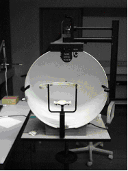
A visual field test is an eye examination that can detect dysfunction in central and peripheral vision which may be caused by various medical conditions such as glaucoma, stroke, pituitary disease, brain tumours or other neurological deficits. Visual field testing can be performed clinically by keeping the subject's gaze fixed while presenting objects at various places within their visual field. Simple manual equipment can be used such as in the tangent screen test or the Amsler grid. When dedicated machinery is used it is called a perimeter. The exam may be performed by a technician in one of several ways. The test may be performed by a technician directly, with the assistance of a machine, or completely by an automated machine. Machine-based tests aid diagnostics by allowing a detailed printout of the patient's visual field. Other names for this test may include perimetry, Tangent screen exam, Automated perimetry exam, Goldmann visual field exam, or brand names such as the H ... [...More Info...] [...Related Items...] OR: [Wikipedia] [Google] [Baidu] |
Posterior Cerebral Artery
The posterior cerebral artery (PCA) is one of a pair of cerebral arteries that supply oxygenated blood to the occipital lobe, as well as the medial and inferior aspects of the temporal lobe of the human brain. The two arteries originate from the distal end of the basilar artery, where it bifurcates into the left and right posterior cerebral arteries. These anastomose with the middle cerebral artery, middle cerebral arteries and internal carotid artery, internal carotid arteries via the posterior communicating arteries. Structure The posterior cerebral artery is subdivided into 4 segments: P1: pre-communicating segment * Originated at the termination of the basilar artery * May give rise to the artery of Percheron if present P2: post-communicating segment * From the PCOM around the midbrain * Terminates as it enters the quadrigeminal ganglion * Gives rise to the choroidal branches (medial and lateral posterior choroidal arteries) P3: quadrigeminal segment * Courses poster ... [...More Info...] [...Related Items...] OR: [Wikipedia] [Google] [Baidu] |


