|
Lowe Syndrome
Oculocerebrorenal syndrome (also called Lowe syndrome) is a rare X-linked recessive disorder characterized by congenital cataracts, hypotonia, intellectual disability, proximal tubular acidosis, aminoaciduria and low-molecular-weight proteinuria. Lowe syndrome can be considered a cause of Fanconi syndrome (bicarbonaturia, renal tubular acidosis, potassium loss and sodium loss). Signs and symptoms Boys with Lowe syndrome are born with cataracts in both eyes; glaucoma is present in about half of the individuals with Lowe syndrome, though usually not at birth. While not present at birth, kidney problems develop in many affected boys at about one year of age. Renal pathology is characterized by an abnormal loss of certain substances into the urine, including bicarbonate, sodium, potassium, amino acids, organic acids, albumin, calcium and L-carnitine. This problem is known as Fanconi-type renal tubular dysfunction. Genetics This syndrome is caused by mutations in the '' OCRL'' ... [...More Info...] [...Related Items...] OR: [Wikipedia] [Google] [Baidu] |
OCRL
Inositol polyphosphate 5-phosphatase OCRL-1, also known as Lowe oculocerebrorenal syndrome protein, is an enzyme encoded by the ''OCRL'' gene located on the X chromosome in humans. This gene encodes an inositol polyphosphate 5-phosphatase. The responsible gene locus is at Xq26.1. This phosphatase enzyme is in part responsible for regulating membrane trafficking actin polymerization, and is located in several subcellular parts of the trans-Golgi network. Deficiencies in OCRL-1 are associated with oculocerebrorenal syndrome and also have been linked to Dent's disease Dent's disease (or Dent disease) is a rare X-linked recessive inherited condition that affects the proximal renal tubules of the kidney. It is one cause of Fanconi syndrome, and is characterized by tubular proteinuria, hypercalciuria, excess calciu .... References Further reading * * * * * * * * * * * * * * * * * * * * External links GeneReviews/NCBI/NIH/UW entry on Lowe Syndrome* PDBe-KB provides an over ... [...More Info...] [...Related Items...] OR: [Wikipedia] [Google] [Baidu] |
Amino Acids
Amino acids are organic compounds that contain both amino and carboxylic acid functional groups. Although over 500 amino acids exist in nature, by far the most important are the Proteinogenic amino acid, 22 α-amino acids incorporated into proteins. Only these 22 appear in the genetic code of life. Amino acids can be classified according to the locations of the core structural functional groups (Alpha and beta carbon, alpha- , beta- , gamma- (γ-) amino acids, etc.); other categories relate to Chemical polarity, polarity, ionization, and side-chain group type (aliphatic, Open-chain compound, acyclic, aromatic, Chemical polarity, polar, etc.). In the form of proteins, amino-acid ''Residue (chemistry)#Biochemistry, residues'' form the second-largest component (water being the largest) of human muscles and other tissue (biology), tissues. Beyond their role as residues in proteins, amino acids participate in a number of processes such as neurotransmitter transport and biosynthesi ... [...More Info...] [...Related Items...] OR: [Wikipedia] [Google] [Baidu] |
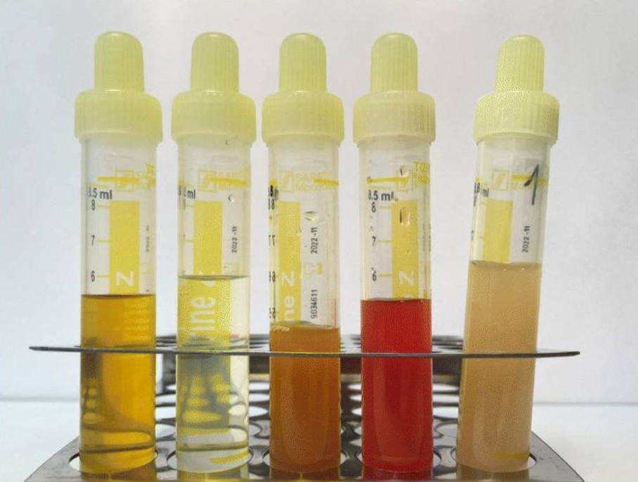
Urinalysis
Urinalysis, a portmanteau of the words ''urine'' and ''analysis'', is a Test panel, panel of medical tests that includes physical (macroscopic) examination of the urine, chemical evaluation using urine test strips, and #Microscopic examination, microscopic examination. Macroscopic examination targets parameters such as color, clarity, odor, and specific gravity; urine test strips measure chemical properties such as pH, glucose concentration, and protein levels; and microscopy is performed to identify elements such as Cell (biology), cells, urinary casts, Crystalluria, crystals, and organisms. Background Urine is produced by the filtration of blood in the kidneys. The formation of urine takes place in microscopic structures called nephrons, about one million of which are found in a normal human kidney. Blood enters the kidney though the renal artery and flows through the kidney's vasculature into the Glomerulus (kidney), glomerulus, a tangled knot of capillaries surrounded by Bow ... [...More Info...] [...Related Items...] OR: [Wikipedia] [Google] [Baidu] |
Genetic Testing
Genetic testing, also known as DNA testing, is used to identify changes in DNA sequence or chromosome structure. Genetic testing can also include measuring the results of genetic changes, such as RNA analysis as an output of gene expression, or through biochemical analysis to measure specific protein output. In a medical setting, genetic testing can be used to diagnose or rule out suspected genetic disorders, predict risks for specific conditions, or gain information that can be used to customize medical treatments based on an individual's genetic makeup. Genetic testing can also be used to determine biological relatives, such as a child's biological parentage (genetic mother and father) through DNA paternity testing, or be used to broadly predict an individual's ancestry. Genetic testing of plants and animals can be used for similar reasons as in humans (e.g. to assess relatedness/ancestry or predict/diagnose genetic disorders), to gain information used for selective breed ... [...More Info...] [...Related Items...] OR: [Wikipedia] [Google] [Baidu] |
Nephron
The nephron is the minute or microscopic structural and functional unit of the kidney. It is composed of a renal corpuscle and a renal tubule. The renal corpuscle consists of a tuft of capillaries called a glomerulus and a cup-shaped structure called Bowman's capsule. The renal tubule extends from the capsule. The capsule and tubule are connected and are composed of epithelial cells with a lumen. A healthy adult has 1 to 1.5 million nephrons in each kidney. Blood is filtered as it passes through three layers: the endothelial cells of the capillary wall, its basement membrane, and between the podocyte foot processes of the lining of the capsule. The tubule has adjacent peritubular capillaries that run between the descending and ascending portions of the tubule. As the fluid from the capsule flows down into the tubule, it is processed by the epithelial cells lining the tubule: water is reabsorbed and substances are exchanged (some are added, others are removed); first wit ... [...More Info...] [...Related Items...] OR: [Wikipedia] [Google] [Baidu] |
Fibroblast
A fibroblast is a type of cell (biology), biological cell typically with a spindle shape that synthesizes the extracellular matrix and collagen, produces the structural framework (Stroma (tissue), stroma) for animal Tissue (biology), tissues, and plays a critical role in wound healing. Fibroblasts are the most common cells of connective tissue in animals. Structure Fibroblasts have a branched cytoplasm surrounding an elliptical, speckled cell nucleus, nucleus having two or more nucleoli. Active fibroblasts can be recognized by their abundant rough endoplasmic reticulum (RER). Inactive fibroblasts, called 'fibrocytes', are smaller, spindle-shaped, and have less RER. Although disjointed and scattered when covering large spaces, fibroblasts often locally align in parallel clusters when crowded together. Unlike the epithelial cells lining the body structures, fibroblasts do not form flat monolayers and are not restricted by a polarizing attachment to a basal lamina on one side, a ... [...More Info...] [...Related Items...] OR: [Wikipedia] [Google] [Baidu] |
Retinal Pigment Epithelial
The pigmented layer of retina or retinal pigment epithelium (RPE) is the pigmented cell layer just outside the neurosensory retina that nourishes retinal visual cells, and is firmly attached to the underlying choroid and overlying retinal visual cells. History The RPE was known in the 18th and 19th centuries as the pigmentum nigrum, referring to the observation that the RPE is dark (black in many animals, brown in humans); and as the tapetum nigrum, referring to the observation that in animals with a tapetum lucidum, in the region of the tapetum lucidum the RPE is not pigmented. Anatomy The RPE is composed of a single layer of hexagonal cells that are densely packed with pigment granules. When viewed from the outer surface, these cells are smooth and hexagonal in shape. When seen in section, each cell consists of an outer non-pigmented part containing a large oval nucleus and an inner pigmented portion which extends as a series of straight thread-like processes between the rod ... [...More Info...] [...Related Items...] OR: [Wikipedia] [Google] [Baidu] |
Cilia
The cilium (: cilia; ; in Medieval Latin and in anatomy, ''cilium'') is a short hair-like membrane protrusion from many types of eukaryotic cell. (Cilia are absent in bacteria and archaea.) The cilium has the shape of a slender threadlike projection that extends from the surface of the much larger cell body. Eukaryotic flagella found on sperm cells and many protozoans have a similar structure to motile cilia that enables swimming through liquids; they are longer than cilia and have a different undulating motion. There are two major classes of cilia: ''motile'' and ''non-motile'' cilia, each with two subtypes, giving four types in all. A cell will typically have one primary cilium or many motile cilia. The structure of the cilium core, called the axoneme, determines the cilium class. Most motile cilia have a central pair of single microtubules surrounded by nine pairs of double microtubules called a 9+2 axoneme. Most non-motile cilia have a 9+0 axoneme that lacks the central pai ... [...More Info...] [...Related Items...] OR: [Wikipedia] [Google] [Baidu] |
Protein Domain
In molecular biology, a protein domain is a region of a protein's Peptide, polypeptide chain that is self-stabilizing and that Protein folding, folds independently from the rest. Each domain forms a compact folded Protein tertiary structure, three-dimensional structure. Many proteins consist of several domains, and a domain may appear in a variety of different proteins. Molecular evolution uses domains as building blocks and these may be recombined in different arrangements to create proteins with different functions. In general, domains vary in length from between about 50 amino acids up to 250 amino acids in length. The shortest domains, such as zinc fingers, are stabilized by metal ions or Disulfide bond, disulfide bridges. Domains often form functional units, such as the calcium-binding EF-hand, EF hand domain of calmodulin. Because they are independently stable, domains can be "swapped" by genetic engineering between one protein and another to make chimera (protein), chimeric ... [...More Info...] [...Related Items...] OR: [Wikipedia] [Google] [Baidu] |
Rab (G-protein)
The Rab family of proteins is a member of the Ras superfamily of small G proteins. Approximately 70 types of Rabs have now been identified in humans. Rab proteins generally possess a GTPase fold, which consists of a six-stranded beta sheet which is flanked by five alpha helices. Rab GTPases regulate many steps of membrane trafficking, including vesicle formation, vesicle movement along actin and tubulin networks, and membrane fusion. These processes make up the route through which cell surface proteins are trafficked from the Golgi to the plasma membrane and are recycled. Surface protein recycling returns proteins to the surface whose function involves carrying another protein or substance inside the cell, such as the transferrin receptor, or serves as a means of regulating the number of a certain type of protein molecules on the surface. Function Rab proteins are peripheral membrane proteins, anchored to a membrane via a lipid group covalently linked to an amino acid. Spec ... [...More Info...] [...Related Items...] OR: [Wikipedia] [Google] [Baidu] |
Inositol Polyphosphate-5-phosphatase
The enzyme inositol-polyphosphate 5-phosphatase (), systematic name 1D-''myo''-inositol-1,4,5-trisphosphate 5-phosphohydrolase, catalyses the following reactions: : (1) D-''myo''-inositol 1,4,5-trisphosphate + H2O \rightleftharpoons ''myo''-inositol 1,4-bisphosphate + phosphate : (2) 1D-''myo''-inositol 1,3,4,5-tetrakisphosphate + H2O \rightleftharpoons 1D-''myo''-inositol 1,3,4-trisphosphate + phosphate Ten mammalian isoforms A protein isoform, or "protein variant", is a member of a set of highly similar proteins that originate from a single gene and are the result of genetic differences. While many perform the same or similar biological roles, some isoforms have uniqu ... are known. Other names of this enzyme include type I inositol-polyphosphate phosphatase, inositol trisphosphate phosphomonoesterase, InsP3/Ins(1,3,4,5)P4 5-phosphatase, inosine triphosphatase, D-''myo''-inositol 1,4,5-triphosphate 5-phosphatase, D-''myo''-inositol 1,4,5-trisphosphate 5-phosphatase, L-''myo' ... [...More Info...] [...Related Items...] OR: [Wikipedia] [Google] [Baidu] |
L-carnitine
Carnitine is a quaternary ammonium compound involved in metabolism in most mammals, plants, and some bacteria. In support of energy metabolism, carnitine transports long-chain fatty acids from the cytosol into mitochondria to be oxidized for free energy production, and also participates in removing products of metabolism from cells. Given its key metabolic roles, carnitine is concentrated in tissues like skeletal and cardiac muscle that metabolize fatty acids as an energy source. Generally individuals, including strict vegetarians, synthesize enough L-carnitine in vivo. Carnitine exists as one of two stereoisomers: the two enantiomers -carnitine (''S''-(+)-) and -carnitine (''R''-(−)-). Both are biologically active, but only -carnitine naturally occurs in animals, and -carnitine is toxic as it inhibits the activity of the -form. At room temperature, pure carnitine is a whiteish powder, and a water-soluble zwitterion with relatively low toxicity. Derived from amino acids, carnit ... [...More Info...] [...Related Items...] OR: [Wikipedia] [Google] [Baidu] |


