|
Lingual Nerve
The lingual nerve carries sensory innervation from the anterior two-thirds of the tongue. It contains fibres from both the mandibular division of the trigeminal nerve (CN V) and from the facial nerve (CN VII). The fibres from the trigeminal nerve are for touch, pain and temperature (general sensation), and the ones from the facial nerve are for taste (special sensation). Structure Origin The lingual nerve arises from the posterior trunk of mandibular nerve (CN V) within the infratemporal fossa. Course The lingual nerve first courses deep to the lateral pterygoid muscle and superior to the tensor veli palatini muscle; while passing between these two muscle, it is joined by the chorda tympani, and often by a communicating branch from the inferior alveolar nerve. The nerve then comes to pass inferoanteriorly upon the medial pterygoid muscle towards the medial aspect of the ramus of mandible, eventually meeting the mandible at the junction of the ramus and body of mandibl ... [...More Info...] [...Related Items...] OR: [Wikipedia] [Google] [Baidu] |
Maxillary Nerve
In neuroanatomy, the maxillary nerve (V) is one of the three branches or divisions of the trigeminal nerve, the fifth (CN V) cranial nerve. It comprises the principal functions of Sense, sensation from the maxilla, nasal cavity, Sinus (anatomy), sinuses, the palate and subsequently that of the mid-face, and is intermediate, both in position and size, between the ophthalmic nerve and the mandibular nerve.Illustrated Anatomy of the Head and Neck, Fehrenbach and Herring, Elsevier, 2012, page 180 Structure It begins at the middle of the trigeminal ganglion as a flattened plexiform band then it passes through the lateral wall of the cavernous sinus. It leaves the skull through the foramen rotundum, where it becomes more cylindrical in form, and firmer in texture. After leaving foramen rotundum it gives two branches to the pterygopalatine ganglion. It then crosses the pterygopalatine fossa, inclines lateralward on the back of the maxilla, and enters the orbit through the inferior orb ... [...More Info...] [...Related Items...] OR: [Wikipedia] [Google] [Baidu] |
Hyoglossus
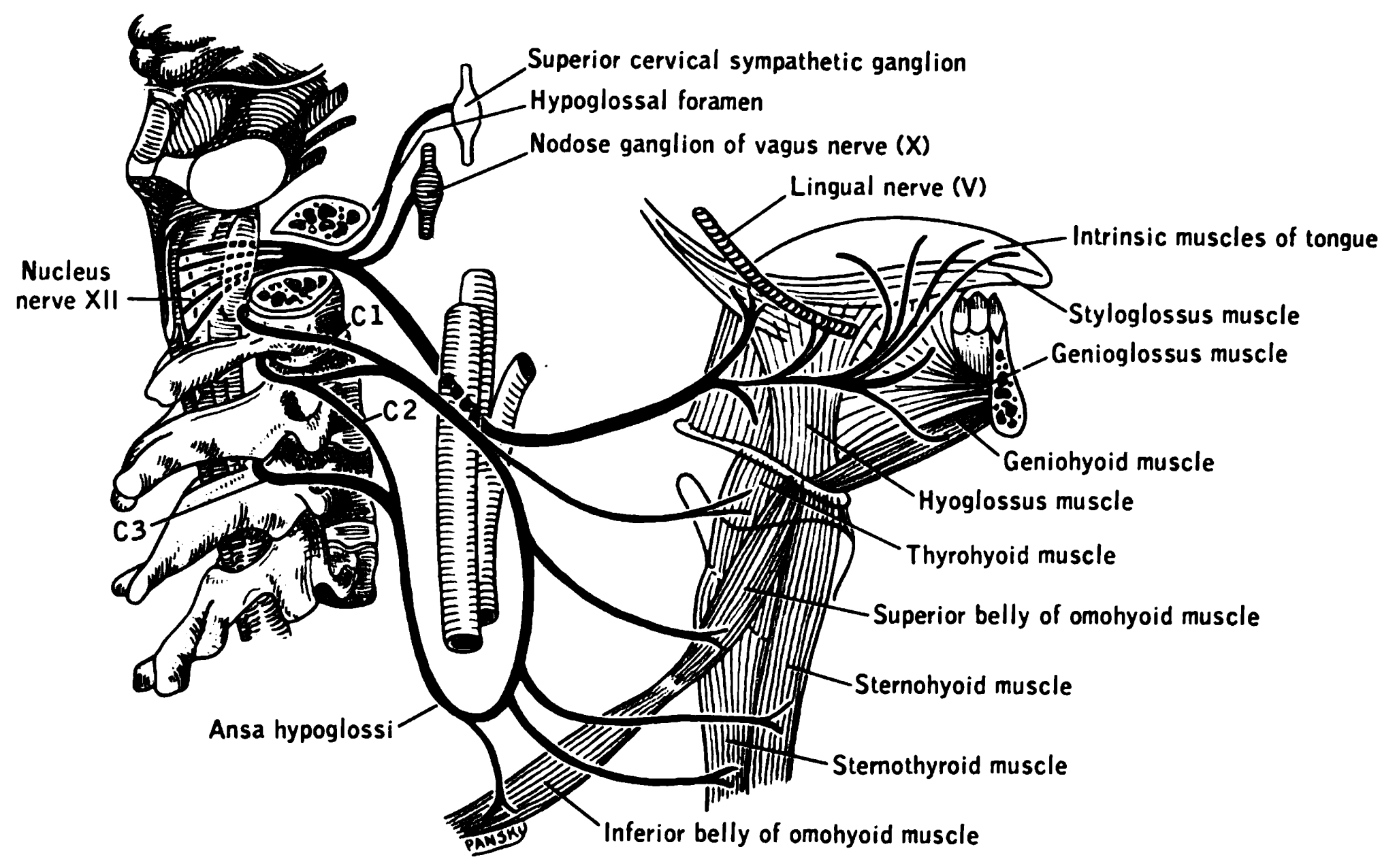
The hyoglossus is a thin and quadrilateral extrinsic muscle of the tongue. It originates from the hyoid bone; it inserts onto the side of the tongue. It is innervated by the hypoglossal nerve (cranial nerve XII). It acts to depress and retract the tongue. Structure It forms a part of the floor of submandibular triangle. Origin from the side of the body and from the whole length of the greater cornu of the hyoid bone. The fibers arising from the body of the hyoid bone overlap those from the greater cornu. Insertion Its fibres pass almost vertically upward to enter the side of the tongue, inserting between the styloglossus and the inferior longitudinal muscle of the tongue. Relations Structures that are medial/deep to the hyoglossus are the glossopharyngeal nerve (CN IX), the stylohyoid ligament and the lingual artery and lingual vein. The lingual vein passes medial to the hyoglossus. The lingual artery passes deep to the hyoglossus. Laterally, in between the hyo ... [...More Info...] [...Related Items...] OR: [Wikipedia] [Google] [Baidu] |
Lingual Branches Of Hypoglossal Nerve
The hypoglossal nerve, also known as the twelfth cranial nerve, cranial nerve XII, or simply CN XII, is a cranial nerve that innervates all the extrinsic and intrinsic muscles of the tongue except for the palatoglossus, which is innervated by the vagus nerve. CN XII is a nerve with a sole motor function. The nerve arises from the hypoglossal nucleus in the medulla as a number of small rootlets, pass through the hypoglossal canal and down through the neck, and eventually passes up again over the tongue muscles it supplies into the tongue. The nerve is involved in controlling tongue movements required for speech and swallowing, including sticking out the tongue and moving it from side to side. Damage to the nerve or the neural pathways which control it can affect the ability of the tongue to move and its appearance, with the most common sources of damage being injury from trauma or surgery, and motor neuron disease. The first recorded description of the nerve was by Herop ... [...More Info...] [...Related Items...] OR: [Wikipedia] [Google] [Baidu] |
Wisdom Tooth
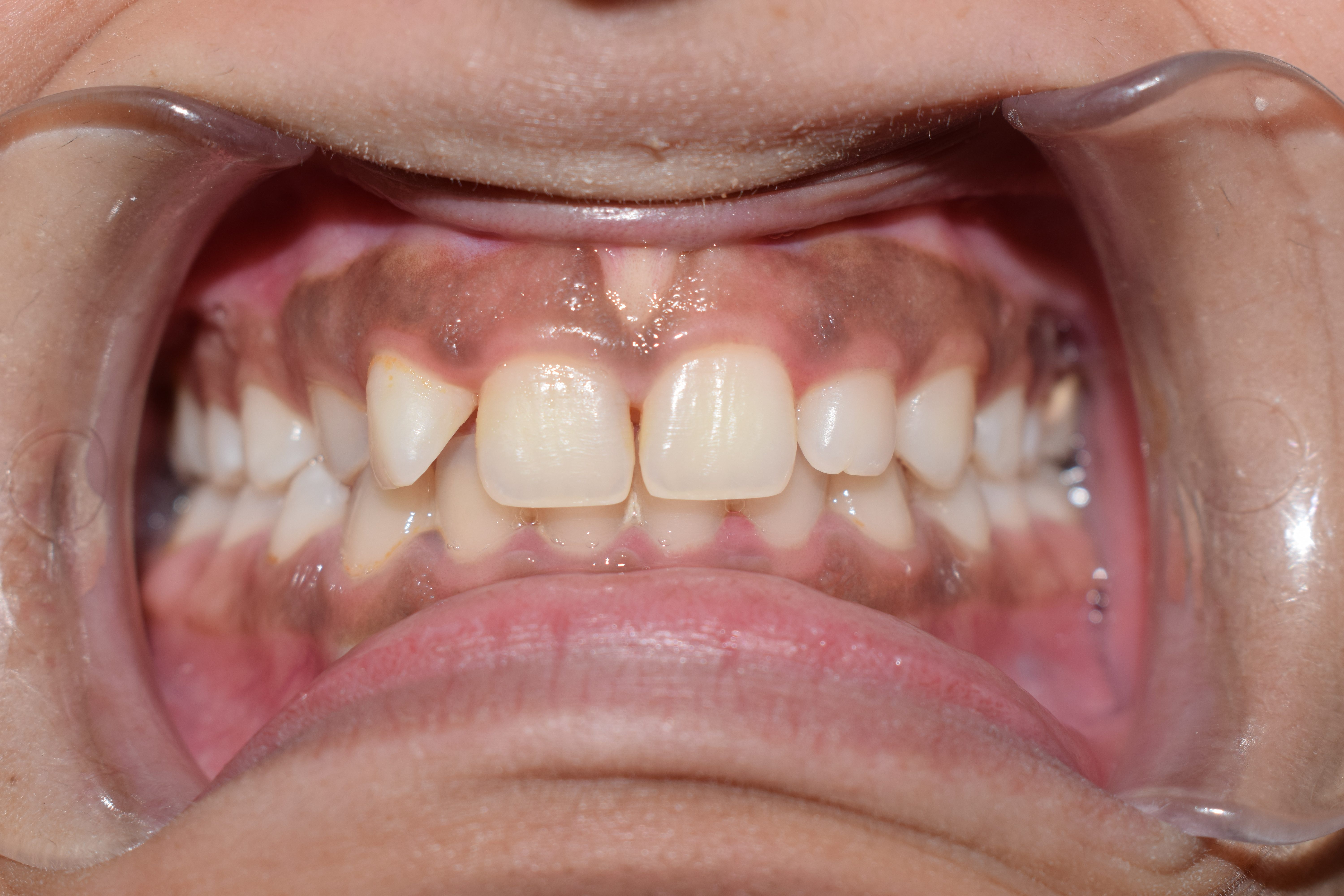
The third molar, commonly called wisdom tooth, is the most posterior of the three molars in each quadrant of the human dentition. The age at which wisdom teeth come through ( erupt) is variable, but this generally occurs between late teens and early twenties. Most adults have four wisdom teeth, one in each of the four quadrants, but it is possible to have none, fewer, or more, in which case the extras are called supernumerary teeth. Wisdom teeth may become stuck ( impacted) and not erupt fully, if there is not enough space for them to come through normally. Impacted wisdom teeth are still sometimes removed for orthodontic treatment, believing that they move the other teeth and cause crowding, though this is disputed. Impacted wisdom teeth may suffer from tooth decay if oral hygiene becomes more difficult. Wisdom teeth which are partially erupted through the gum may also cause inflammation and infection in the surrounding gum tissues, termed pericoronitis. More conservativ ... [...More Info...] [...Related Items...] OR: [Wikipedia] [Google] [Baidu] |
Sympathetic Nervous System
The sympathetic nervous system (SNS or SANS, sympathetic autonomic nervous system, to differentiate it from the somatic nervous system) is one of the three divisions of the autonomic nervous system, the others being the parasympathetic nervous system and the enteric nervous system. The enteric nervous system is sometimes considered part of the autonomic nervous system, and sometimes considered an independent system. The autonomic nervous system functions to regulate the body's unconscious actions. The sympathetic nervous system's primary process is to stimulate the body's fight or flight response. It is, however, constantly active at a basic level to maintain homeostasis. The sympathetic nervous system is described as being antagonistic to the parasympathetic nervous system. The latter stimulates the body to "feed and breed" and to (then) "rest-and-digest". The SNS has a major role in various physiological processes such as blood glucose levels, body temperature, cardiac output, ... [...More Info...] [...Related Items...] OR: [Wikipedia] [Google] [Baidu] |
Parasympathetic
The parasympathetic nervous system (PSNS) is one of the three divisions of the autonomic nervous system, the others being the sympathetic nervous system and the enteric nervous system. The autonomic nervous system is responsible for regulating the body's unconscious actions. The parasympathetic system is responsible for stimulation of "rest-and-digest" or "feed-and-breed" activities that occur when the body is at rest, especially after eating, including sexual arousal, salivation, lacrimation (tears), urination, digestion, and defecation. Its action is described as being complementary to that of the sympathetic nervous system, which is responsible for stimulating activities associated with the fight-or-flight response. Nerve fibres of the parasympathetic nervous system arise from the central nervous system. Specific nerves include several cranial nerves, specifically the oculomotor nerve, facial nerve, glossopharyngeal nerve, and vagus nerve. Three spinal nerves in ... [...More Info...] [...Related Items...] OR: [Wikipedia] [Google] [Baidu] |
Chorda Tympani Nerve
Chorda tympani is a branch of the facial nerve that carries gustatory (taste) sensory innervation from the front of the tongue and parasympathetic ( secretomotor) innervation to the submandibular and sublingual salivary glands. Chorda tympani has a complex course from the brainstem, through the temporal bone and middle ear, into the infratemporal fossa, and ending in the oral cavity. Structure Chorda tympani fibers emerge from the pons of the brainstem as part of the intermediate nerve of the facial nerve. The facial nerve exits the cranial cavity through the internal acoustic meatus and enters the facial canal. In the facial canal, the chorda tympani branches off the facial nerve and enters the lateral wall of the tympanic cavity inside the middle ear where it runs across the tympanic membrane (from posterior to anterior) and medial to the neck of the malleus. The chorda then exits the skull by descending through the petrotympanic fissure into the infratemporal fossa ju ... [...More Info...] [...Related Items...] OR: [Wikipedia] [Google] [Baidu] |
Gingiva
The gums or gingiva (: gingivae) consist of the mucosal tissue that lies over the mandible and maxilla inside the mouth. Gum health and disease can have an effect on general health. Structure The gums are part of the soft tissue lining of the mouth. They surround the teeth and provide a seal around them. Unlike the soft tissue linings of the lips and cheeks, most of the gums are tightly bound to the underlying bone which helps resist the friction of food passing over them. Thus when healthy, it presents an effective barrier to the barrage of periodontal insults to deeper tissue. Healthy gums are usually coral pink in light skinned people, and may be naturally darker with melanin pigmentation. Changes in color, particularly increased redness, together with swelling and an increased tendency to bleed, suggest an inflammation that is possibly due to the accumulation of bacterial plaque. Overall, the clinical appearance of the tissue reflects the underlying histology, both in hea ... [...More Info...] [...Related Items...] OR: [Wikipedia] [Google] [Baidu] |
Oral Cavity
A mouth also referred to as the oral is the body orifice through which many animals ingest food and vocalize. The body cavity immediately behind the mouth opening, known as the oral cavity (or in Latin), is also the first part of the alimentary canal, which leads to the pharynx and the gullet. In tetrapod vertebrates, the mouth is bounded on the outside by the lips and cheeks — thus the oral cavity is also known as the buccal cavity (from Latin ', meaning "cheek") — and contains the tongue on the inside. Except for some groups like birds and lissamphibians, vertebrates usually have teeth in their mouths, although some fish species have pharyngeal teeth instead of oral teeth. Most bilaterian phyla, including arthropods, molluscs and chordates, have a two-opening gut tube with a mouth at one end and an anus at the other. Which end forms first in ontogeny is a criterion used to classify bilaterian animals into protostomes and deuterostomes. Development In t ... [...More Info...] [...Related Items...] OR: [Wikipedia] [Google] [Baidu] |
Glossopharyngeal Nerve
The glossopharyngeal nerve (), also known as the ninth cranial nerve, cranial nerve IX, or simply CN IX, is a cranial nerve that exits the brainstem from the sides of the upper Medulla oblongata, medulla, just anterior (closer to the nose) to the vagus nerve. Being a mixed nerve (sensorimotor), it carries afferent sensory and efferent motor information. The motor division of the glossopharyngeal nerve is derived from the Basal plate (neural tube), basal plate of the embryonic medulla oblongata, whereas the sensory division originates from the cranial neural crest. Structure From the anterior portion of the medulla oblongata, the glossopharyngeal nerve passes laterally across or below the Flocculus (cerebellar), flocculus, and leaves the skull through the central part of the jugular foramen. From the superior and inferior ganglia in jugular foramen, it has its own sheath of dura mater. The inferior ganglion on the inferior surface of petrous part of temporal is related with a tri ... [...More Info...] [...Related Items...] OR: [Wikipedia] [Google] [Baidu] |
General Somatic Afferent
The general somatic afferent fibers (GSA or somatic sensory fibers) are afferent fibers that arise from neurons in sensory ganglia and are found in all the spinal nerves, except occasionally the first cervical. General somatic afferents conduct impulses of pain, touch and temperature from the surface of the body through the dorsal roots to the spinal cord The spinal cord is a long, thin, tubular structure made up of nervous tissue that extends from the medulla oblongata in the lower brainstem to the lumbar region of the vertebral column (backbone) of vertebrate animals. The center of the spinal c ..., and impulses of muscle sense, tendon sense and joint sense from the deeper structures. See also * Afferent nerve * General visceral afferent fiber (GVA) * Special somatic afferent fiber (SSA) * Special visceral afferent fiber (SVA) References Spinal cord {{Portal bar, Anatomy ... [...More Info...] [...Related Items...] OR: [Wikipedia] [Google] [Baidu] |
Submandibular Ganglion
The submandibular ganglion (or submaxillary ganglion in older texts) is part of the human autonomic nervous system. It is one of four parasympathetic ganglia of the head and neck. (The others are the otic ganglion, pterygopalatine ganglion, and ciliary ganglion). Location and relations The submandibular ganglion is small and fusiform in shape. It is situated above the deep portion of the submandibular gland, on the hyoglossus muscle, near the posterior border of the mylohyoid muscle. The ganglion 'hangs' by two nerve filaments from the lower border of the lingual nerve (itself a branch of the mandibular nerve, CN V3). It is suspended from the lingual nerve by two filaments, one anterior and one posterior. Through the posterior of these it receives a branch from the chorda tympani nerve which runs in the sheath of the lingual nerve. Fibers Like other parasympathetic ganglia of the head and neck, the submandibular ganglion is the site of synapse for parasympathetic fibers ... [...More Info...] [...Related Items...] OR: [Wikipedia] [Google] [Baidu] |


