|
Leptosillia Mimosae
''Leptosillia'' is a fungal genus in the monogeneric family Leptosilliaceae. The genus was established in 1928 by the Austrian mycologist Franz Xaver Rudolf von Höhnel and was originally thought to contain only one species for many decades. The bark-dwelling fungi of ''Leptosillia'' primarily live as harmless residents inside tree tissues, occasionally forming partnerships with microscopic algae, though one species can cause disease in pistachio trees. They produce tiny black, flask-shaped fruiting bodies on tree bark and are found mainly in temperate regions of Europe, though related species probably occur worldwide. Taxonomy The genus was circumscribed by Austrian mycologist Franz Xaver Rudolf von Höhnel in 1928. As the genus name suggests, ''Leptosillia'' was considered to be closely related to the diaporthalean genus '' Sillia'' (in the Stilbosporaceae family, Diaporthales order). The genus was monotypic for a long time, containing only the type species, '' Leptosillia ... [...More Info...] [...Related Items...] OR: [Wikipedia] [Google] [Baidu] |
Franz Xaver Rudolf Von Höhnel
Franz Xaver Rudolf von Höhnel (24 September 1852 – 11 November 1920) was an Austrian bryologist, mycologist, and algologist, brother of explorer Ludwig von Höhnel (1857–1942).Ronald E. Coons and Pascal James Imperato, eds. ''Over Land and Sea: Memoir of an Austrian rear Admiral's Life in Europe and Africa, 1857-1909.'' Holmes and Meier, New York, 2000. He obtained his PhD in Strasbourg in 1877, and was a professor of botany in the Vienna University of Technology from 1884 to 1920. Höhnel described roughly 250 new genera and 500 species of fungi, and was known for his contributions to the taxonomy of the Coelomycetes ( asexual fungi that form conidia in a cavity ( pycnidia) or a mat-like cushion of hyphae). He died in Vienna Vienna ( ; ; ) is the capital city, capital, List of largest cities in Austria, most populous city, and one of Federal states of Austria, nine federal states of Austria. It is Austria's primate city, with just over two million inhabita ... [...More Info...] [...Related Items...] OR: [Wikipedia] [Google] [Baidu] |
Lichen
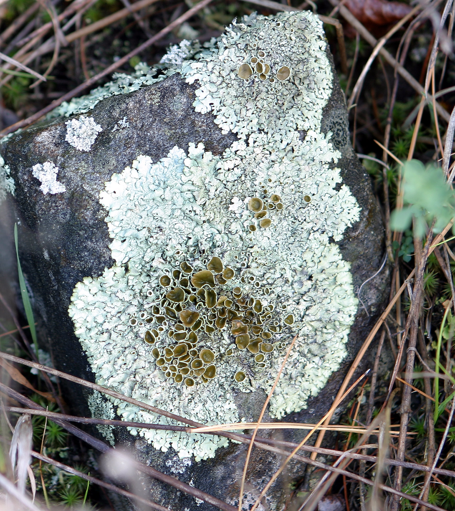
A lichen ( , ) is a hybrid colony (biology), colony of algae or cyanobacteria living symbiotically among hypha, filaments of multiple fungus species, along with yeasts and bacteria embedded in the cortex or "skin", in a mutualism (biology), mutualistic relationship.Introduction to Lichens – An Alliance between Kingdoms . University of California Museum of Paleontology. . Lichens are the lifeform that first brought the term symbiosis (as ''Symbiotismus'') into biological context. Lichens have since been recognized as important actors in nutrient cycling and producers which many higher trophic feeders feed on, such as reindeer, gastropods, nematodes, mites, and springtails. Lichens have properties different from those of their component organisms. They come in man ... [...More Info...] [...Related Items...] OR: [Wikipedia] [Google] [Baidu] |
Pycnidia
A pycnidium (plural pycnidia) is an asexual fruiting body produced by mitosporic fungi, for instance in the order Sphaeropsidales ( Deuteromycota, Coelomycetes) or order Pleosporales (Ascomycota, Dothideomycetes). It is often spherical or inversely pearshaped ( obpyriform) and its internal cavity is lined with conidiophore A conidium ( ; : conidia), sometimes termed an asexual chlamydospore or chlamydoconidium (: chlamydoconidia), is an Asexual reproduction, asexual, non-motility, motile spore of a fungus. The word ''conidium'' comes from the Ancient Greek word f ...s. When ripe, an opening generally appears at the top, through which the pycnidiospores escape. References {{reflist Further reading *Kulik, Martin M. "Symptomless infection, persistence, and production of pycnidia in host and non-host plants by Phomopsis batatae, Phomopsis phaseoli, and Phomopsis sojae, and the taxonomic implications." Mycologia(1984): 274–291. *Calpouzos, L., and D. B. Lapis. "Effects of l ... [...More Info...] [...Related Items...] OR: [Wikipedia] [Google] [Baidu] |
Asexual Reproduction
Asexual reproduction is a type of reproduction that does not involve the fusion of gametes or change in the number of chromosomes. The offspring that arise by asexual reproduction from either unicellular or multicellular organisms inherit the full set of genes of their single parent and thus the newly created individual is genetically and physically similar to the parent or an exact clone of the parent. Asexual reproduction is the primary form of reproduction for single-celled organisms such as archaea and eubacteria, bacteria. Many Eukaryote, eukaryotic organisms including plants, animals, and Fungus, fungi can also reproduce asexually. In Vertebrate, vertebrates, the most common form of asexual reproduction is parthenogenesis, which is typically used as an alternative to sexual reproduction in times when reproductive opportunities are limited. Some Monitor lizard, monitor lizards, including Komodo dragons, can reproduce asexually. While all prokaryotes reproduce without the fo ... [...More Info...] [...Related Items...] OR: [Wikipedia] [Google] [Baidu] |
Septum
In biology, a septum (Latin language, Latin for ''something that encloses''; septa) is a wall, dividing a Body cavity, cavity or structure into smaller ones. A cavity or structure divided in this way may be referred to as septate. Examples Human anatomy * Interatrial septum, the wall of tissue that is a sectional part of the left and right atria of the heart * Interventricular septum, the wall separating the left and right ventricles of the heart * Lingual septum, a vertical layer of fibrous tissue that separates the halves of the tongue *Nasal septum: the cartilage wall separating the nostrils of the nose * Alveolar septum: the thin wall which separates the Pulmonary alveolus, alveoli from each other in the lungs * Orbital septum, a palpebral ligament in the upper and lower eyelids * Septum pellucidum or septum lucidum, a thin structure separating two fluid pockets in the brain * Uterine septum, a malformation of the uterus * Septum of the penis, Penile septum, a fibrous w ... [...More Info...] [...Related Items...] OR: [Wikipedia] [Google] [Baidu] |
Hyaline
A hyaline substance is one with a glassy appearance. The word is derived from , and . Histopathology Hyaline cartilage is named after its glassy appearance on fresh gross pathology. On light microscopy of H&E stained slides, the extracellular matrix of hyaline cartilage looks homogeneously pink, and the term "hyaline" is used to describe similarly homogeneously pink material besides the cartilage. Hyaline material is usually acellular and proteinaceous. For example, arterial hyaline is seen in aging, high blood pressure, diabetes mellitus and in association with some drugs (e.g. calcineurin inhibitors). It is bright pink with PAS staining. Ichthyology and entomology In ichthyology and entomology Entomology (from Ancient Greek ἔντομον (''éntomon''), meaning "insect", and -logy from λόγος (''lógos''), meaning "study") is the branch of zoology that focuses on insects. Those who study entomology are known as entomologists. In ..., ''hyaline'' denotes a ... [...More Info...] [...Related Items...] OR: [Wikipedia] [Google] [Baidu] |
Ascospore
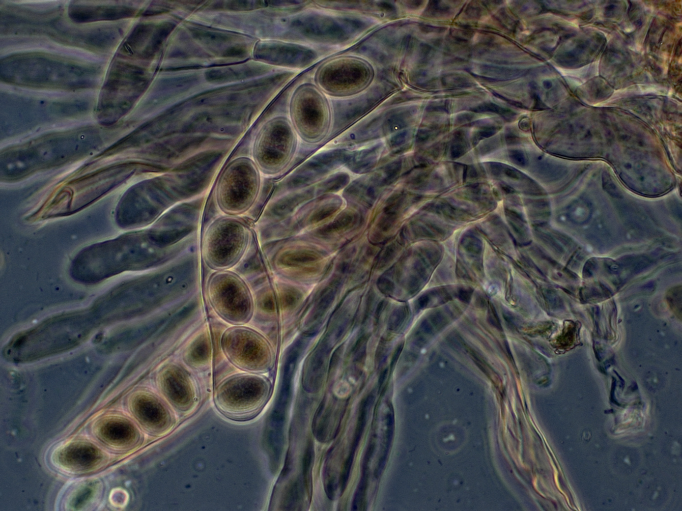
In fungi, an ascospore is the sexual spore formed inside an ascus—the sac-like cell that defines the division Ascomycota, the largest and most diverse Division (botany), division of fungi. After two parental cell nucleus, nuclei fuse, the ascus undergoes meiosis (halving of genetic material) followed by a mitosis (cell division), ordinarily producing eight genetically distinct haploid spores; most yeasts stop at four ascospores, whereas some moulds carry out extra post-meiotic divisions to yield dozens. Many asci build turgor, internal pressure and shoot their spores clear of the calm boundary layer, thin layer of still air enveloping the fruit body, whereas subterranean truffles depend on animals for biological dispersal, dispersal. Ontogeny, Development shapes both form and endurance of ascospores. A hook-shaped crozier aligns the paired nuclei; a double-biological membrane, membrane system then parcels each daughter nucleus, and successive wall layers of β-glucan, chitosan ... [...More Info...] [...Related Items...] OR: [Wikipedia] [Google] [Baidu] |
Amyloid (mycology)
In mycology a tissue (biology), tissue or feature is said to be amyloid if it has a positive amyloid reaction when subjected to a crude chemical test using iodine as an ingredient of either Melzer's reagent or Lugol's solution, producing a blue to blue-black staining. The term "amyloid" is derived from the Latin ''amyloideus'' ("starch-like"). It refers to the fact that starch gives a similar reaction, also called an amyloid reaction. The test can be on microscopic features, such as spore walls or hyphae, hyphal walls, or the apical apparatus or entire ascus wall of an ascus, or be a macroscopic reaction on tissue where a drop of the reagent is applied. Negative reactions, called inamyloid or nonamyloid, are for structures that remain pale yellow-brown or clear. A reaction producing a deep reddish to reddish-brown staining is either termed a dextrinoid reaction (pseudoamyloid is a synonym) or a hemiamyloid reaction. Melzer's reagent reactions Hemiamyloidity Hemiamyloidity in mycol ... [...More Info...] [...Related Items...] OR: [Wikipedia] [Google] [Baidu] |
Staining
Staining is a technique used to enhance contrast in samples, generally at the Microscope, microscopic level. Stains and dyes are frequently used in histology (microscopic study of biological tissue (biology), tissues), in cytology (microscopic study of cell (biology), cells), and in the medical fields of histopathology, hematology, and cytopathology that focus on the study and diagnoses of diseases at the microscopic level. Stains may be used to define biological tissues (highlighting, for example, muscle fibers or connective tissue), cell (biology), cell populations (classifying different blood cells), or organelles within individual cells. In biochemistry, it involves adding a class-specific (DNA, proteins, lipids, carbohydrates) dye to a substrate to qualify or quantify the presence of a specific compound. Staining and fluorescent tagging can serve similar purposes. Biological staining is also used to mark cells in flow cytometry, and to flag proteins or nucleic acids in gel ... [...More Info...] [...Related Items...] OR: [Wikipedia] [Google] [Baidu] |
Ascus
An ascus (; : asci) is the sexual spore-bearing cell produced in ascomycete fungi. Each ascus usually contains eight ascospores (or octad), produced by meiosis followed, in most species, by a mitotic cell division. However, asci in some genera or species can occur in numbers of one (e.g. '' Monosporascus cannonballus''), two, four, or multiples of four. In a few cases, the ascospores can bud off conidia that may fill the asci (e.g. '' Tympanis'') with hundreds of conidia, or the ascospores may fragment, e.g. some '' Cordyceps'', also filling the asci with smaller cells. Ascospores are nonmotile, usually single celled, but not infrequently may be coenocytic (lacking a septum), and in some cases coenocytic in multiple planes. Mitotic divisions within the developing spores populate each resulting cell in septate ascospores with nuclei. The term ocular chamber, or oculus, refers to the epiplasm (the portion of cytoplasm not used in ascospore formation) that is surrounded by the ... [...More Info...] [...Related Items...] OR: [Wikipedia] [Google] [Baidu] |
Paraphyses
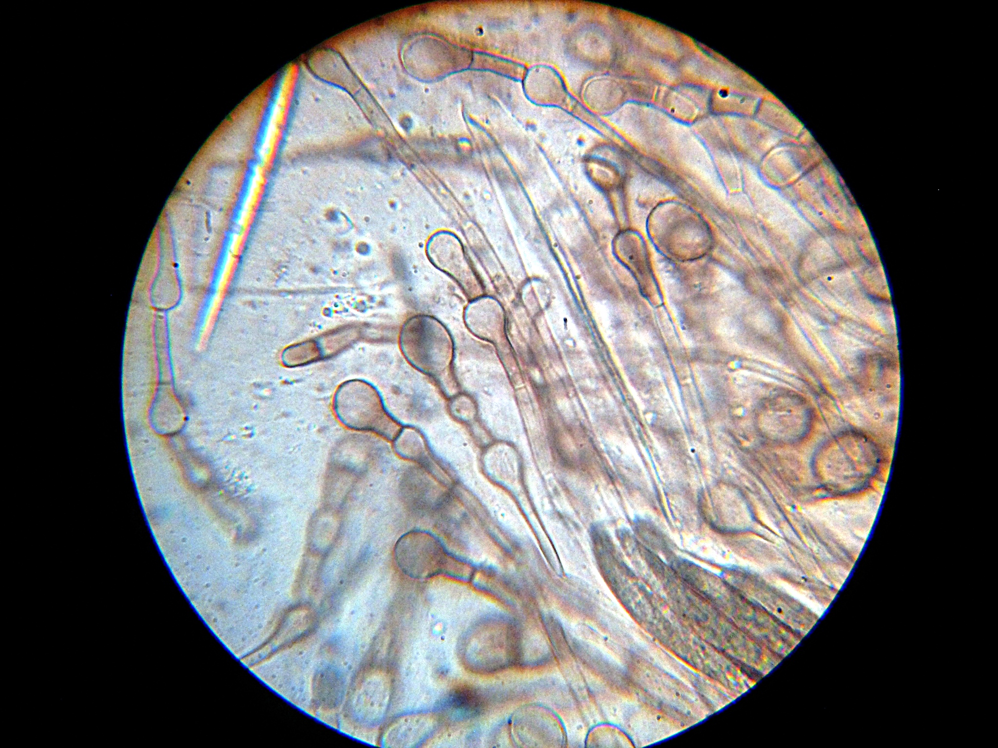
Paraphyses are erect sterile filament-like support structures occurring among the reproductive apparatuses of fungi, ferns, bryophytes and some thallophytes. The singular form of the word is paraphysis. In certain fungi, they are part of the fertile spore-bearing layer. More specifically, paraphyses are sterile filamentous hyphal end cells composing part of the hymenium of Ascomycota and Basidiomycota interspersed among either the asci or basidia respectively, and not sufficiently differentiated to be called cystidia A cystidium (: cystidia) is a relatively large cell found on the sporocarp of a basidiomycete (for example, on the surface of a mushroom gill), often between clusters of basidia. Since cystidia have highly varied and distinct shapes that are o ..., which are specialized, swollen, often protruding cells. The tips of paraphyses may contain the pigments which colour the hymenium. In ferns and mosses, they are filament-like structures that are found on sporangi ... [...More Info...] [...Related Items...] OR: [Wikipedia] [Google] [Baidu] |
Ostiole
An ''ostiole'' is a small hole or opening through which algae or fungi release their mature spores. The word is a diminutive of wikt:ostium, "ostium", "opening". The term is also used in higher plants, for example to denote the opening of the involuted syconium (fig inflorescence) through which fig wasps enter to Pollination, pollinate and breed. The species pharamacosycea have an arrangement interlocking pattern but there is an exception because of insipdia because it is partly cover the ostiole. On the adaxial side of the bracts is made out of cubic cells, that has a staining reactions and contain phenolic compounds. Sometimes a stomatal aperture is called an "ostiole"."Synergistic Pectin Degradation and Guard Cell Pressurization Underlie Stomatal Pore Formation", See also *Ostium (other) References Castro-Cárdenas, N., Vázquez-Santana, S., Teixeira, S. P., & Ibarra-Manríquez, G. (2023). Correction to: The roles of the ostiole in the fig-fig wasp mutu ... [...More Info...] [...Related Items...] OR: [Wikipedia] [Google] [Baidu] |


