|
Lentigo Maligna
Lentigo maligna is where melanocyte cells have become malignant and grow continuously along the stratum basale of the skin, but have not invaded below the epidermis. Lentigo maligna is not the same as lentigo maligna melanoma, as detailed below. It typically progresses very slowly and can remain in a non-invasive form for years. It is normally found in the elderly (peak incidence in the 9th decade), on skin areas with high levels of sun exposure like the face and forearms. Incidence of evolution to lentigo maligna melanoma is low, about 2.2% to 5% in elderly patients. It is also known as "Hutchinson's melanotic freckle". This is named for Jonathan Hutchinson. The word lentiginous comes from the latin for freckle. Relation to melanoma Lentigo maligna is a histopathological variant of melanoma ''in situ''. Lentigo maligna is sometimes classified as a very early melanoma, and sometimes as a precursor to melanoma. When malignant melanocytes from a lentigo maligna have invaded be ... [...More Info...] [...Related Items...] OR: [Wikipedia] [Google] [Baidu] |
Dermatology
Dermatology is the branch of medicine dealing with the Human skin, skin.''Random House Webster's Unabridged Dictionary.'' Random House, Inc. 2001. Page 537. . It is a speciality with both medical and surgical aspects. A List of dermatologists, dermatologist is a specialist medical doctor who manages diseases related to skin. Etymology Attested in English in 1819, the word "dermatology" derives from the Ancient Greek, Greek δέρματος (''dermatos''), genitive of δέρμα (''derma''), "skin" (itself from δέρω ''dero'', "to flay") and -λογία ''wikt:-logia, -logia''. Neo-Latin ''dermatologia'' was coined in 1630, an anatomical term with various French and German uses attested from the 1730s. History In 1708, the first great school of dermatology became a reality at the famous Hôpital Saint-Louis in Paris, and the first textbooks (Willan's, 1798–1808) and atlases (Jean-Louis-Marc Alibert, Alibert's, 1806–1816) appeared in print around the same time.Freedber ... [...More Info...] [...Related Items...] OR: [Wikipedia] [Google] [Baidu] |
Dermatoscopy
Dermatoscopy, also known as dermoscopy or epiluminescence microscopy, is the examination of skin lesions with a dermatoscope. It is a tool similar to a camera to allow for inspection of skin lesions unobstructed by skin surface reflections. The dermatoscope consists of a magnifier, a light source (polarized or non-polarized), a transparent plate and sometimes a liquid medium between the instrument and the skin. The dermatoscope is often handheld, although there are stationary cameras allowing the capture of whole body images in a single shot. When the images or video clips are digitally captured or processed, the instrument can be referred to as a digital epiluminescence dermatoscope. The image is then analyzed automatically and given a score indicating how dangerous it is. This technique is useful to dermatologists and skin cancer practitioners in distinguishing benign from malignant (cancerous) lesions, especially in the diagnosis of melanoma. Types There are two main types o ... [...More Info...] [...Related Items...] OR: [Wikipedia] [Google] [Baidu] |
Imiquimod
Imiquimod, sold under the brand name Aldara among others, is a medication that acts as an immune response modifier that is used to treat genital warts, superficial basal cell carcinoma, and actinic keratosis. Scientists at 3M's pharmaceuticals division discovered the drug and 3M obtained the first FDA approval in 1997. As of 2015, imiquimod is generic and is available worldwide under many brands. In 2021, it was the 290th most commonly prescribed medication in the United States, with more than 600,000 prescriptions. Medical uses Imiquimod is a patient-applied cream prescribed to treat genital warts, Bowens disease (squamous cell carcinoma in situ), and, secondary to surgery, for basal cell carcinoma, as well as actinic keratosis. Imiquimod 5% cream is indicated for the topical treatment of: * external genital and perianal warts (condylomata acuminata) in adults; * small superficial basal-cell carcinomas (sBCCs) in adults; * clinically typical, non-hyperkeratotic, non-hypert ... [...More Info...] [...Related Items...] OR: [Wikipedia] [Google] [Baidu] |
Mohs Surgery
Mohs surgery, developed in 1938 by general surgeon Frederic E. Mohs, is microscopically controlled surgery used to treat both common and rare types of skin cancer. During the surgery, after each removal of tissue and while the patient waits, the tissue is examined for cancer cells. That examination dictates the decision for additional tissue removal. Mohs surgery is the gold standard method for obtaining complete margin control during removal of a skin cancer (complete circumferential peripheral and deep margin assessment or CCPDMA) using frozen section histology. CCPDMA or Mohs surgery allows for the removal of a skin cancer with very narrow surgical margin and a high cure rate. The cure rate with Mohs surgery cited by most studies is between 97% and 99.8% for primary basal-cell carcinoma, the most common type of skin cancer. Mohs procedure is also used for squamous cell skin cancer, squamous cell carcinoma, but with a lower cure rate. Recurrent basal-cell cancer has a lower cure ... [...More Info...] [...Related Items...] OR: [Wikipedia] [Google] [Baidu] |
Skin Graft
Skin grafting, a type of graft (surgery), graft surgery, involves the organ transplant, transplantation of skin without a defined circulation. The transplanted biological tissue, tissue is called a skin graft. Surgeons may use skin grafting to treat: * extensive wounding or physical trauma, trauma * Burn (injury), burns * areas of extensive skin loss due to infection such as necrotizing fasciitis or purpura fulminans * specific surgeries that may require skin grafts for healing to occur – most commonly removal of skin cancers Skin grafting often takes place after serious injuries when some of the body's skin is damaged. Surgical removal (excision or debridement) of the damaged skin is followed by skin grafting. The grafting serves two purposes: reducing the course of treatment needed (and time in the hospital), and improving the function and appearance of the area of the body which receives the skin graft. There are two types of skin grafts: * Partial-thickness: The more co ... [...More Info...] [...Related Items...] OR: [Wikipedia] [Google] [Baidu] |
Skin Flap
The terms free flap, free autologous tissue transfer and microvascular free tissue transfer are synonymous terms used to describe the "transplantation" of tissue from one site of the body to another, in order to reconstruct an existing defect. "Free" implies that the tissue is completely detached from its blood supply at the original location ("donor site") and then transferred to another location ("recipient site") and the circulation in the tissue re-established by anastomosis of artery(s) and vein(s). This is in contrast to a "pedicled" flap in which the tissue is left partly attached to the donor site ("pedicle") and simply transposed to a new location; keeping the "pedicle" intact as a conduit to supply the tissue with blood. Various types of tissue may be transferred as a "free flap" including skin and fat, muscle, nerve, bone, cartilage (or any combination of these), lymph nodes and intestinal segments. An example of "free flap" could be a "free toe transfer" in which ... [...More Info...] [...Related Items...] OR: [Wikipedia] [Google] [Baidu] |
Scars From Skin Flap
SCARS or S.C.A.R.S. is an acronym that may refer to: * SCARS (military) (Special Combat Aggressive Reactionary System), an American combat fighting system * Severe cutaneous adverse reactions Severe cutaneous adverse reactions (SCARs) are a group of potentially lethal adverse drug reactions that involve the skin and mucous membranes of various body openings such as the eyes, ears, and inside the nose, mouth, and lips. In more sever ..., often abbreviated as SCARs, i.e. the last letter is lower case * ''S.C.A.R.S.'' (video game) (''Super Computer Animal Racing Simulator''), a video game featuring cars that are shaped like animals See also * Scar (other) * SCAR (other) * Scarred (other) {{disambig ... [...More Info...] [...Related Items...] OR: [Wikipedia] [Google] [Baidu] |
Resection Margin
A resection margin or surgical margin is the edge or "margin" of apparently non-tumorous tissue around a tumor that has been surgically removed, called " resected", in surgical oncology. The resection is an attempt to remove a cancer tumor so that no portion of the malignant growth extends past the edges or margin of the removed tumor and surrounding tissue. These are retained after the surgery and examined microscopically by a pathologist to see if the margin is indeed free from tumor cells (called "negative"). If cancerous cells are found at the edges (called "positive") the operation is much less likely to achieve the desired results. The size of the margin is an important issue in areas that are functionally important (i.e., large vessels like the aorta or vital organs) or in areas for which the extent of surgery is minimized due to aesthetic concerns (i.e., melanoma of the face or squamous cell carcinoma of the penis). The desired size of margin around the tumour can vary. ... [...More Info...] [...Related Items...] OR: [Wikipedia] [Google] [Baidu] |
Pleomorphism (cytology)
Pleomorphism is a term used in histology and cytopathology to describe variability in the size, shape and staining of cells and/or their nuclei. Several key determinants of cell and nuclear size, like ploidy and the regulation of cellular metabolism, are commonly disrupted in tumors. Therefore, cellular and nuclear pleomorphism is one of the earliest hallmarks of cancer progression and a feature characteristic of malignant neoplasms and dysplasia. Certain benign cell types may also exhibit pleomorphism, e.g. neuroendocrine cells, Arias-Stella reaction. A rare type of rhabdomyosarcoma that is found in adults is known as pleomorphic rhabdomyosarcoma. Despite the prevalence of pleomorphism in human pathology, its role in disease progression is unclear. In epithelial tissue, pleomorphism in cellular size can induce packing defects and disperse aberrant cells. But the consequence of atypical cell and nuclear morphology in other tissues is unknown. See also *Anaplasia *Cell grow ... [...More Info...] [...Related Items...] OR: [Wikipedia] [Google] [Baidu] |
Melanocytes
Melanocytes are melanin-producing neural crest-derived cells located in the bottom layer (the stratum basale) of the skin's epidermis, the middle layer of the eye (the uvea), the inner ear, vaginal epithelium, meninges, bones, and heart found in many mammals and birds. Melanin is a dark pigment primarily responsible for skin color. Once synthesized, melanin is contained in special organelles called melanosomes which can be transported to nearby keratinocytes to induce pigmentation. Thus darker skin tones have more melanosomes present than lighter skin tones. Functionally, melanin serves as protection against UV radiation. Melanocytes also have a role in the immune system. Function Through a process called melanogenesis, melanocytes produce melanin, which is a pigment found in the skin, eyes, hair, nasal cavity, and inner ear. This melanogenesis leads to a long-lasting pigmentation, which is in contrast to the pigmentation that originates from oxidation of al ... [...More Info...] [...Related Items...] OR: [Wikipedia] [Google] [Baidu] |
SOX10
Transcription factor SOX-10 is a protein that in humans is encoded by the ''SOX10'' gene. Function This gene encodes a member of the SOX gene family, SOX (Testis-determining factor, SRY-related HMG-box) family of transcription factors involved in the regulation of embryonic development and determination of cell fate determination, cell fate. The encoded protein acts as a transcriptional activator after forming a protein complex with other proteins. This protein acts as a nucleocytoplasmic shuttle protein and is important for neural crest and peripheral nervous system development. In melanocyte, melanocytic cells, there is evidence that SOX10 gene expression may be regulated by Microphthalmia-associated transcription factor, MITF. Mutations Mutations in this gene are associated with Waardenburg–Shah syndrome and uveal melanoma. Immunostain SOX10 is used as an immunohistochemistry marker, being positive in: Topic Completed: 1 February 2014. Revised: 20 September 2019 *Neuro ... [...More Info...] [...Related Items...] OR: [Wikipedia] [Google] [Baidu] |
Immunohistochemistry
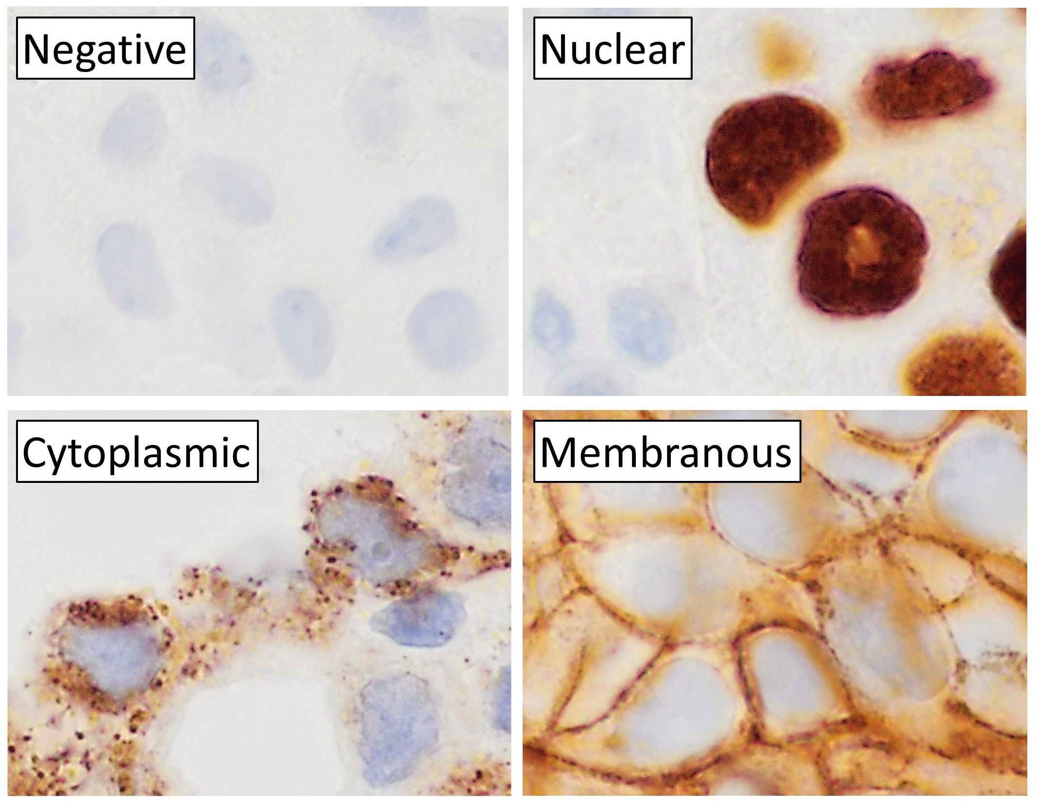
Immunohistochemistry is a form of immunostaining. It involves the process of selectively identifying antigens in cells and tissue, by exploiting the principle of Antibody, antibodies binding specifically to antigens in biological tissues. Albert Coons, Albert Hewett Coons, Ernst Berliner, Ernest Berliner, Norman Jones and Hugh J Creech was the first to develop immunofluorescence in 1941. This led to the later development of immunohistochemistry. Immunohistochemical staining is widely used in the diagnosis of abnormal cells such as those found in cancerous tumors. In some cancer cells certain tumor antigens are expressed which make it possible to detect. Immunohistochemistry is also widely used in basic research, to understand the distribution and localization of biomarkers and differentially expressed proteins in different parts of a biological tissue. Sample preparation Immunohistochemistry can be performed on tissue that has been fixed and embedded in Paraffin wax, paraffin, ... [...More Info...] [...Related Items...] OR: [Wikipedia] [Google] [Baidu] |


