|
Lamina (anatomy)
Lamina is a general anatomical term meaning "plate" or "layer". It is used in both gross anatomy and microscopic anatomy to describe structures. Some examples include: * The laminae of the thyroid cartilage: two leaf-like plates of cartilage that make up the walls of the structure. * The vertebral laminae: plates of bone that form the posterior walls of each vertebra, enclosing the spinal cord. * The laminae of the thalamus: the layers of thalamus tissue. * The lamina propria: a connective tissue layer under the epithelium of an organ. * The nuclear lamina: a dense fiber network inside the nucleus of cells. * The lamina affixa: a layer of epithelium growing on the surface of the thalamus. * The lamina of Drosophila is the most peripheral neuropil Neuropil (or "neuropile") is any area in the nervous system composed of mostly unmyelinated axons, dendrites and glial cell processes that forms a synaptically dense region containing a relatively low number of cell bodies. The ... [...More Info...] [...Related Items...] OR: [Wikipedia] [Google] [Baidu] |
Anatomy
Anatomy () is the branch of morphology concerned with the study of the internal structure of organisms and their parts. Anatomy is a branch of natural science that deals with the structural organization of living things. It is an old science, having its beginnings in prehistoric times. Anatomy is inherently tied to developmental biology, embryology, comparative anatomy, evolutionary biology, and phylogeny, as these are the processes by which anatomy is generated, both over immediate and long-term timescales. Anatomy and physiology, which study the structure and function of organisms and their parts respectively, make a natural pair of related disciplines, and are often studied together. Human anatomy is one of the essential basic sciences that are applied in medicine, and is often studied alongside physiology. Anatomy is a complex and dynamic field that is constantly evolving as discoveries are made. In recent years, there has been a significant increase in the use of ... [...More Info...] [...Related Items...] OR: [Wikipedia] [Google] [Baidu] |
Gross Anatomy
Gross anatomy is the study of anatomy at the visible or macroscopic level. The counterpart to gross anatomy is the field of histology, which studies microscopic anatomy. Gross anatomy of the human body or other animals seeks to understand the relationship between components of an organism in order to gain a greater appreciation of the roles of those components and their relationships in maintaining the functions of life. The study of gross anatomy can be performed on deceased organisms using dissection or on living organisms using medical imaging. Education in the gross anatomy of humans is included training for most health professionals. Techniques of study Gross anatomy is studied using both invasive and noninvasive methods with the goal of obtaining information about the macroscopic structure and organisation of organs and organ systems. Among the most common methods of study is dissection, in which the corpse of an animal or a human cadaver is surgically opened and i ... [...More Info...] [...Related Items...] OR: [Wikipedia] [Google] [Baidu] |
Lamina Of The Vertebral Arch
Each vertebra (: vertebrae) is an irregular bone with a complex structure composed of bone and some hyaline cartilage, that make up the vertebral column or spine, of vertebrates. The proportions of the vertebrae differ according to their spinal segment and the particular species. The basic configuration of a vertebra varies; the vertebral body (also ''centrum'') is of bone and bears the load of the vertebral column. The upper and lower surfaces of the vertebra body give attachment to the intervertebral discs. The posterior part of a vertebra forms a vertebral arch, in eleven parts, consisting of two pedicles (pedicle of vertebral arch), two laminae, and seven processes. The laminae give attachment to the ligamenta flava (ligaments of the spine). There are vertebral notches formed from the shape of the pedicles, which form the intervertebral foramina when the vertebrae articulate. These foramina are the entry and exit conduits for the spinal nerves. The body of the vertebra ... [...More Info...] [...Related Items...] OR: [Wikipedia] [Google] [Baidu] |
Thalamus
The thalamus (: thalami; from Greek language, Greek Wikt:θάλαμος, θάλαμος, "chamber") is a large mass of gray matter on the lateral wall of the third ventricle forming the wikt:dorsal, dorsal part of the diencephalon (a division of the forebrain). Nerve fibers project out of the thalamus to the cerebral cortex in all directions, known as the thalamocortical radiations, allowing hub (network science), hub-like exchanges of information. It has several functions, such as the relaying of sensory neuron, sensory and motor neuron, motor signals to the cerebral cortex and the regulation of consciousness, sleep, and alertness. Anatomically, the thalami are paramedian symmetrical structures (left and right), within the vertebrate brain, situated between the cerebral cortex and the midbrain. It forms during embryonic development as the main product of the diencephalon, as first recognized by the Swiss embryologist and anatomist Wilhelm His Sr. in 1893. Anatomy The thalami ar ... [...More Info...] [...Related Items...] OR: [Wikipedia] [Google] [Baidu] |
Lamina Propria
The lamina propria is a thin layer of connective tissue that forms part of the moist linings known as mucous membranes or mucosae, which line various tubes in the body, such as the respiratory tract, the gastrointestinal tract, and the urogenital tract. The lamina propria is a thin layer of loose (areolar) connective tissue, which lies beneath the epithelium, and together with the epithelium and basement membrane constitutes the mucosa. As its Latin name indicates, it is a characteristic component of the mucosa, or the mucosa's "own special layer." Thus, the term mucosa or mucous membrane refers to the combination of the epithelium and the lamina propria. The connective tissue of the lamina propria is loose and rich in cells. The cells of the lamina propria are variable and can include fibroblasts, lymphocytes, plasma cells, macrophages, eosinophilic leukocytes, and mast cells. It provides support and nutrition to the epithelium, as well as the means to bind to the underlying t ... [...More Info...] [...Related Items...] OR: [Wikipedia] [Google] [Baidu] |
Nuclear Lamina
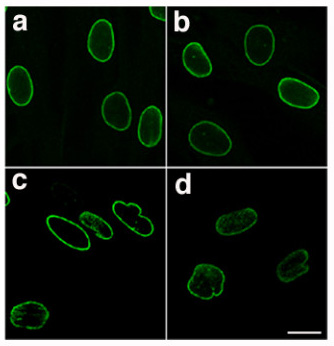
The nuclear lamina is a dense (~30 to 100 nm thick) fibrillar network inside the nucleus of eukaryote cells. It is composed of intermediate filaments and membrane associated proteins. Besides providing mechanical support, the nuclear lamina regulates important cellular events such as DNA replication and cell division. Additionally, it participates in chromatin organization and it anchors the nuclear pore complexes embedded in the nuclear envelope. The nuclear lamina is associated with the inner face of the inner nuclear membrane of the nuclear envelope, whereas the outer face of the outer nuclear membrane is continuous with the endoplasmic reticulum. The nuclear lamina is similar in structure to the nuclear matrix, that extends throughout the nucleoplasm. Structure and composition The nuclear lamina consists of two components, lamins and nuclear lamin-associated membrane proteins. The lamins are type V intermediate filaments which can be categorized as either A-type ... [...More Info...] [...Related Items...] OR: [Wikipedia] [Google] [Baidu] |
Lamina Affixa
Lamina affixa is a layer of epithelium growing on the surface of the thalamus and forming the floor of the central part of lateral ventricle, on whose medial margin is attached the choroid plexus of the lateral ventricle; it covers the superior thalamostriate vein and the superior choroid vein. The torn edge of this plexus is called the tela choroidea. On the surface of the terminal vein is a narrow white band, named the lamina affixa. GDF-15 Growth/differentiation factor 15 is a protein that in humans is encoded by the GDF15 gene. GDF15 was first identified as Macrophage inhibitory cytokine-1 or MIC-1. It is a protein belonging to the transforming growth factor beta superfamily. U .../MIC-1 has been observed in lamina affixa cells. References External links * https://web.archive.org/web/20071024195305/http://www.univie.ac.at/anatomie2/plastinatedbrain/surfaceanatomy/surface-2-text.html Thalamus {{neuroanatomy-stub ... [...More Info...] [...Related Items...] OR: [Wikipedia] [Google] [Baidu] |
Epithelium
Epithelium or epithelial tissue is a thin, continuous, protective layer of cells with little extracellular matrix. An example is the epidermis, the outermost layer of the skin. Epithelial ( mesothelial) tissues line the outer surfaces of many internal organs, the corresponding inner surfaces of body cavities, and the inner surfaces of blood vessels. Epithelial tissue is one of the four basic types of animal tissue, along with connective tissue, muscle tissue and nervous tissue. These tissues also lack blood or lymph supply. The tissue is supplied by nerves. There are three principal shapes of epithelial cell: squamous (scaly), columnar, and cuboidal. These can be arranged in a singular layer of cells as simple epithelium, either simple squamous, simple columnar, or simple cuboidal, or in layers of two or more cells deep as stratified (layered), or ''compound'', either squamous, columnar or cuboidal. In some tissues, a layer of columnar cells may appear to be stratified d ... [...More Info...] [...Related Items...] OR: [Wikipedia] [Google] [Baidu] |
Lamina (neuropil)
The lamina is the most peripheral neuropil of the insect visual system. There are twelve distinct neuron classes in the lamina: the lamina monopolar cells L1-L5, two GABA GABA (gamma-aminobutyric acid, γ-aminobutyric acid) is the chief inhibitory neurotransmitter in the developmentally mature mammalian central nervous system. Its principal role is reducing neuronal excitability throughout the nervous system. GA ...ergic feedback neurons (C2 and C3), two wide-field feedback neurons (Lawf1 and Lawf2), lamina intrinsic amacrine neurons (Lai) and the T1 basket cell. The outer photoreceptors, R1-R6, terminate in the lamina, where they form tetrad synapses with L1, L2, L3, and Lai. The lamina was the first portion of the nervous system of Drosophila to be reconstructed, thus starting the field of Drosophila connectomics. However the methods used were largely manual and further progress awaited more automated techniques. References Insect anatomy Visual perception ... [...More Info...] [...Related Items...] OR: [Wikipedia] [Google] [Baidu] |
Neuropil
Neuropil (or "neuropile") is any area in the nervous system composed of mostly unmyelinated axons, dendrites and glial cell processes that forms a synaptically dense region containing a relatively low number of cell bodies. The most prevalent anatomical region of neuropil is the brain which, although not completely composed of neuropil, does have the largest and highest synaptically concentrated areas of neuropil in the body. For example, the neocortex and olfactory bulb both contain neuropil. White matter, which is mostly composed of myelinated axons (hence its white color) and glial cells, is generally not considered to be a part of the neuropil. Neuropil (pl. neuropils) comes from the Greek: ''neuro'', meaning "tendon, sinew; nerve" and ''pilos'', meaning "felt". The term's origin can be traced back to the late 19th century. Location Neuropil has been found in the following regions: outer neocortex layer, barrel cortex, inner plexiform layer and outer plexiform lay ... [...More Info...] [...Related Items...] OR: [Wikipedia] [Google] [Baidu] |
Insect Visual System
The optic(al) lobe of arthropods is a structure of the protocerebrum that sits behind the arthropod eye (mostly compound eyes) and is responsible for the processing of the visual information. It is made up of three layers: ;Lamina (ganglionaris): responsible for contrast enhancement through lateral inhibition ;Medulla: processes movement and shows movement direction sensitivity. Possesses local motion detectors ;Lobula: integrates information from large areas of the visual field to abstract visual information and object recognition Object recognition – technology in the field of computer vision for finding and identifying objects in an image or video sequence. Humans recognize a multitude of objects in images with little effort, despite the fact that the image of the ... :;Lobula plate: wide-field motion vision References {{insect-stub Arthropod anatomy Animal nervous system ... [...More Info...] [...Related Items...] OR: [Wikipedia] [Google] [Baidu] |


