|
Lambdoidal
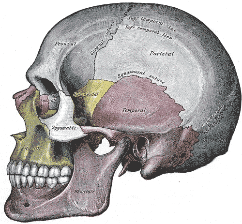
The lambdoid suture, or lambdoidal suture, is a dense, fibrous connective tissue joint on the posterior aspect of the skull that connects the parietal bones with the occipital bone. It is continuous with the occipitomastoid suture. Structure The lambdoid suture is between the paired parietal bones and the occipital bone of the skull. It runs from the asterion on each side. Nerve supply The lambdoid suture may be supplied by a branch of the supraorbital nerve, a branch of the frontal branch of the trigeminal nerve. Clinical significance At birth, the bones of the skull do not meet. If certain bones of the skull grow too fast, then craniosynostosis (premature closure of the sutures) may occur. This can result in skull deformities. If the lambdoid suture closes too soon on one side, the skull will appear twisted and asymmetrical, a condition called "plagiocephaly". Plagiocephaly refers to the shape and not the condition. The condition is craniosynostosis. The lambdoid su ... [...More Info...] [...Related Items...] OR: [Wikipedia] [Google] [Baidu] |
Occipital Bone
The occipital bone () is a neurocranium, cranial dermal bone and the main bone of the occiput (back and lower part of the skull). It is trapezoidal in shape and curved on itself like a shallow dish. The occipital bone lies over the occipital lobes of the cerebrum. At the base of the skull in the occipital bone, there is a large oval opening called the foramen magnum, which allows the passage of the spinal cord. Like the other cranial bones, it is classed as a flat bone. Due to its many attachments and features, the occipital bone is described in terms of separate parts. From its front to the back is the basilar part of occipital bone, basilar part, also called the basioccipital, at the sides of the foramen magnum are the lateral parts of occipital bone, lateral parts, also called the exoccipitals, and the back is named as the squamous part of occipital bone, squamous part. The basilar part is a thick, somewhat quadrilateral piece in front of the foramen magnum and directed toward ... [...More Info...] [...Related Items...] OR: [Wikipedia] [Google] [Baidu] |
Parietal Bone
The parietal bones ( ) are two bones in the skull which, when joined at a fibrous joint known as a cranial suture, form the sides and roof of the neurocranium. In humans, each bone is roughly quadrilateral in form, and has two surfaces, four borders, and four angles. It is named from the Latin ''paries'' (''-ietis''), wall. Surfaces External The external surface [Fig. 1] is convex, smooth, and marked near the center by an eminence, the parietal eminence (''tuber parietale''), which indicates the point where ossification commenced. Crossing the middle of the bone in an arched direction are two curved lines, the superior and inferior temporal lines; the former gives attachment to the temporal fascia, and the latter indicates the upper limit of the muscular origin of the temporal muscle. Above these lines the bone is covered by a tough layer of fibrous tissue – the epicranial aponeurosis; below them it forms part of the temporal fossa, and affords attachment to the temporal mu ... [...More Info...] [...Related Items...] OR: [Wikipedia] [Google] [Baidu] |
Occipitomastoid Suture
The occipitomastoid suture, or occipitotemporal suture, is the cranial suture between the occipital bone and the mastoid portion of the temporal bone. It is continuous with the lambdoidal suture. See also * Jugular foramen Additional images File:Occipitomastoid suture - animation03.gif, Animation. Occipitomastoid suture shown in red. File:Occipitomastoid suture - animation06.gif, Temporal bones and occipital bone, seen from inside. File:Occipitomastoid suture - skull - inferior view.png, Base of skull The base of skull, also known as the cranial base or the cranial floor, is the most Anatomical terms of location#Superior and inferior, inferior area of the human skull, skull. It is composed of the endocranium and the lower parts of the Calvaria .... Inferior surface. File:Sobo 1909 42 - Occipitomastoid suture.png, Base of skull. Inferior surface. References External links * * * Bones of the head and neck Cranial sutures Human head and neck Joi ... [...More Info...] [...Related Items...] OR: [Wikipedia] [Google] [Baidu] |
Parietal Bone
The parietal bones ( ) are two bones in the skull which, when joined at a fibrous joint known as a cranial suture, form the sides and roof of the neurocranium. In humans, each bone is roughly quadrilateral in form, and has two surfaces, four borders, and four angles. It is named from the Latin ''paries'' (''-ietis''), wall. Surfaces External The external surface [Fig. 1] is convex, smooth, and marked near the center by an eminence, the parietal eminence (''tuber parietale''), which indicates the point where ossification commenced. Crossing the middle of the bone in an arched direction are two curved lines, the superior and inferior temporal lines; the former gives attachment to the temporal fascia, and the latter indicates the upper limit of the muscular origin of the temporal muscle. Above these lines the bone is covered by a tough layer of fibrous tissue – the epicranial aponeurosis; below them it forms part of the temporal fossa, and affords attachment to the temporal mu ... [...More Info...] [...Related Items...] OR: [Wikipedia] [Google] [Baidu] |
Skull
The skull, or cranium, is typically a bony enclosure around the brain of a vertebrate. In some fish, and amphibians, the skull is of cartilage. The skull is at the head end of the vertebrate. In the human, the skull comprises two prominent parts: the neurocranium and the facial skeleton, which evolved from the first pharyngeal arch. The skull forms the frontmost portion of the axial skeleton and is a product of cephalization and vesicular enlargement of the brain, with several special senses structures such as the eyes, ears, nose, tongue and, in fish, specialized tactile organs such as barbels near the mouth. The skull is composed of three types of bone: cranial bones, facial bones and ossicles, which is made up of a number of fused flat and irregular bones. The cranial bones are joined at firm fibrous junctions called sutures and contains many foramina, fossae, processes, and sinuses. In zoology, the openings in the skull are called fenestrae, the most ... [...More Info...] [...Related Items...] OR: [Wikipedia] [Google] [Baidu] |
Plagiocephaly
Plagiocephaly, also known as flat head syndrome, is a condition characterized by an asymmetrical distortion (flattening of one side) of the skull. A mild and widespread form is characterized by a flat spot on the back or one side of the head caused by remaining in a supine position for prolonged periods. Plagiocephaly is a diagonal asymmetry across the head shape. Often it is a flattening which is to one side at the back of the head, and there is often some facial asymmetry. Depending on whether synostosis is involved, plagiocephaly divides into two groups: synostotic, with one or more fused cranial sutures, and non-synostotic (deformational). Surgical treatment of these groups includes the deference method; however, the treatment of deformational plagiocephaly is controversial. Brachycephaly describes a very wide head shape with a flattening across the whole back of the head. Causes Slight plagiocephaly is routinely diagnosed at birth and may be the result of a restrictive int ... [...More Info...] [...Related Items...] OR: [Wikipedia] [Google] [Baidu] |
Joints Of The Head And Neck
A joint or articulation (or articular surface) is the connection made between bones, ossicles, or other hard structures in the body which link an animal's skeletal system into a functional whole.Saladin, Ken. Anatomy & Physiology. 7th ed. McGraw-Hill Connect. Webp.274/ref> They are constructed to allow for different degrees and types of movement. Some joints, such as the knee, elbow, and shoulder, are self-lubricating, almost frictionless, and are able to withstand compression and maintain heavy loads while still executing smooth and precise movements. Other joints such as suture (joint), sutures between the bones of the skull permit very little movement (only during birth) in order to protect the brain and the sense organs. The connection between a tooth and the jawbone is also called a joint, and is described as a fibrous joint known as a gomphosis. Joints are classified both structurally and functionally. Joints play a vital role in the human body, contributing to movement, sta ... [...More Info...] [...Related Items...] OR: [Wikipedia] [Google] [Baidu] |
Human Head And Neck
Humans (''Homo sapiens'') or modern humans are the most common and widespread species of primate, and the last surviving species of the genus ''Homo''. They are great apes characterized by their hairlessness, bipedalism, and high intelligence. Humans have large brains, enabling more advanced cognitive skills that facilitate successful adaptation to varied environments, development of sophisticated tools, and formation of complex social structures and civilizations. Humans are highly social, with individual humans tending to belong to a multi-layered network of distinct social groups — from families and peer groups to corporations and political states. As such, social interactions between humans have established a wide variety of values, social norms, languages, and traditions (collectively termed institutions), each of which bolsters human society. Humans are also highly curious: the desire to understand and influence phenomena has motivated humanity's deve ... [...More Info...] [...Related Items...] OR: [Wikipedia] [Google] [Baidu] |
Cranial Sutures
In anatomy, fibrous joints are joints connected by Fibrous connective tissue, fibrous tissue, consisting mainly of collagen. These are fixed joints where bones are united by a layer of white fibrous tissue of varying thickness. In the skull, the joints between the bones are called Suture (anatomy), sutures. Such immovable joints are also referred to as synarthrosis, synarthroses. Types Most fibrous joints are also called "fixed" or "immovable". These joints have no joint cavity and are connected via fibrous connective tissue. * Suture (anatomy), Sutures: The skull bones are connected by fibrous joints called ''#Sutures, sutures''. In fetus, fetal skulls, the sutures are wide to allow slight movement during birth. They later become rigid (synarthrosis, synarthrodial). * Syndesmosis: Some of the long bones in the body such as the radius (bone), radius and ulna in the forearm are joined by a ''#Syndesmosis, syndesmosis'' (along the interosseous membrane of forearm, interosseous mem ... [...More Info...] [...Related Items...] OR: [Wikipedia] [Google] [Baidu] |
Bones Of The Head And Neck
A bone is a rigid organ that constitutes part of the skeleton in most vertebrate animals. Bones protect the various other organs of the body, produce red and white blood cells, store minerals, provide structure and support for the body, and enable mobility. Bones come in a variety of shapes and sizes and have complex internal and external structures. They are lightweight yet strong and hard and serve multiple functions. Bone tissue (osseous tissue), which is also called bone in the uncountable sense of that word, is hard tissue, a type of specialised connective tissue. It has a honeycomb-like matrix internally, which helps to give the bone rigidity. Bone tissue is made up of different types of bone cells. Osteoblasts and osteocytes are involved in the formation and mineralisation of bone; osteoclasts are involved in the resorption of bone tissue. Modified (flattened) osteoblasts become the lining cells that form a protective layer on the bone surface. The minerali ... [...More Info...] [...Related Items...] OR: [Wikipedia] [Google] [Baidu] |
Wormian Bones
Wormian bones, also known as intrasutural bones or sutural bones, are extra bone pieces that can occur within a suture (joint) in the skull. These are irregular isolated bones that can appear in addition to the usual centres of ossification of the skull and, although unusual, are not rare. They occur most frequently in the course of the lambdoid suture, which is more tortuous than other sutures. They are also occasionally seen within the sagittal and coronal sutures. A large Wormian bone at lambda is often called an Inca bone (''os incae''), due to the relatively high frequency of occurrence in Peruvian mummies. Another specific Wormian bone, the pterion ossicle, sometimes exists between the sphenoidal angle of the parietal bone and the great wing of the sphenoid bone. They tend to vary in size and can be found on either side of the skull. Usually, not more than several are found in a single individual, but more than one hundred have been once found in the skull of a hydrocephal ... [...More Info...] [...Related Items...] OR: [Wikipedia] [Google] [Baidu] |


