|
LGN
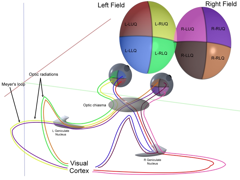
In neuroanatomy, the lateral geniculate nucleus (LGN; also called the lateral geniculate body or lateral geniculate complex) is a structure in the thalamus and a key component of the mammalian visual pathway. It is a small, ovoid, ventral projection of the thalamus where the thalamus connects with the optic nerve. There are two LGNs, one on the left and another on the right side of the thalamus. In humans, both LGNs have six layers of neurons (grey matter) alternating with optic fibers (white matter). The LGN receives information directly from the ascending retinal ganglion cells via the optic tract and from the reticular activating system. Neurons of the LGN send their axons through the optic radiation, a direct pathway to the primary visual cortex. In addition, the LGN receives many strong feedback connections from the primary visual cortex. In humans as well as other mammals, the two strongest pathways linking the eye to the brain are those projecting to the dorsal part of the ... [...More Info...] [...Related Items...] OR: [Wikipedia] [Google] [Baidu] |
Visual Pathway
The visual system comprises the sensory organ (the eye) and parts of the central nervous system (the retina containing photoreceptor cells, the optic nerve, the optic tract and the visual cortex) which gives organisms the sense of sight (the ability to detect and process visible light) as well as enabling the formation of several non-image photo response functions. It detects and interprets information from the optical spectrum perceptible to that species to "build a representation" of the surrounding environment. The visual system carries out a number of complex tasks, including the reception of light and the formation of monocular neural representations, colour vision, the neural mechanisms underlying stereopsis and assessment of distances to and between objects, the identification of a particular object of interest, motion perception, the analysis and integration of visual information, pattern recognition, accurate motor coordination under visual guidance, and more. The ... [...More Info...] [...Related Items...] OR: [Wikipedia] [Google] [Baidu] |
Visual System
The visual system comprises the sensory organ (the eye) and parts of the central nervous system (the retina containing photoreceptor cells, the optic nerve, the optic tract and the visual cortex) which gives organisms the sense of sight (the ability to detect and process visible light) as well as enabling the formation of several non-image photo response functions. It detects and interprets information from the optical spectrum perceptible to that species to "build a representation" of the surrounding environment. The visual system carries out a number of complex tasks, including the reception of light and the formation of monocular neural representations, colour vision, the neural mechanisms underlying stereopsis and assessment of distances to and between objects, the identification of a particular object of interest, motion perception, the analysis and integration of visual information, pattern recognition, accurate motor coordination under visual guidance, and m ... [...More Info...] [...Related Items...] OR: [Wikipedia] [Google] [Baidu] |
Magnocellular Cells
Magnocellular cells, also called M-cells, are neurons located within the Adina magnocellular layer of the lateral geniculate nucleus of the thalamus. The cells are part of the visual system. They are termed "magnocellular" since they are characterized by their relatively large size compared to parvocellular cells. Structure The full details of the flow of signaling from the eye to the visual cortex of the brain that result in the experience of vision are incompletely understood. Many aspects are subject to active controversy and the disruption of new evidence. In the visual system, signals mostly travel from the retina to the lateral geniculate nucleus (LGN) and then to the visual cortex. In humans the LGN is normally described as having six distinctive layers. The inner two layers, (1 and 2) are magnocellular cell (M cell) layers, while the outer four layers, (3,4,5 and 6), are parvocellular cell (P cell) layers. An additional set of neurons, known as the koniocellular cell ... [...More Info...] [...Related Items...] OR: [Wikipedia] [Google] [Baidu] |
Magnocellular Cell
Magnocellular cells, also called M-cells, are neurons located within the Adina magnocellular layer of the lateral geniculate nucleus of the thalamus. The cells are part of the visual system. They are termed "magnocellular" since they are characterized by their relatively large size compared to parvocellular cells. Structure The full details of the flow of signaling from the eye to the visual cortex of the brain that result in the experience of vision are incompletely understood. Many aspects are subject to active controversy and the disruption of new evidence. In the visual system, signals mostly travel from the retina to the lateral geniculate nucleus (LGN) and then to the visual cortex. In humans the LGN is normally described as having six distinctive layers. The inner two layers, (1 and 2) are magnocellular cell (M cell) layers, while the outer four layers, (3,4,5 and 6), are parvocellular cell (P cell) layers. An additional set of neurons, known as the koniocellular ... [...More Info...] [...Related Items...] OR: [Wikipedia] [Google] [Baidu] |
Primary Visual Cortex
The visual cortex of the brain is the area of the cerebral cortex that processes visual information. It is located in the occipital lobe. Sensory input originating from the eyes travels through the lateral geniculate nucleus in the thalamus and then reaches the visual cortex. The area of the visual cortex that receives the sensory input from the lateral geniculate nucleus is the primary visual cortex, also known as visual area 1 ( V1), Brodmann area 17, or the striate cortex. The extrastriate areas consist of visual areas 2, 3, 4, and 5 (also known as V2, V3, V4, and V5, or Brodmann area 18 and all Brodmann area 19). Both hemispheres of the brain include a visual cortex; the visual cortex in the left hemisphere receives signals from the right visual field, and the visual cortex in the right hemisphere receives signals from the left visual field. Introduction The primary visual cortex (V1) is located in and around the calcarine fissure in the occipital lobe. Each hemispher ... [...More Info...] [...Related Items...] OR: [Wikipedia] [Google] [Baidu] |
Parvocellular Cells
Parvocellular cells, also called P-cells, are neurons located within the parvocellular layers of the lateral geniculate nucleus (LGN) of the thalamus. "''Parvus''" is Latin for "small", and the name "parvocellular" refers to the small size of the cell compared to the larger magnocellular cells. Phylogenetically, parvocellular neurons are more modern than magnocellular ones. Function The parvocellular neurons of the visual system receive their input from midget cells, a type of retinal ganglion cell, whose axons are exiting the optic tract. These synapses occur in one of the four dorsal parvocellular layers of the lateral geniculate nucleus. The information from each eye is kept separate at this point, and continues to be segregated until processing in the visual cortex. The electrically-encoded visual information leaves the parvocellular cells via relay cells in the optic radiations, traveling to the primary visual cortex layer 4C-β. The parvocellular neurons are sensi ... [...More Info...] [...Related Items...] OR: [Wikipedia] [Google] [Baidu] |
Parvocellular Cell
Parvocellular cells, also called P-cells, are neurons located within the parvocellular layers of the lateral geniculate nucleus (LGN) of the thalamus. "''Parvus''" is Latin for "small", and the name "parvocellular" refers to the small size of the cell compared to the larger magnocellular cells. Phylogenetically, parvocellular neurons are more modern than magnocellular ones. Function The parvocellular neurons of the visual system receive their input from midget cells, a type of retinal ganglion cell, whose axons are exiting the optic tract. These synapses occur in one of the four dorsal parvocellular layers of the lateral geniculate nucleus. The information from each eye is kept separate at this point, and continues to be segregated until processing in the visual cortex. The electrically-encoded visual information leaves the parvocellular cells via relay cells in the optic radiations, traveling to the primary visual cortex layer 4C-β. The parvocellular neurons are s ... [...More Info...] [...Related Items...] OR: [Wikipedia] [Google] [Baidu] |
Neuron
A neuron, neurone, or nerve cell is an membrane potential#Cell excitability, electrically excitable cell (biology), cell that communicates with other cells via specialized connections called synapses. The neuron is the main component of nervous tissue in all Animalia, animals except sponges and placozoa. Non-animals like plants and fungi do not have nerve cells. Neurons are typically classified into three types based on their function. Sensory neurons respond to Stimulus (physiology), stimuli such as touch, sound, or light that affect the cells of the Sense, sensory organs, and they send signals to the spinal cord or brain. Motor neurons receive signals from the brain and spinal cord to control everything from muscle contractions to gland, glandular output. Interneurons connect neurons to other neurons within the same region of the brain or spinal cord. When multiple neurons are connected together, they form what is called a neural circuit. A typical neuron consists of a cell bo ... [...More Info...] [...Related Items...] OR: [Wikipedia] [Google] [Baidu] |
Retinal Ganglion Cell
A retinal ganglion cell (RGC) is a type of neuron located near the inner surface (the ganglion cell layer) of the retina of the eye. It receives visual information from photoreceptors via two intermediate neuron types: bipolar cells and retina amacrine cells. Retina amacrine cells, particularly narrow field cells, are important for creating functional subunits within the ganglion cell layer and making it so that ganglion cells can observe a small dot moving a small distance. Retinal ganglion cells collectively transmit image-forming and non-image forming visual information from the retina in the form of action potential to several regions in the thalamus, hypothalamus, and mesencephalon, or midbrain. Retinal ganglion cells vary significantly in terms of their size, connections, and responses to visual stimulation but they all share the defining property of having a long axon that extends into the brain. These axons form the optic nerve, optic chiasm, and optic tract. ... [...More Info...] [...Related Items...] OR: [Wikipedia] [Google] [Baidu] |
Primate
Primates are a diverse order (biology), order of mammals. They are divided into the Strepsirrhini, strepsirrhines, which include the lemurs, galagos, and lorisids, and the Haplorhini, haplorhines, which include the Tarsiiformes, tarsiers and the Simiiformes, simians (monkeys and apes, the latter including humans). Primates arose 85–55 million years ago first from small Terrestrial animal, terrestrial mammals, which adapted to living in the trees of tropical forests: many primate characteristics represent adaptations to life in this challenging environment, including large brains, visual acuity, color vision, a shoulder girdle allowing a large degree of movement in the shoulder joint, and dextrous hands. Primates range in size from Madame Berthe's mouse lemur, which weighs , to the eastern gorilla, weighing over . There are 376–524 species of living primates, depending on which classification is used. New primate species continue to be discovered: over 25 species were d ... [...More Info...] [...Related Items...] OR: [Wikipedia] [Google] [Baidu] |
Human
Humans (''Homo sapiens'') are the most abundant and widespread species of primate, characterized by bipedalism and exceptional cognitive skills due to a large and complex brain. This has enabled the development of advanced tools, culture, and language. Humans are highly social and tend to live in complex social structures composed of many cooperating and competing groups, from families and kinship networks to political states. Social interactions between humans have established a wide variety of values, social norms, and rituals, which bolster human society. Its intelligence and its desire to understand and influence the environment and to explain and manipulate phenomena have motivated humanity's development of science, philosophy, mythology, religion, and other fields of study. Although some scientists equate the term ''humans'' with all members of the genus ''Homo'', in common usage, it generally refers to ''Homo sapiens'', the only Extant taxon, extant member. A ... [...More Info...] [...Related Items...] OR: [Wikipedia] [Google] [Baidu] |


.jpg)
