|
Knee-joint
In humans and other primates, the knee joins the thigh with the leg and consists of two joints: one between the femur and tibia (tibiofemoral joint), and one between the femur and patella (patellofemoral joint). It is the largest joint in the human body. The knee is a modified hinge joint, which permits flexion and extension (kinesiology), extension as well as slight internal and external rotation. The knee is vulnerable to injury and to the development of osteoarthritis. It is often termed a ''compound joint'' having tibiofemoral and patellofemoral components. (The fibular collateral ligament is often considered with tibiofemoral components.) Structure The knee is a modified hinge joint, a type of synovial joint, which is composed of three functional compartments: the patellofemoral articulation, consisting of the patella, or "kneecap", and the patellar groove on the front of the femur through which it slides; and the medial and lateral tibiofemoral articulations linking the ... [...More Info...] [...Related Items...] OR: [Wikipedia] [Google] [Baidu] |
Tibia
The tibia (; : tibiae or tibias), also known as the shinbone or shankbone, is the larger, stronger, and anterior (frontal) of the two Leg bones, bones in the leg below the knee in vertebrates (the other being the fibula, behind and to the outside of the tibia); it connects the knee with the ankle bones, ankle. The tibia is found on the anatomical terms of location#Medial, medial side of the leg next to the fibula and closer to the median plane. The tibia is connected to the fibula by the interosseous membrane of leg, forming a type of fibrous joint called a syndesmosis with very little movement. The tibia is named for the flute ''aulos, tibia''. It is the second largest bone in the human body, after the femur. The leg bones are the strongest long bones as they support the rest of the body. Structure In human anatomy, the tibia is the second largest bone next to the femur. As in other vertebrates the tibia is one of two bones in the lower leg, the other being the fibula, and is a ... [...More Info...] [...Related Items...] OR: [Wikipedia] [Google] [Baidu] |
Articular Capsule Of The Knee Joint
The articular capsule of the knee joint is the wide and lax joint capsule of the knee. It is thin in front and at the side, and contains the patella, ligaments, menisci, and bursae of the knee.Platzer (2004), p 206 The capsule consists of an inner synovial membrane, and an outer fibrous membrane separated by fatty deposits anteriorly and posteriorly.Platzer (2004), p 210 Synovial membrane Anteriorly, the reflection of the synovial membrane lies on the femur; located at some distance from the cartilage because of the presence of the suprapatellar bursa. Above, the reflection appears lifted from the bone by underlying periosteal connective tissue. In a standing posture, the suprapatellar bursa is seemingly redundant. It is however also referred to as the ''suprapatellar synovial recess'' as it gradually unfolds as the knee is flexed; to open up completely when the knee is flexed 130 degrees.''Thieme Atlas of Anatomy'', pp 400-401 The suprapatellar bursa is prevented from ... [...More Info...] [...Related Items...] OR: [Wikipedia] [Google] [Baidu] |
Lateral Condyle Of Femur
The lateral condyle is one of the two projections on the lower extremity of the femur. The other one is the medial condyle. The lateral condyle is the more prominent and is broader both in its front-to-back and transverse diameters. Clinical significance The most common injury to the lateral femoral condyle is an osteochondral fracture combined with a patellar dislocation. The osteochondral fracture occurs on the weight-bearing portion of the lateral condyle. Typically, the condyle will fracture (and the patella may dislocate) as a result of severe impaction from activities such as downhill skiing and parachuting. Open reduction and internal fixation surgery is typically used to repair an osteochondral fracture. For a AO Type B1 partial articular fracture of the lateral condyle, interfragmentary lag screws are used to secure the bone back together. Supplementation of buttress screws or a buttress plate is used if the fracture extends to the proximal metaphysis or distal diaphysi ... [...More Info...] [...Related Items...] OR: [Wikipedia] [Google] [Baidu] |
Fibular Collateral Ligament
The lateral collateral ligament (LCL, long external lateral ligament or fibular collateral ligament) is an extrinsic ligament of the knee located on the lateral side of the knee. Its superior attachment is at the lateral epicondyle of the femur (superoposterior to the popliteal groove); its inferior attachment is at the lateral aspect of the head of fibula (anterior to the apex). The LCL is not fused with the joint capsule. Inferiorly, the LCL splits the tendon of insertion of the biceps femoris muscle. Structure The LCL measures some 5 cm in length. It is rounded, and is more narrow and less broad compared to the medial collateral ligament. It extends obliquely inferoposteriorly from its superior attachment to its inferior attachment. In contrast to the medial collateral ligament, it is not fused with either the capsular ligament nor the lateral meniscus. Because of this, the LCL is more flexible than its medial counterpart, and is therefore less susceptible to injury. ... [...More Info...] [...Related Items...] OR: [Wikipedia] [Google] [Baidu] |
Femur
The femur (; : femurs or femora ), or thigh bone is the only long bone, bone in the thigh — the region of the lower limb between the hip and the knee. In many quadrupeds, four-legged animals the femur is the upper bone of the hindleg. The Femoral head, top of the femur fits into a socket in the pelvis called the hip joint, and the bottom of the femur connects to the shinbone (tibia) and kneecap (patella) to form the knee. In humans the femur is the largest and thickest bone in the body. Structure The femur is the only bone in the upper Human leg, leg. The two femurs converge Anatomical terms of location, medially toward the knees, where they articulate with the Anatomical terms of location, proximal ends of the tibiae. The angle at which the femora converge is an important factor in determining the femoral-tibial angle. In females, thicker pelvic bones cause the femora to converge more than in males. In the condition genu valgum, ''genu valgum'' (knock knee), the femurs conve ... [...More Info...] [...Related Items...] OR: [Wikipedia] [Google] [Baidu] |
Medial Condyle Of Femur
The medial condyle is one of the two projections on the lower extremity of femur, the other being the lateral condyle. The medial condyle is larger than the lateral (outer) condyle due to more weight bearing caused by the centre of mass being medial to the knee. On the posterior surface of the condyle the linea aspera The linea aspera () is a ridge of roughened surface on the posterior surface of the shaft of the femur. It is the site of attachments of muscles and the intermuscular septum. Its margins diverge above and below. The linea aspera is a prominent ... (a ridge with two lips: medial and lateral; running down the posterior shaft of the femur) turns into the medial and lateral supracondylar ridges, respectively. The outermost protrusion on the medial surface of the medial condyle is referred to as the "medial epicondyle" and can be palpated by running fingers medially from the patella with the knee in flexion. It is important to take into consideration the diff ... [...More Info...] [...Related Items...] OR: [Wikipedia] [Google] [Baidu] |
Osteoarthritis
Osteoarthritis is a type of degenerative joint disease that results from breakdown of articular cartilage, joint cartilage and underlying bone. A form of arthritis, it is believed to be the fourth leading cause of disability in the world, affecting 1 in 7 adults in the United States alone. The most common symptoms are joint pain and Joint stiffness, stiffness. Usually the symptoms progress slowly over years. Other symptoms may include joint effusion, joint swelling, decreased range of motion, and, when the back is affected, weakness or numbness of the arms and legs. The most commonly involved joints are the two near the ends of the fingers and the joint at the base of the thumbs, the knee and hip joints, and the joints of the neck and lower back. The symptoms can interfere with work and normal daily activities. Unlike some other types of arthritis, only the joints, not internal organs, are affected. Possible causes include previous joint injury, abnormal joint or limb development ... [...More Info...] [...Related Items...] OR: [Wikipedia] [Google] [Baidu] |
Condyle (anatomy)
A condyle (; in Merriam-Webster Online Dictionary '. , from ; κόνδυλος knuckle) is the round prominence at the end of a , most often part of a joint – an articulation with another bone. It is one of the markings or features of bones, and can refer to: * On the , in the joint: ** [...More Info...] [...Related Items...] OR: [Wikipedia] [Google] [Baidu] |
Sesamoid Bone
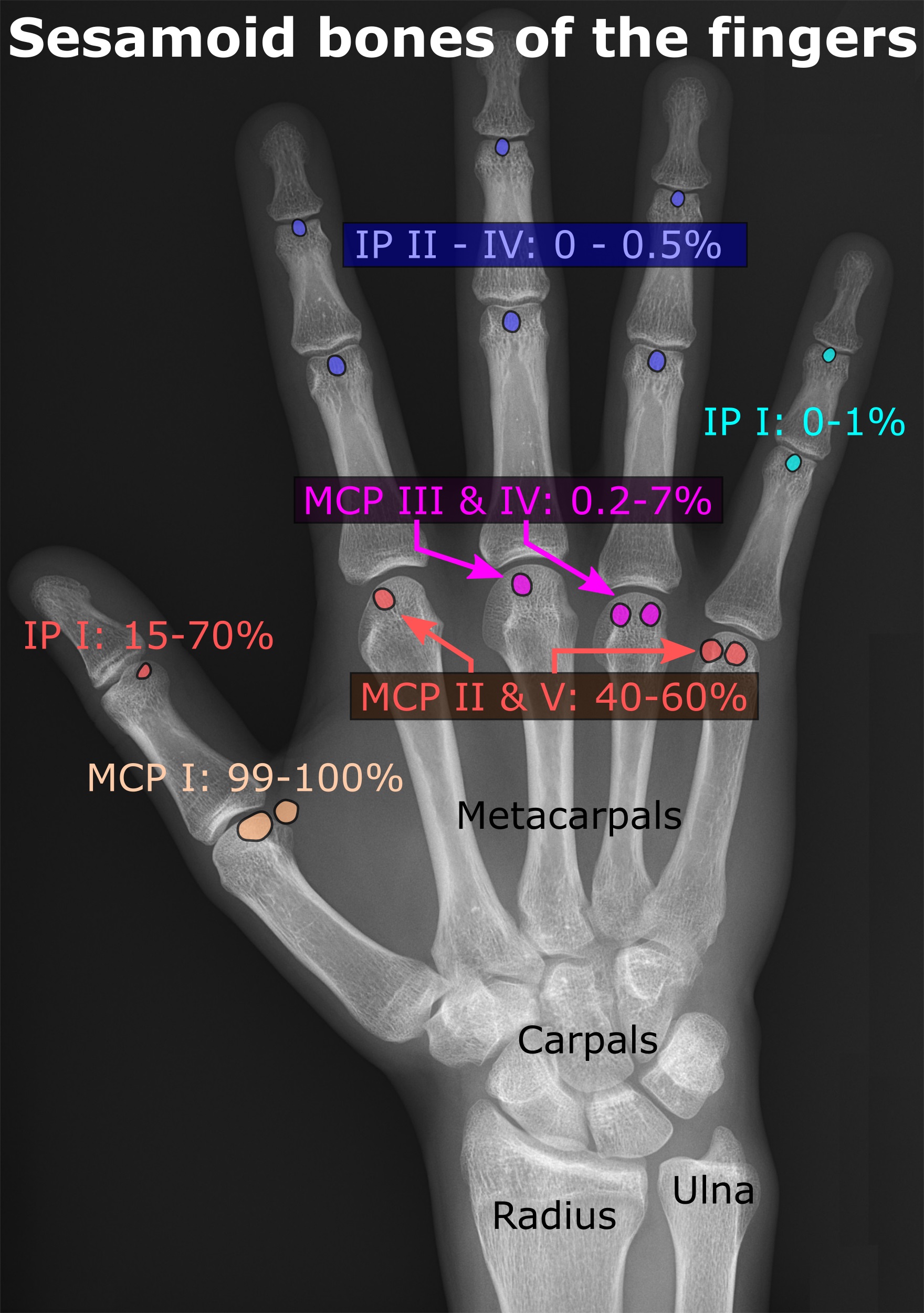
In anatomy, a sesamoid bone () is a bone embedded within a tendon or a muscle. Its name is derived from the Greek word for 'sesame seed', indicating the small size of most sesamoids. Often, these bones form in response to strain, or can be present as a anatomical variation, normal variant. The patella is the largest sesamoid bone in the body. Sesamoids act like pulleys, providing a smooth surface for tendons to slide over, increasing the tendon's ability to transmit Muscle#Function, muscular forces. Structure Sesamoid bones can be found on joints throughout the human body, including: * In the knee—the patella (within the quadriceps tendon). This is the largest sesamoid bone. * In the hand—two sesamoid bones are commonly found in the Anatomical terms of location#Arms, distal portions of the first metacarpal bone (within the tendons of adductor pollicis and flexor pollicis brevis). There is also commonly a sesamoid bone in distal portions of the second metacarpal bone and fif ... [...More Info...] [...Related Items...] OR: [Wikipedia] [Google] [Baidu] |
Synovial Membrane
Synovial () may refer to: * Synovial fluid * Synovial joint A synovial joint, also known as diarthrosis, joins bones or cartilage with a fibrous joint capsule that is continuous with the periosteum of the joined bones, constitutes the outer boundary of a synovial cavity, and surrounds the bones' articulati ... * Synovial membrane * Synovial bursa {{disambiguation ... [...More Info...] [...Related Items...] OR: [Wikipedia] [Google] [Baidu] |
Thieme Medical Publishers
Thieme Medical Publishers is a German academic publishing, medical and science publisher in the Thieme Publishing Group. It produces professional journals, textbooks, atlases, monographs and reference books in both German and English covering a variety of medical specialties, including neurosurgery, orthopaedics, endocrinology, urology, radiology, anatomy, chemistry, otolaryngology, ophthalmology, audiology and speech-language pathology, complementary medicine, complementary and alternative medicine. Thieme has more than 1,000 employees and maintains offices in seven cities worldwide, including New York City, Beijing, Delhi, Stuttgart, and three other cities in Germany. History Georg Thieme Verlag was founded in 1886 in Leipzig, Germany, by Georg Thieme when he was 26 years old. Thieme remains privately held and family-owned. The company received some early success in 1896 by publishing Wilhelm Röntgen's famous picture of his wife's hand in what is still one of Thieme's and ... [...More Info...] [...Related Items...] OR: [Wikipedia] [Google] [Baidu] |
Bone
A bone is a rigid organ that constitutes part of the skeleton in most vertebrate animals. Bones protect the various other organs of the body, produce red and white blood cells, store minerals, provide structure and support for the body, and enable mobility. Bones come in a variety of shapes and sizes and have complex internal and external structures. They are lightweight yet strong and hard and serve multiple functions. Bone tissue (osseous tissue), which is also called bone in the uncountable sense of that word, is hard tissue, a type of specialised connective tissue. It has a honeycomb-like matrix internally, which helps to give the bone rigidity. Bone tissue is made up of different types of bone cells. Osteoblasts and osteocytes are involved in the formation and mineralisation of bone; osteoclasts are involved in the resorption of bone tissue. Modified (flattened) osteoblasts become the lining cells that form a protective layer on the bone surface. The mine ... [...More Info...] [...Related Items...] OR: [Wikipedia] [Google] [Baidu] |


