|
Keipert Syndrome
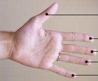
Nasodigitoacoustic syndrome, also called Keipert syndrome, is a rare congenital syndrome first described by J.A. Keipert and colleagues in 1973. The syndrome is characterized by a misshaped nose, broad thumbs and halluces (the big toes), brachydactyly, sensorineural hearing loss, facial features such as hypertelorism (unusually wide-set eyes), and developmental delay. It is believed to be inherited in an X-linked recessive manner, which means a genetic mutation causing the disorder is located on the X chromosome, and while two copies of the mutated gene must be inherited for a female to be born with the disorder, just one copy is sufficient to cause a male to be born with the disorder. Nasodigitoacoustic syndrome is likely caused by a mutated gene located on the X chromosome between positions Xq22.2–q28. The incidence of the syndrome has not been determined, but it is considered to affect less than 200,000 people in the United States, and no greater than 1 per 2,000 in Euro ... [...More Info...] [...Related Items...] OR: [Wikipedia] [Google] [Baidu] |
Rare Disease
A rare disease is any disease that affects a small percentage of the population. In some parts of the world, the term orphan disease describes a rare disease whose rarity results in little or no funding or research for treatments, without financial incentives from governments or other agencies. Orphan drugs are medications targeting orphan diseases. Most rare diseases are genetic in origin and thus are present throughout the person's entire life, even if symptoms do not immediately appear. Many rare diseases appear early in life, and about 30% of children with rare diseases will die before reaching their fifth birthdays. Fields condition is considered the rarest known disease, affecting three known individuals, two of whom are identical twins. With four diagnosed patients in 27 years, ribose-5-phosphate isomerase deficiency is considered the second rarest. While no single number has been agreed upon for which a disease is considered rare, several efforts have been undertaken to ... [...More Info...] [...Related Items...] OR: [Wikipedia] [Google] [Baidu] |
Teunissen–Cremers Syndrome
Teunissen–Cremers syndrome is a genetic disorder that presents with skeleton defects some of which can include the bones of the inner ear, fingers and toes. This can result in conductive hearing loss Conductive hearing loss (CHL) is a type of hearing impairment that occurs when sound waves are unable to efficiently travel through the outer ear, tympanic membrane (eardrum), or middle ear structures such as the ossicles. This blockage or dysfun ... and finger deformities. References Bert Teunissen, Cor Cremers. An autosomal dominant inherited syndrome with congenital stapes ankylosis. Laryngoscope 100: April 1990, 380-384 External links Genetic syndromes Deafness Skeletal disorders Syndromes {{Genetic-disorder-stub} ... [...More Info...] [...Related Items...] OR: [Wikipedia] [Google] [Baidu] |
Radiograph
Radiography is an imaging technique using X-rays, gamma rays, or similar ionizing radiation and non-ionizing radiation to view the internal form of an object. Applications of radiography include medical ("diagnostic" radiography and "therapeutic radiography") and industrial radiography. Similar techniques are used in airport security, (where "body scanners" generally use backscatter X-ray). To create an image in conventional radiography, a beam of X-rays is produced by an X-ray generator and it is projected towards the object. A certain amount of the X-rays or other radiation are absorbed by the object, dependent on the object's density and structural composition. The X-rays that pass through the object are captured behind the object by a detector (either photographic film or a digital detector). The generation of flat two-dimensional images by this technique is called projectional radiography. In computed tomography (CT scanning), an X-ray source and its associated de ... [...More Info...] [...Related Items...] OR: [Wikipedia] [Google] [Baidu] |
Clinodactyly
Clinodactyly is a medical term describing the curvature of a digit (a finger or toe) in the plane of the palm, most commonly the fifth finger (the "little finger") towards the adjacent fourth finger (the "ring finger"). It is a fairly common isolated anomaly which often goes unnoticed, but also occurs in combination with other abnormalities in certain genetic syndromes. The term comes . Genetics Clinodactyly is an autosomal dominant trait that has variable expressiveness and incomplete penetrance. Clinodactyly can be passed through inheritance and presents as either an isolated anomaly or a component manifestation of a genetic syndrome. Many syndromes are associated with clinodactyly, including those listed below. But the phenotype, by itself, is not a sensitive or specific diagnostic test for these syndromes (it is present in up to 18% of the normal population). * Down syndrome * Turner syndrome Turner syndrome (TS), commonly known as 45,X, or 45,X0,Also written as 45, ... [...More Info...] [...Related Items...] OR: [Wikipedia] [Google] [Baidu] |
Distal Phalanges
The phalanges (: phalanx ) are digital bones in the hands and feet of most vertebrates. In primates, the thumbs and big toes have two phalanges while the other digits have three phalanges. The phalanges are classed as long bones. Structure The phalanges are the bones that make up the fingers of the hand and the toes of the foot. There are 56 phalanges in the human body, with fourteen on each hand and foot. Three phalanges are present on each finger and toe, with the exception of the thumb and big toe, which possess only two. The middle and far phalanges of the fifth toes are often fused together (symphalangism). The phalanges of the hand are commonly known as the finger bones. The phalanges of the foot differ from the hand in that they are often shorter and more compressed, especially in the proximal phalanges, those closest to the torso. A phalanx is named according to whether it is proximal, middle, or distal and its associated finger or toe. The proximal phalanges ... [...More Info...] [...Related Items...] OR: [Wikipedia] [Google] [Baidu] |
Digit (anatomy)
A digit is one of several most distal parts of a limb, such as fingers or toes, present in many vertebrates. Names Some languages have different names for hand and foot digits (English: respectively "finger" and " toe", German: "Finger" and "Zeh", French: "doigt" and "orteil"). In other languages, e.g. Arabic, Russian, Polish, Spanish, Portuguese, Italian, Czech, Tagalog, Turkish, Bulgarian, and Persian, there are no specific one-word names for fingers and toes; these are called "digit of the hand" or "digit of the foot" instead. In Japanese, yubi (指) can mean either, depending on context. Human digits Humans normally have five digits on each extremity. Each digit is formed by several bones called phalanges, surrounded by soft tissue. Human fingers normally have a nail at the distal phalanx. The phenomenon of polydactyly occurs when extra digits are present; fewer digits than normal are also possible, for instance in ectrodactyly. Whether such a mutation can ... [...More Info...] [...Related Items...] OR: [Wikipedia] [Google] [Baidu] |
Retrognathism
Retrognathia is a type of malocclusion which refers to an abnormal posterior positioning of the maxilla or mandible, particularly the mandible, relative to the facial skeleton and soft tissues. A retrognathic mandible is commonly referred to as an overbite, though this terminology is not used medically. See also * Micrognathism * Prognathism Prognathism is a positional relationship of the mandible or maxilla to the skeletal base where either of the jaws protrudes beyond a predetermined imaginary line in the coronal plane of the skull. In the case of ''mandibular'' prognathism (nev ... References External links Diagram at brooksideorthodontics.com - see Classification of Face:Class 2 section Jaw disorders {{disease-stub ... [...More Info...] [...Related Items...] OR: [Wikipedia] [Google] [Baidu] |
Maxilla
In vertebrates, the maxilla (: maxillae ) is the upper fixed (not fixed in Neopterygii) bone of the jaw formed from the fusion of two maxillary bones. In humans, the upper jaw includes the hard palate in the front of the mouth. The two maxillary bones are fused at the intermaxillary suture, forming the anterior nasal spine. This is similar to the mandible (lower jaw), which is also a fusion of two mandibular bones at the mandibular symphysis. The mandible is the movable part of the jaw. Anatomy Structure The maxilla is a paired bone - the two maxillae unite with each other at the intermaxillary suture. The maxilla consists of: * The body of the maxilla: pyramid-shaped; has an orbital, a nasal, an infratemporal, and a facial surface; contains the maxillary sinus. * Four processes: ** the zygomatic process ** the frontal process ** the alveolar process ** the palatine process It has three surfaces: * the anterior, posterior, medial Features of the maxilla include: * t ... [...More Info...] [...Related Items...] OR: [Wikipedia] [Google] [Baidu] |
Maxillary Hypoplasia
Maxillary hypoplasia, or maxillary deficiency, is an underdevelopment of the bones of the upper jaw. It is associated with Crouzon syndrome, Angelman syndrome, as well as Fetal alcohol syndrome. It can also be associated with Cleft lip and cleft palate. Some people could develop it due to poor dental extractions. Signs and symptoms The underdevelopment of the bones in the upper jaw, which gives the middle of the face a sunken look. This same underdevelopment can make it difficult to eat and can lead to complications such as Nasopharyngeal airway restriction. This restriction causes forward head posture which can then lead to back pain, neck pain, and numbness in the hands and arms. The nasopharyngeal airway restriction can also lead to Sleep apnea and snoring. Sleep apnea can lead to heart problems, endocrine problems, increased weight, and cognition problems, among other issues. Cause Although the exact genetic link for isolated maxillary hypoplasia has not been identified ... [...More Info...] [...Related Items...] OR: [Wikipedia] [Google] [Baidu] |
Cupid's Bow
The Cupid's bow is a facial feature where the double curve of a human upper lip is said to resemble a recurve bow of the sort used in ancient Greece or Rome. The name is taken from Cupid, the bow-wielding Roman god of erotic love equivalent to the Greek Eros. The peaks of the bow coincide with the philtrum, philtral columns giving a prominent bow appearance to the lip. It is outlined with the vermilion border, which connects the lip skin to the facial skin. Celebrities with a pronounced cupid's bow include Taylor Swift and Rihanna. See also * Philtrum *White roll References External links Facial features Words and phrases derived from Greek mythology Cupid Lips {{anatomy-stub ... [...More Info...] [...Related Items...] OR: [Wikipedia] [Google] [Baidu] |
Epicanthic Fold
An epicanthic fold or epicanthus is a skin fold of the upper eyelid that covers the inner corner (medial canthus) of the Human eye, eye. However, variation occurs in the nature of this feature and the presence of "partial epicanthic folds" or "slight epicanthic folds" is noted in the relevant literature. Various factors influence whether epicanthic folds form, including ancestry, age, and certain medical conditions. The primary cause of the epicanthic fold is the hypertrophy of the preseptal portion of the orbicularis oculi muscle. Etymology ''Epicanthus'' means 'above the canthus', with epi-canthus being the Latinized form of the Ancient Greek : 'corner of the eye'. Classification Variation in the shape of the epicanthic fold has led to four types being recognised: * ''Epicanthus supraciliaris'' runs from the brow, curving downwards towards the lachrymal sac. * ''Epicanthus palpebralis'' begins above the upper Tarsus (eyelids), tarsus and extends to the inferior orbita ... [...More Info...] [...Related Items...] OR: [Wikipedia] [Google] [Baidu] |
Supraorbital Ridge
The brow ridge, or supraorbital ridge known as superciliary arch in medicine, is a bony ridge located above the eye sockets of all primates and some other animals. In humans, the Eyebrow, eyebrows are located on their lower margin. Structure The brow ridge is a nodule or crest of bone situated on the frontal bone of the skull. It forms the separation between the forehead portion itself (the squama frontalis) and the roof of the eye sockets (the Orbital part of frontal bone, pars orbitalis). Normally, in humans, the ridges arch over each eye, offering mechanical protection. In other primates, the ridge is usually continuous and often straight rather than arched. The ridges are separated from the frontal eminences by a shallow groove. The ridges are most prominent medially, and are joined to one another by a smooth elevation named the glabella. Typically, the arches are more prominent in men than in women, and vary between different human populations. Behind the ridges, deeper in t ... [...More Info...] [...Related Items...] OR: [Wikipedia] [Google] [Baidu] |


