|
Juxtaglomerular Cell Tumor
Juxtaglomerular cell tumor (JCT, JGCT, also reninoma) is an extremely rare kidney tumour of the juxtaglomerular cells, with fewer than 100 cases reported in literature. This tumor typically secretes renin, hence the former name of reninoma. It often causes severe hypertension that is difficult to control, in adults and children, although among causes of secondary hypertension it is rare. It develops most commonly in young adults, but can be diagnosed much later in life. It is generally considered benign, but its malignant potential is uncertain. Pathophysiology By hypersecretion of renin, JCT causes hypertension, often severe and usually sustained but occasionally paroxysmal, and secondary hyperaldosteronism inducing hypokalemia, though the later can be mild despite high renin. Both of these conditions may be corrected by surgical removal of the tumor. Asymptomatic cases have been reported. Histopathology JCT is morphologically characterized by multiple foci malignant mesenchym ... [...More Info...] [...Related Items...] OR: [Wikipedia] [Google] [Baidu] |
Kidney Tumour
Kidney tumours are tumours, or growths, on or in the kidney. These growths can be benign or malignant ( kidney cancer). Presentation Kidney tumours may be discovered on medical imaging incidentally (i.e. an incidentaloma), or may be present in patients as an abdominal mass or kidney cyst, hematuria, abdominal pain, or manifest first in a paraneoplastic syndrome that seems unrelated to the kidney. Other markers or complications that may arise from kidney tumours can appear to be more subtle, including; low hemoglobin, fatigue, nausea, constipation, and/or hyperglycemia. Diagnosis A CT scan is the first choice modality for workup of solid masses in the kidneys. Nevertheless, hemorrhagic cysts can resemble renal cell carcinomas on CT, but they are easily distinguished with Doppler ultrasonography (Doppler US). In renal cell carcinomas, Doppler US often shows vessels with high velocities caused by neovascularization and arteriovenous shunting. Some renal cell carcinomas are hypov ... [...More Info...] [...Related Items...] OR: [Wikipedia] [Google] [Baidu] |
CD117
Proto-oncogene c-KIT is the gene encoding the receptor tyrosine kinase protein known as tyrosine-protein kinase KIT, CD117 ( cluster of differentiation 117) or mast/stem cell growth factor receptor (SCFR). Multiple transcript variants encoding different isoforms have been found for this gene. KIT was first described by the German biochemist Axel Ullrich in 1987 as the cellular homolog of the feline sarcoma viral oncogene v-kit. Function KIT is a cytokine receptor expressed on the surface of hematopoietic stem cells as well as other cell types. Altered forms of this receptor may be associated with some types of cancer. KIT is a receptor tyrosine kinase type III, which binds to stem cell factor, also known as "steel factor" or "c-kit ligand". When this receptor binds to stem cell factor (SCF) it forms a dimer that activates its intrinsic tyrosine kinase activity, that in turn phosphorylates and activates signal transduction molecules that propagate the signal in the cell. ... [...More Info...] [...Related Items...] OR: [Wikipedia] [Google] [Baidu] |
Karyotyping
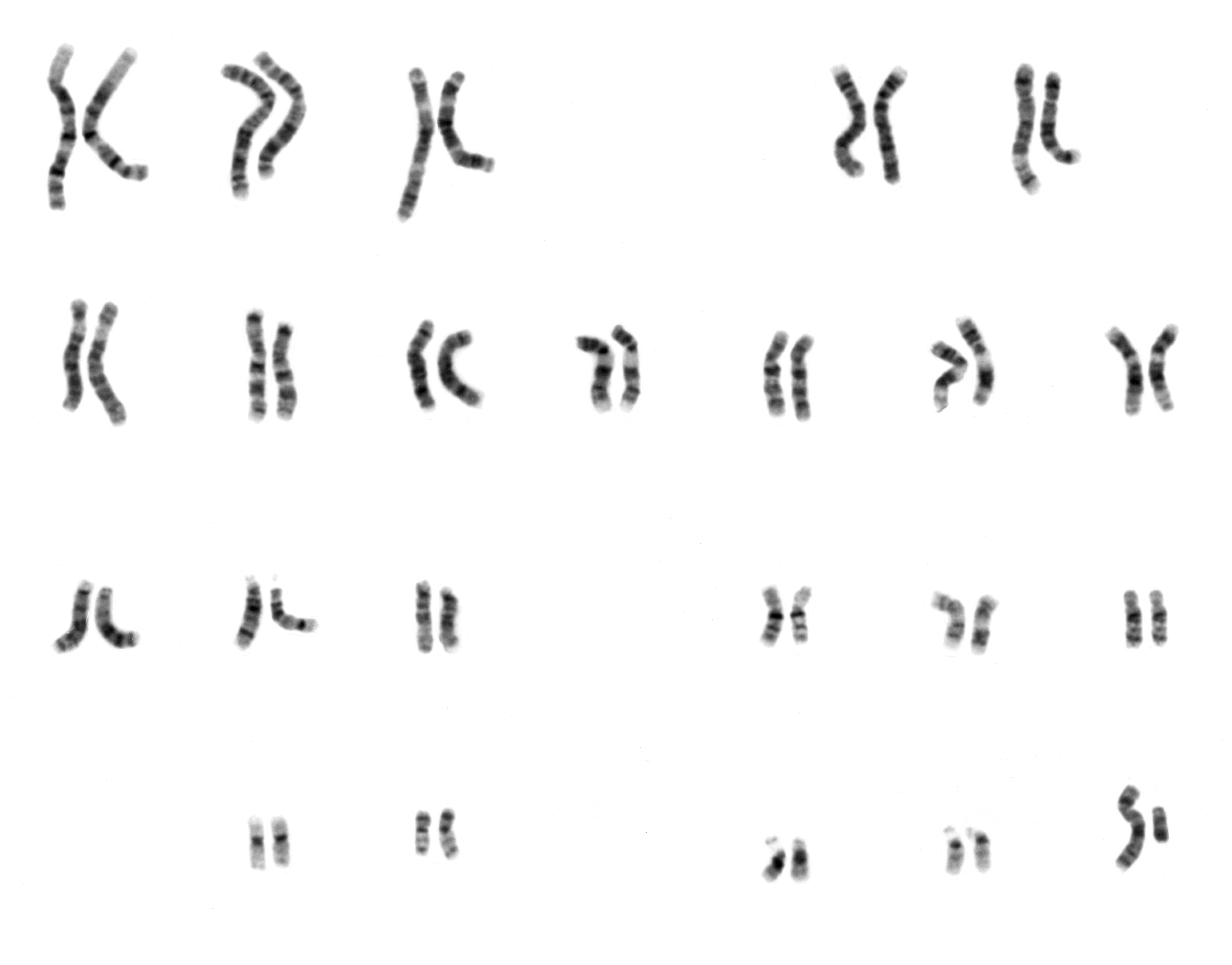
A karyotype is the general appearance of the complete set of chromosomes in the cells of a species or in an individual organism, mainly including their sizes, numbers, and shapes. Karyotyping is the process by which a karyotype is discerned by determining the chromosome complement of an individual, including the number of chromosomes and any abnormalities. A karyogram or idiogram is a graphical depiction of a karyotype, wherein chromosomes are generally organized in pairs, ordered by size and position of centromere for chromosomes of the same size. Karyotyping generally combines light microscopy and photography in the metaphase of the cell cycle, and results in a photomicrographic (or simply micrographic) karyogram. In contrast, a schematic karyogram is a designed graphic representation of a karyotype. In schematic karyograms, just one of the sister chromatids of each chromosome is generally shown for brevity, and in reality they are generally so close together that they look as ... [...More Info...] [...Related Items...] OR: [Wikipedia] [Google] [Baidu] |
American Journal Of Clinical Pathology
The ''American Journal of Clinical Pathology'' is a monthly peer-reviewed medical journal covering clinical pathology. It was established in 1931 and is published by Oxford University Press. It is the official journal of the American Society for Clinical Pathology and the Academy of Clinical Laboratory Physicians and Scientists. The editor-in-chief is Steven H. Kroft ( Medical College of Wisconsin). According to the ''Journal Citation Reports'', the journal has a 2020 impact factor The impact factor (IF) or journal impact factor (JIF) of an academic journal is a type of journal ranking. Journals with higher impact factor values are considered more prestigious or important within their field. The Impact Factor of a journa ... of 2.493. References External links * Clinical pathology Pathology journals Oxford University Press academic journals Academic journals established in 1931 Monthly journals Academic journals associated with learned and professional societies ... [...More Info...] [...Related Items...] OR: [Wikipedia] [Google] [Baidu] |
Solitary Fibrous Tumor
Solitary fibrous tumor (SFT), also known as fibrous tumor of the pleura, is a rare mesenchymal tumor originating in the pleuraTravis WD, Brambilla E, Muller-Hermelink HK, Harris CC (Eds.): World Health Organization Classification of Tumours. Pathology and Genetics of Tumours of the Lung, Pleura, Thymus and Heart. IARC Press: Lyon 2004. or at virtually any site in the soft tissue including the seminal vesicle. Approximately 78% to 88% of SFT's are benign and 12% to 22% are malignant.Robinson LA. Solitary fibrous tumor of the pleura. Cancer Control 2006;13:264-9. The World Health Organization (2020) classified SFT as a specific type of tumor in the category of malignant fibroblastic and myofibroblastic tumors. Signs and symptoms About 80% of pleural SFTs originate in the visceral pleura, while 20% arise from parietal pleura. Although they are often very large tumors—up to in diameter—over half are asymptomatic at diagnosis.Briselli M, Mark EJ, Dickersin GR. Solitary fibrous tum ... [...More Info...] [...Related Items...] OR: [Wikipedia] [Google] [Baidu] |
Wilms' Tumor
Wilms' tumor or Wilms tumor, also known as nephroblastoma, is a cancer of the kidneys that typically occurs in children (rarely in adults), and occurs most commonly as a renal tumor in child patients. It is named after Max Wilms, the German surgeon (1867–1918) who first described it. Approximately 650 cases are diagnosed in the U.S. annually. The majority of cases occur in children with no associated genetic syndromes; however, a minority of children with Wilms' tumor have a congenital abnormality. It is highly responsive to treatment, with about 90 percent of children being cured. Signs and symptoms Typical signs and symptoms of Wilms' tumor include the following: * a painless, palpable abdominal mass * loss of appetite * abdominal pain * fever * nausea and vomiting * blood in the urine (in about 20% of cases) * high blood pressure in some cases (especially if synchronous or metachronous bilateral kidney involvement) * Rarely as varicoceleErginel B, Vural S, Akın M ... [...More Info...] [...Related Items...] OR: [Wikipedia] [Google] [Baidu] |
Angiomyolipoma
Angiomyolipomas are the most common benign tumour of the kidney. Although regarded as benign, angiomyolipomas may grow such that kidney function is impaired or the blood vessels may dilate and burst, leading to bleeding. Angiomyolipomas are strongly associated with the genetic disease tuberous sclerosis, in which most individuals have several angiomyolipomas affecting both kidneys. They are also commonly found in women with the rare lung disease lymphangioleiomyomatosis. Angiomyolipomas are less commonly found in the liver and rarely in other organs. Whether associated with these diseases or sporadic, angiomyolipomas are caused by mutations in either the ''TSC1'' or ''TSC2 ''genes, which govern cell growth and proliferation. They are composed of blood vessels, smooth muscle cells, and fat cells. Large angiomyolipomas can be treated with embolisation. Drug therapy for angiomyolipomas is at the research stage. The Tuberous Sclerosis Alliance has published guidelines on diagno ... [...More Info...] [...Related Items...] OR: [Wikipedia] [Google] [Baidu] |
Metanephric Adenoma
Metanephric adenoma (MA) is a rare, benign tumour of the kidney, that can have a microscopic appearance similar to a nephroblastoma (Wilms tumours), or a papillary renal cell carcinoma. It should not be confused with the pathologically unrelated, yet similar sounding, '' mesonephric adenoma''. Symptoms The symptoms may be similar to those classically associated with renal cell carcinoma, and may include polycythemia, abdominal pain, hematuria Hematuria or haematuria is defined as the presence of blood or red blood cells in the urine. "Gross hematuria" occurs when urine appears red, brown, or tea-colored due to the presence of blood. Hematuria may also be subtle and only detectable with ... and a palpable mass. Mean age at onset is around 40 years with a range of 5 to 83 years and the mean size of the tumour is 5.5 cm with a range 0.3 to 15 cm (1). Polycythemia is more frequent in MA than in any other type of renal tumour. Of further relevance is that this tumour is mo ... [...More Info...] [...Related Items...] OR: [Wikipedia] [Google] [Baidu] |
Glomus Tumor
:''Glomus tumor was also the name formerly (and incorrectly) used for a tumor now called a paraganglioma.'' A glomus tumor (also known as a "solitary glomus tumor") is a rare neoplasm arising from the glomus body and mainly found under the nail, on the fingertip or in the foot.Freedberg, et al. (2003). ''Fitzpatrick's Dermatology in General Medicine''. (6th ed.). McGraw-Hill. . They account for less than 2% of all soft tissue tumors. The majority of glomus tumors are benign, but they can also show malignant features. Glomus tumors were first described by Hoyer in 1877 while the first complete clinical description was given by Masson in 1924. Histologically, glomus tumors are made up of an afferent arteriole, anastomotic vessel, and collecting venule. Glomus tumors are modified smooth muscle cells that control the thermoregulatory function of dermal glomus bodies. As stated above, these lesions should not be confused with paragangliomas, which were formerly also called glomus tu ... [...More Info...] [...Related Items...] OR: [Wikipedia] [Google] [Baidu] |
Hemangiopericytoma
A hemangiopericytoma is a type of soft-tissue sarcoma that originates in the pericytes in the walls of capillaries. When inside the nervous system, although not strictly a meningioma tumor, it is a meningeal tumor with a special aggressive behavior. It was first characterized in 1942. Signs and symptoms Symptoms of hemangiopericytoma vary greatly depending on both tumor stage and affected organs. Most patients report pain and mass-related symptoms, while others also report vascular disease-related symptoms, and some have no symptoms until late in the disease process. Hemangiopericytomas are most commonly found in the meninges, lower extremities, retroperitoneum, pelvis, lungs, and pleura. Histopathology Hemangiopericytomas are tumors that are derived from specialized spindle shaped cells called pericytes, which line capillaries. Diagnosis Computerized tomography and magnetic resonance imaging are not effective methods for diagnosis of hemangiocytomas. In practice, a presum ... [...More Info...] [...Related Items...] OR: [Wikipedia] [Google] [Baidu] |
Differential Diagnosis
In healthcare, a differential diagnosis (DDx) is a method of analysis that distinguishes a particular disease or condition from others that present with similar clinical features. Differential diagnostic procedures are used by clinicians to diagnose the specific disease in a patient, or, at least, to consider any imminently life-threatening conditions. Often, each possible disease is called a differential diagnosis (e.g., acute bronchitis could be a differential diagnosis in the evaluation of a cough, even if the final diagnosis is common cold). More generally, a differential diagnostic procedure is a systematic diagnostic method used to identify the presence of a disease entity where multiple alternatives are possible. This method may employ algorithms, akin to the process of elimination, or at least a process of obtaining information that decreases the "probabilities" of candidate conditions to negligible levels, by using evidence such as symptoms, patient history, and medi ... [...More Info...] [...Related Items...] OR: [Wikipedia] [Google] [Baidu] |
Computed Tomography
A computed tomography scan (CT scan), formerly called computed axial tomography scan (CAT scan), is a medical imaging technique used to obtain detailed internal images of the body. The personnel that perform CT scans are called radiographers or radiology technologists. CT scanners use a rotating X-ray tube and a row of detectors placed in a gantry to measure X-ray attenuations by different tissues inside the body. The multiple X-ray measurements taken from different angles are then processed on a computer using tomographic reconstruction algorithms to produce tomographic (cross-sectional) images (virtual "slices") of a body. CT scans can be used in patients with metallic implants or pacemakers, for whom magnetic resonance imaging (MRI) is contraindicated. Since its development in the 1970s, CT scanning has proven to be a versatile imaging technique. While CT is most prominently used in medical diagnosis, it can also be used to form images of non-living objects. The 1979 N ... [...More Info...] [...Related Items...] OR: [Wikipedia] [Google] [Baidu] |


