|
Juvenile Hyaline Fibromatosis
Juvenile hyaline fibromatosis (also known as "Fibromatosis hyalinica multiplex juvenilis," "Murray–Puretic–Drescher syndrome") is a very rare, autosomal recessive disease due to mutations in capillary morphogenesis protein-2 (CMG-2 gene). It occurs from early childhood to adulthood, and presents as slow-growing, pearly white or skin-colored dermal or subcutaneous papules or nodules on the face, scalp, and back, which may be confused clinically with neurofibromatosis.Freedberg, et al. (2003). ''Fitzpatrick's Dermatology in General Medicine''. (6th ed.). Page 989. McGraw-Hill. . The World Health Organization, 2020, reclassified the papules and nodules that occur in juvenile hyaline firbromatosis as one of the specific benign types of tumors in the category of fibroblastic and myofibroblastic tumors. Presentation This condition is characterised by abnormal growth of hyalinized fibrous tissue with cutaneous, mucosal, osteoarticular and systemic involvement. Clinical features i ... [...More Info...] [...Related Items...] OR: [Wikipedia] [Google] [Baidu] |
Rare Disease
A rare disease is any disease that affects a small percentage of the population. In some parts of the world, an orphan disease is a rare disease whose rarity means there is a lack of a market large enough to gain support and resources for discovering treatments for it, except by the government granting economically advantageous conditions to creating and selling such treatments. Orphan drugs are ones so created or sold. Most rare diseases are genetic and thus are present throughout the person's entire life, even if symptoms do not immediately appear. Many rare diseases appear early in life, and about 30% of children with rare diseases will die before reaching their fifth birthdays. With only four diagnosed patients in 27 years, ribose-5-phosphate isomerase deficiency is considered the rarest known genetic disease. No single cut-off number has been agreed upon for which a disease is considered rare. A disease may be considered rare in one part of the world, or in a particular gr ... [...More Info...] [...Related Items...] OR: [Wikipedia] [Google] [Baidu] |
Face
The face is the front of an animal's head that features the eyes, nose and mouth, and through which animals express many of their emotions. The face is crucial for human identity, and damage such as scarring or developmental deformities may affect the psyche adversely. Structure The front of the human head is called the face. It includes several distinct areas, of which the main features are: *The forehead, comprising the skin beneath the hairline, bordered laterally by the temples and inferiorly by eyebrows and ears *The eyes, sitting in the orbit and protected by eyelids and eyelashes * The distinctive human nose shape, nostrils, and nasal septum *The cheeks, covering the maxilla and mandibula (or jaw), the extremity of which is the chin *The mouth, with the upper lip divided by the philtrum, sometimes revealing the teeth Facial appearance is vital for human recognition and communication. Facial muscles in humans allow expression of emotions. The face is itself a hi ... [...More Info...] [...Related Items...] OR: [Wikipedia] [Google] [Baidu] |
Chromosome 4
Chromosome 4 is one of the 23 pairs of chromosomes in humans. People normally have two copies of this chromosome. Chromosome 4 spans more than 186 million base pairs (the building material of DNA) and represents between 6 and 6.5 percent of the total DNA in cell (biology), cells. Genomics The chromosome is ~191 megabases in length. In a 2012 paper, 775 protein-encoding genes were identified on this chromosome.Chen LC, Liu MY, Hsiao YC, Choong WK, Wu HY, Hsu WL, Liao PC, Sung TY, Tsai SF, Yu JS, Chen YJ (2012) Decoding the disease-associated proteins encoded in the human chromosome 4. J Proteome Res 211 (27.9%) of these coding sequences did not have any experimental evidence at the protein level, in 2012. 271 appear to be membrane proteins. 54 have been classified as cancer-associated proteins. Genes Number of genes The following are some of the gene count estimates of human chromosome 4. Because researchers use different approaches to genome annotation their predictions of the num ... [...More Info...] [...Related Items...] OR: [Wikipedia] [Google] [Baidu] |
ANTXR2
Anthrax toxin receptor 2 (ANTXR2 also known as Capillary Morphogenesis Gene 2 or CMG2) is a protein that in humans is encoded by the ''ANTXR2'' gene. See also * Anthrax toxin Anthrax toxin is a three-protein exotoxin secreted by virulent strains of the bacterium, ''Bacillus anthracis''—the causative agent of anthrax. The toxin was first discovered by Harry Smith in 1954. Anthrax toxin is composed of a cell-bindin ... References External links * Further reading * * * * * * * * * * * * * * * * * {{gene-4-stub ... [...More Info...] [...Related Items...] OR: [Wikipedia] [Google] [Baidu] |
Fibroblastic And Myofibroblastic Tumors
Fibroblastic and myofibroblastic tumors (FMTs) develop from the mesenchymal stem cells which differentiate into fibroblasts (the most common cell type in connective tissue) and/or the myocytes/ myoblasts that differentiate into muscle cells. FMTs are a heterogeneous group of soft tissue neoplasms (i.e. abnormal and excessive tissue growths). The World Health Organization (2020) defined tumors as being FMTs based on their morphology and, more importantly, newly discovered abnormalities in the expression levels of key gene products made by these tumors' neoplastic cells. Histopathologically, FMTs consist of neoplastic connective tissue cells which have differented into cells that have microscopic appearances resembling fibroblasts and/or myofibroblasts. The fibroblastic cells are characterized as spindle-shaped cells with inconspicuous nucleoli that express vimentin, an intracellular protein typically found in mesenchymal cells, and CD34, a cell surface membrane glycopr ... [...More Info...] [...Related Items...] OR: [Wikipedia] [Google] [Baidu] |
Neurofibromatosis
Neurofibromatosis (NF) is a group of three conditions in which tumors grow in the nervous system. The three types are neurofibromatosis type I (NF1), neurofibromatosis type II (NF2), and schwannomatosis. In NF1 symptoms include light brown spots on the skin, freckles in the armpit and groin, small bumps within nerves, and scoliosis. In NF2, there may be hearing loss, cataracts at a young age, balance problems, flesh colored skin flaps, and muscle wasting. In schwannomatosis there may be pain either in one location or in wide areas of the body. The tumors in NF are generally non-cancerous. The cause is a genetic mutation in certain oncogenes. These can be inherited from a person's parents, or in about half of cases spontaneously occur during early development. Different mutations result in the three types of NF. Neurofibromatosis arise from the supporting cells of the nervous system rather than the neurons themselves. In NF1, the tumors are neurofibromas (tumors of ... [...More Info...] [...Related Items...] OR: [Wikipedia] [Google] [Baidu] |
Human Back
The human back, also called the dorsum, is the large posterior area of the human body, rising from the top of the buttocks to the back of the neck. It is the surface of the body opposite from the chest and the abdomen. The vertebral column runs the length of the back and creates a central area of recession. The breadth of the back is created by the shoulders at the top and the pelvis at the bottom. Back pain is a common medical condition, generally benign in origin. Structure The central feature of the human back is the vertebral column, specifically the length from the top of the thoracic vertebrae to the bottom of the lumbar vertebrae, which houses the spinal cord in its spinal canal, and which generally has some curvature that gives shape to the back. The ribcage extends from the spine at the top of the back (with the top of the ribcage corresponding to the T1 vertebra), more than halfway down the length of the back, leaving an area with less protection between the bott ... [...More Info...] [...Related Items...] OR: [Wikipedia] [Google] [Baidu] |
Scalp
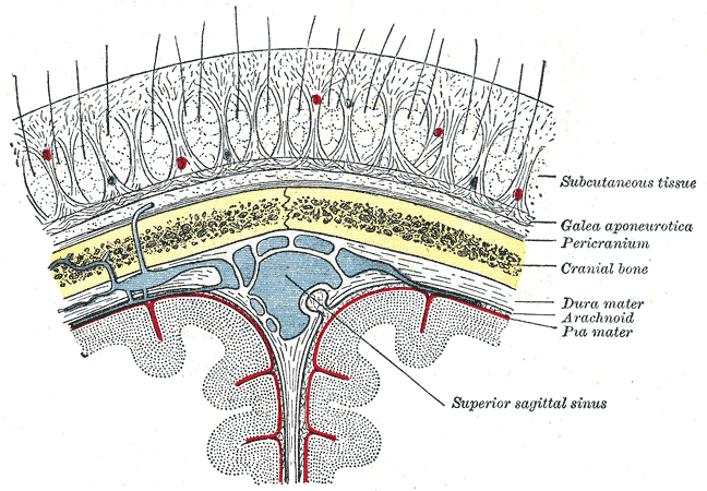
The scalp is the anatomical area bordered by the human face at the front, and by the neck at the sides and back. Structure The scalp is usually described as having five layers, which can conveniently be remembered as a mnemonic: * S: The skin on the head from which head hair grows. It contains numerous sebaceous glands and hair follicles. * C: Connective tissue. A dense subcutaneous layer of fat and fibrous tissue that lies beneath the skin, containing the nerves and vessels of the scalp. * A: The aponeurosis called epicranial aponeurosis (or galea aponeurotica) is the next layer. It is a tough layer of dense fibrous tissue which runs from the frontalis muscle anteriorly to the occipitalis posteriorly. * L: The loose areolar connective tissue layer provides an easy plane of separation between the upper three layers and the pericranium. In scalping the scalp is torn off through this layer. It also provides a plane of access in craniofacial surgery and neurosurgery. Th ... [...More Info...] [...Related Items...] OR: [Wikipedia] [Google] [Baidu] |
Cutaneous Condition
A skin condition, also known as cutaneous condition, is any medical condition that affects the integumentary system—the organ system that encloses the body and includes skin, nails, and related muscle and glands. The major function of this system is as a barrier against the external environment. Conditions of the human integumentary system constitute a broad spectrum of diseases, also known as dermatoses, as well as many nonpathologic states (like, in certain circumstances, melanonychia and racquet nails). While only a small number of skin diseases account for most visits to the physician, thousands of skin conditions have been described. Classification of these conditions often presents many nosological challenges, since underlying causes and pathogenetics are often not known. Therefore, most current textbooks present a classification based on location (for example, conditions of the mucous membrane), morphology ( chronic blistering conditions), cause (skin conditions ... [...More Info...] [...Related Items...] OR: [Wikipedia] [Google] [Baidu] |
Autosome
An autosome is any chromosome that is not a sex chromosome. The members of an autosome pair in a diploid cell have the same morphology, unlike those in allosomal (sex chromosome) pairs, which may have different structures. The DNA in autosomes is collectively known as atDNA or auDNA. For example, humans have a diploid genome that usually contains 22 pairs of autosomes and one allosome pair (46 chromosomes total). The autosome pairs are labeled with numbers (1–22 in humans) roughly in order of their sizes in base pairs, while allosomes are labelled with their letters. By contrast, the allosome pair consists of two X chromosomes in females or one X and one Y chromosome in males. Unusual combinations of XYY, XXY, XXX, XXXX, XXXXX or XXYY, among other Salome combinations, are known to occur and usually cause developmental abnormalities. Autosomes still contain sexual determination genes even though they are not sex chromosomes. For example, the SRY gene on the Y chr ... [...More Info...] [...Related Items...] OR: [Wikipedia] [Google] [Baidu] |
Papule
A papule is a small, well-defined bump in the skin. It may have a rounded, pointed or flat top, and may have a dip. It can appear with a stalk, be thread-like or look warty. It can be soft or firm and its surface may be rough or smooth. Some have crusts or scales. A papule can be flesh colored, yellow, white, brown, red, blue or purplish. There may be just one or many, and they may occur irregularly in different parts of the body or appear in clusters. It does not contain fluid but may progress to a pustule or vesicle. A papule is smaller than a nodule; it can be as tiny as a pinhead and is typically less than 1 cm in width, according to some sources, and 0.5 cm according to others. When merged together, it appears as a plaque. Its color might indicate its cause, such as white in milia, red in eczema, yellowish in xanthoma and black in melanoma. They may open when scratched and become infected and crusty. Definition A papule is a small, well-defined bump in ... [...More Info...] [...Related Items...] OR: [Wikipedia] [Google] [Baidu] |
Subcutaneous Tissue
The subcutaneous tissue (), also called the hypodermis, hypoderm (), subcutis, superficial fascia, is the lowermost layer of the integumentary system in vertebrates. The types of cells found in the layer are fibroblasts, adipose cells, and macrophages. The subcutaneous tissue is derived from the mesoderm, but unlike the dermis, it is not derived from the mesoderm's dermatome region. It consists primarily of loose connective tissue, and contains larger blood vessels and nerves than those found in the dermis. It is a major site of fat storage in the body. In arthropods, a hypodermis can refer to an epidermal layer of cells that secretes the chitinous cuticle. The term also refers to a layer of cells lying immediately below the epidermis of plants. Structure * Fibrous bands anchoring the skin to the deep fascia * Collagen and elastin fibers attaching it to the dermis * Fat is absent from the eyelids, clitoris, penis, much of pinna, and scrotum * Blood vessels on route to ... [...More Info...] [...Related Items...] OR: [Wikipedia] [Google] [Baidu] |


