|
Jugular Ganglion
The superior ganglion of the vagus nerve (jugular ganglion) is a sensory ganglion of the peripheral nervous system. It is located within the jugular foramen, where the vagus nerve exits the skull. It is smaller than and proximal to the inferior ganglion of the vagus nerve. Structure The neurons in the superior ganglion of the vagus nerve are pseudounipolar and provide sensory innervation ( general somatic afferent) through either the auricular or meningeal branch. The axon An axon (from Greek ἄξων ''áxōn'', axis) or nerve fiber (or nerve fibre: see American and British English spelling differences#-re, -er, spelling differences) is a long, slender cellular extensions, projection of a nerve cell, or neuron, ...s of these neurons synapse in the spinal trigeminal nucleus of the brainstem. Peripherally, the neurons found in the superior ganglion form two branches, the auricular and meningeal branch. Function Auricular branch of the vagus nerve The superior ... [...More Info...] [...Related Items...] OR: [Wikipedia] [Google] [Baidu] |
Glossopharyngeal
The glossopharyngeal nerve (), also known as the ninth cranial nerve, cranial nerve IX, or simply CN IX, is a cranial nerve that exits the brainstem from the sides of the upper Medulla oblongata, medulla, just anterior (closer to the nose) to the vagus nerve. Being a mixed nerve (sensorimotor), it carries afferent sensory and efferent motor information. The motor division of the glossopharyngeal nerve is derived from the Basal plate (neural tube), basal plate of the embryonic medulla oblongata, whereas the sensory division originates from the cranial neural crest. Structure From the anterior portion of the medulla oblongata, the glossopharyngeal nerve passes laterally across or below the Flocculus (cerebellar), flocculus, and leaves the skull through the central part of the jugular foramen. From the superior and inferior ganglia in jugular foramen, it has its own sheath of dura mater. The inferior ganglion on the inferior surface of petrous part of temporal is related with a tri ... [...More Info...] [...Related Items...] OR: [Wikipedia] [Google] [Baidu] |
Inferior Ganglion Of Vagus Nerve
The inferior ganglion of the vagus nerve (also known as the nodose ganglion) is one of the two sensory ganglia of each vagus nerve (cranial nerve X). It contains neuron cell bodies of general visceral afferent fibers and special visceral afferent fibers. It is situated within the jugular fossa just below the skull. It is situated just below the superior ganglion of vagus nerve. Anatomy The inferior ganglion of vagus nerve is elongated. It is larger than the superior ganglion of vagus nerve. It is situated within the jugular fossa, just inferior to the jugular foramen. Structure The inferior ganglion contains the neuron cell bodies of all sensory fibres of the CN X except those of the auricular branch of vagus nerve. The neurons in the inferior ganglion of the vagus nerve are pseudounipolar and provide sensory innervation (general somatic afferent and general visceral afferent). The axons of the neurons which innervate the taste buds of the epiglottis synapse in the ro ... [...More Info...] [...Related Items...] OR: [Wikipedia] [Google] [Baidu] |
Posterior Inferior Cerebellar Artery
The posterior inferior cerebellar artery (PICA) is the largest branch of the vertebral artery. It is one of the three main arteries that supply blood to the cerebellum, a part of the brain. Blockage of the posterior inferior cerebellar artery can result in a type of stroke called lateral medullary syndrome. Supply The PICA supplies blood to the medulla oblongata; the choroid plexus and tela choroidea of the fourth ventricle; the tonsils; the inferior vermis, and the inferior parts of the cerebellum. Course It winds backward around the upper part of the medulla oblongata, passing between the origins of the vagus nerve and the accessory nerve, over the inferior cerebellar peduncle to the undersurface of the cerebellum, where it divides into two branches. The medial branch continues backward to the notch between the two hemispheres of the cerebellum; while the lateral supplies the under surface of the cerebellum, as far as its lateral border, where it anastomoses with the ant ... [...More Info...] [...Related Items...] OR: [Wikipedia] [Google] [Baidu] |
Ear Pain
Ear pain, also known as earache or otalgia, is pain in the ear. Primary ear pain is pain that originates from the ear. Secondary ear pain is a type of referred pain, meaning that the source of the pain differs from the location where the pain is felt. Most causes of ear pain are non-life-threatening. Primary ear pain is more common than secondary ear pain, and it is often due to infection or injury. The conditions that cause secondary (referred) ear pain are broad and range from temporomandibular joint syndrome to inflammation of the throat. In general, the reason for ear pain can be discovered by taking a thorough history of all symptoms and performing a physical examination, without need for imaging tools like a CT scan. However, further testing may be needed if red flags are present like hearing loss, dizziness, ringing in the ear or unexpected weight loss. Management of ear pain depends on the cause. If there is a bacterial infection, antibiotics are sometimes recommended an ... [...More Info...] [...Related Items...] OR: [Wikipedia] [Google] [Baidu] |
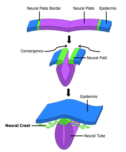
Neural Crest
The neural crest is a ridge-like structure that is formed transiently between the epidermal ectoderm and neural plate during vertebrate development. Neural crest cells originate from this structure through the epithelial-mesenchymal transition, and in turn give rise to a diverse cell lineage—including melanocytes, craniofacial cartilage and bone, smooth muscle, dentin, peripheral and enteric neurons, adrenal medulla and glia. After gastrulation, the neural crest is specified at the border of the neural plate and the non-neural ectoderm. During neurulation, the borders of the neural plate, also known as the neural folds, converge at the dorsal midline to form the neural tube. Subsequently, neural crest cells from the roof plate of the neural tube undergo an epithelial to mesenchymal transition, delaminating from the neuroepithelium and migrating through the periphery, where they differentiate into varied cell types. The emergence of the neural crest was important in v ... [...More Info...] [...Related Items...] OR: [Wikipedia] [Google] [Baidu] |
Eardrum
In the anatomy of humans and various other tetrapods, the eardrum, also called the tympanic membrane or myringa, is a thin, cone-shaped membrane that separates the external ear from the middle ear. Its function is to transmit changes in pressure of sound from the air to the ossicles inside the middle ear, and thence to the oval window in the fluid-filled cochlea. The ear thereby converts and amplifies vibration in the air to vibration in cochlear fluid. The malleus bone bridges the gap between the eardrum and the other ossicles. Rupture or perforation of the eardrum can lead to conductive hearing loss. Collapse or retraction of the eardrum can cause conductive hearing loss or cholesteatoma. Structure Orientation and relations The tympanic membrane is oriented obliquely in the anteroposterior, mediolateral, and superoinferior planes. Consequently, its superoposterior end lies lateral to its anteroinferior end. Anatomically, it relates superiorly to the middle crani ... [...More Info...] [...Related Items...] OR: [Wikipedia] [Google] [Baidu] |
Ear Canal
The ear canal (external acoustic meatus, external auditory meatus, EAM) is a pathway running from the outer ear to the middle ear. The adult human ear canal extends from the auricle to the eardrum and is about in length and in diameter. Structure The human ear canal is divided into two parts. The elastic cartilage part forms the outer third of the canal; its anterior and lower wall are cartilaginous, whereas its superior and back wall are fibrous. The cartilage is the continuation of the cartilage framework of auricle. The cartilaginous portion of the ear canal contains small hairs and specialized sweat glands, called apocrine glands, which produce cerumen ( ear wax). The bony part forms the inner two thirds. The bony part is much shorter in children and is only a ring (''annulus tympanicus'') in the newborn. The layer of epithelium encompassing the bony portion of the ear canal is much thinner and therefore, more sensitive in comparison to the cartilaginous portion. Size ... [...More Info...] [...Related Items...] OR: [Wikipedia] [Google] [Baidu] |
Auricle (anatomy)
The auricle or auricula is the visible part of the ear that is outside the head. It is also called the pinna (Latin for 'wing' or ' fin', : pinnae), a term that is used more in zoology. Structure The diagram shows the shape and location of most of these components: * ''antihelix'' forms a 'Y' shape where the upper parts are: ** ''Superior crus'' (to the left of the ''fossa triangularis'' in the diagram) ** ''Inferior crus'' (to the right of the ''fossa triangularis'' in the diagram) * ''Antitragus'' is below the ''tragus'' * ''Aperture'' is the entrance to the ear canal * ''Auricular sulcus'' is the depression behind the ear next to the head * ''Concha'' is the hollow next to the ear canal * Conchal angle is the angle that the back of the ''concha'' makes with the side of the head * ''Crus'' of the helix is just above the ''tragus'' * ''Cymba conchae'' is the narrowest end of the ''concha'' * External auditory meatus is the ear canal * ''Fossa triangularis'' is the depression ... [...More Info...] [...Related Items...] OR: [Wikipedia] [Google] [Baidu] |
Spinal Trigeminal Nucleus
The spinal trigeminal nucleus is a nucleus in the medulla that receives information about deep/crude touch, pain, and temperature from the ipsilateral face. In addition to the trigeminal nerve (CN V), the facial (CN VII), glossopharyngeal (CN IX), and vagus nerves (CN X) also convey pain information from their areas to the spinal trigeminal nucleus. Thus the spinal trigeminal nucleus receives afferents from V, VII, [...More Info...] [...Related Items...] OR: [Wikipedia] [Google] [Baidu] |
Axon
An axon (from Greek ἄξων ''áxōn'', axis) or nerve fiber (or nerve fibre: see American and British English spelling differences#-re, -er, spelling differences) is a long, slender cellular extensions, projection of a nerve cell, or neuron, in Vertebrate, vertebrates, that typically conducts electrical impulses known as action potentials away from the Soma (biology), nerve cell body. The function of the axon is to transmit information to different neurons, muscles, and glands. In certain sensory neurons (pseudounipolar neurons), such as those for touch and warmth, the axons are called afferent nerve fibers and the electrical impulse travels along these from the peripheral nervous system, periphery to the cell body and from the cell body to the spinal cord along another branch of the same axon. Axon dysfunction can be the cause of many inherited and acquired neurological disorders that affect both the Peripheral nervous system, peripheral and Central nervous system, central ne ... [...More Info...] [...Related Items...] OR: [Wikipedia] [Google] [Baidu] |


