|
Ischiofemoral Ligament
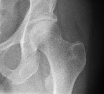
The ischiofemoral ligament (ischiocapsular ligament or ischiocapsular band) consists of a triangular band of strong fibers on the posterior side of the hip joint. It is one of the four ligaments that reinforce the hip joint. It attaches to the posterior surface of the acetabular rim and acetabular labrum, and extends around the circumference of the joint to insert on the anterior aspect of the femur. The ischiofemoral ligament limits the internal rotation and adduction of the hip when it is in a flexed position. Some deeper fibres of the ligament are continuous with the fibres of the zona orbicularis The zona orbicularis or annular ligament is a ligament on the neck of the femur formed by the circular fibers of the articular capsule of the hip joint In vertebrate anatomy, the hip, or coxaLatin ''coxa'' was used by Celsus in the sense "hi ... of the capsule. This ligament is less well-defined than the other two capsular ligaments of the hip joint. Function Studies o ... [...More Info...] [...Related Items...] OR: [Wikipedia] [Google] [Baidu] |
Hip Joint
In vertebrate anatomy, the hip, or coxaLatin ''coxa'' was used by Celsus in the sense "hip", but by Pliny the Elder in the sense "hip bone" (Diab, p 77) (: ''coxae'') in medical terminology, refers to either an anatomical region or a joint on the outer (lateral) side of the pelvis. The hip region is located lateral and anterior to the gluteal region, inferior to the iliac crest, and lateral to the obturator foramen, with muscle tendons and soft tissues overlying the greater trochanter of the femur The femur (; : femurs or femora ), or thigh bone is the only long bone, bone in the thigh — the region of the lower limb between the hip and the knee. In many quadrupeds, four-legged animals the femur is the upper bone of the hindleg. The Femo .... In adults, the three pelvic bones (ilium (bone), ilium, ischium and pubis (bone), pubis) have fused into one hip bone, which forms the superomedial/deep wall of the hip region. The hip joint, scientifically referred to as t ... [...More Info...] [...Related Items...] OR: [Wikipedia] [Google] [Baidu] |
Ischium
The ischium (; : ischia) is a paired bone forming the lower and back part of the hip bone. Situated below the ilium (bone), ilium and behind the pubis (bone), pubis, it is one of three regions whose fusion creates the coxal bone. The superior portion of this region forms approximately one-third of the acetabulum. Structure The ischium is made up of three parts–the body, the superior ramus and the inferior ramus. The body contains a prominent ischial spine, spine, which serves as the origin for the superior gemellus muscle. The indentation inferior to the spine is the lesser sciatic notch. Continuing down the posterior side, the ischial tuberosity is a thick, rough-surfaced prominence below the lesser sciatic notch. This is the portion ...[...More Info...] [...Related Items...] OR: [Wikipedia] [Google] [Baidu] |
Femur
The femur (; : femurs or femora ), or thigh bone is the only long bone, bone in the thigh — the region of the lower limb between the hip and the knee. In many quadrupeds, four-legged animals the femur is the upper bone of the hindleg. The Femoral head, top of the femur fits into a socket in the pelvis called the hip joint, and the bottom of the femur connects to the shinbone (tibia) and kneecap (patella) to form the knee. In humans the femur is the largest and thickest bone in the body. Structure The femur is the only bone in the upper Human leg, leg. The two femurs converge Anatomical terms of location, medially toward the knees, where they articulate with the Anatomical terms of location, proximal ends of the tibiae. The angle at which the femora converge is an important factor in determining the femoral-tibial angle. In females, thicker pelvic bones cause the femora to converge more than in males. In the condition genu valgum, ''genu valgum'' (knock knee), the femurs conve ... [...More Info...] [...Related Items...] OR: [Wikipedia] [Google] [Baidu] |
Acetabulum
The acetabulum (; : acetabula), also called the cotyloid cavity, is a wikt:concave, concave surface of the pelvis. The femur head, head of the femur meets with the pelvis at the acetabulum, forming the Hip#Articulation, hip joint. Structure There are three bones of the ''os coxae'' (hip bone) that come together to form the ''acetabulum''. Contributing a little more than two-fifths of the structure is the ischium, which provides lower and side boundaries to the acetabulum. The Ilium (bone), ilium forms the upper boundary, providing a little less than two-fifths of the structure of the acetabulum. The rest is formed by the Pubis (bone), pubis, near the midline. It is bounded by a prominent uneven rim, thick and strong on top, which serves as the point of attachment for the acetabular labrum. The acetabular labrum reduces the size of the opening of the acetabulum and deepens the surface of the hip joint. At the lower part of the acetabulum is the acetabular notch, which is continuo ... [...More Info...] [...Related Items...] OR: [Wikipedia] [Google] [Baidu] |
Acetabular Labrum
The acetabular labrum (glenoidal labrum of the hip joint or cotyloid ligament in older texts) is a fibrocartilaginous ring which surrounds the circumference of the acetabulum of the hip, deepening the acetabulum. The labrum is attached onto the bony rim and transverse acetabular ligament. It is triangular in cross-section (with the apex represented by the free margin). The labrum contributes to the articular surface of the joint (increasing it by almost 10%). It embraces the femoral head, holding it firmly in the joint socket to stabilise the joint. It thus also seals the joint cavity, facilitating even distribution of synovial fluid Synovial fluid, also called synovia, elp 1/sup> is a viscous, non-Newtonian fluid found in the cavities of synovial joints. With its egg white–like consistency, the principal role of synovial fluid is to reduce friction between the articul ... so that friction is reduced and dissolved nutrients are better distributed. The labrum is about 2 ... [...More Info...] [...Related Items...] OR: [Wikipedia] [Google] [Baidu] |
Flexion
Motion, the process of movement, is described using specific anatomical terminology, anatomical terms. Motion includes movement of Organ (anatomy), organs, joints, Limb (anatomy), limbs, and specific sections of the body. The terminology used describes this motion according to its direction relative to the anatomical position of the body parts involved. Anatomy, Anatomists and others use a unified set of terms to describe most of the movements, although other, more specialized terms are necessary for describing unique movements such as those of the hands, feet, and eyes. In general, motion is classified according to the anatomical plane it occurs in. ''Flexion'' and ''extension'' are examples of ''angular'' motions, in which two axes of a joint are brought closer together or moved further apart. ''Rotational'' motion may occur at other joints, for example the shoulder, and are described as ''internal'' or ''external''. Other terms, such as ''elevation'' and ''depression'', descri ... [...More Info...] [...Related Items...] OR: [Wikipedia] [Google] [Baidu] |
Zona Orbicularis
The zona orbicularis or annular ligament is a ligament on the neck of the femur formed by the circular fibers of the articular capsule of the hip joint In vertebrate anatomy, the hip, or coxaLatin ''coxa'' was used by Celsus in the sense "hip", but by Pliny the Elder in the sense "hip bone" (Diab, p 77) (: ''coxae'') in medical terminology, refers to either an anatomical region or a joint o .... It is also known as the orbicular zone, ring ligament, and zonular band. Structure The zona orbicularis forms a ring around the neck of the femur. The articular capsule is much thicker above and in front of the joint, where the greatest amount of resistance is required, and thin and loose behind and below the joint. The capsule consists of two sets of fibers, circular and longitudinal. The circular fibers, the zona orbicularis, are most abundant at the lower and back part of the capsule where they form a sling or collar around the femoral neck. Anteriorly, they blend with the ... [...More Info...] [...Related Items...] OR: [Wikipedia] [Google] [Baidu] |


