|
Intertubercular Line
A lower transverse plane midway between the upper transverse and the upper border of the pubic symphysis; this is termed the intertubercular plane (or transtubercular), since it practically corresponds to that passing through the iliac tubercles; behind, its plane cuts the body of the fifth lumbar vertebra. Additional images Image:Gray1034.png, Front view of the thoracic and abdominal viscera.a. Median plane.b. Lateral planes.c. Trans tubercular plane.d. Subcostal plane.e. Transpyloric plane. Image:Gray1227.png, Front of abdomen, showing surface markings for arteries and inguinal canal. See also * Quadrants and regions of abdomen * Subcostal plane * Supracristal plane * Transpyloric plane The transpyloric plane, also known as Addison's plane, is an imaginary transverse plane, horizontal plane, located halfway between the suprasternal notch of the manubrium and the upper border of the symphysis pubis at the level of the first lumba ... References External links * http ... [...More Info...] [...Related Items...] OR: [Wikipedia] [Google] [Baidu] |
Thorax
The thorax (: thoraces or thoraxes) or chest is a part of the anatomy of mammals and other tetrapod animals located between the neck and the abdomen. In insects, crustaceans, and the extinct trilobites, the thorax is one of the three main divisions of the body, each in turn composed of multiple segments. The human thorax includes the thoracic cavity and the thoracic wall. It contains organs including the heart, lungs, and thymus gland, as well as muscles and various other internal structures. The chest may be affected by many diseases, of which the most common symptom is chest pain. Etymology The word thorax comes from the Greek θώραξ ''thṓrax'' " breastplate, cuirass, corslet" via . Humans Structure In humans and other hominids, the thorax is the chest region of the body between the neck and the abdomen, along with its internal organs and other contents. It is mostly protected and supported by the rib cage, spine, and shoulder girdle. Contents The ... [...More Info...] [...Related Items...] OR: [Wikipedia] [Google] [Baidu] |
Abdomen
The abdomen (colloquially called the gut, belly, tummy, midriff, tucky, or stomach) is the front part of the torso between the thorax (chest) and pelvis in humans and in other vertebrates. The area occupied by the abdomen is called the abdominal cavity. In arthropods, it is the posterior (anatomy), posterior tagma (biology), tagma of the body; it follows the thorax or cephalothorax. In humans, the abdomen stretches from the thorax at the thoracic diaphragm to the pelvis at the pelvic brim. The pelvic brim stretches from the lumbosacral joint (the intervertebral disc between Lumbar vertebrae, L5 and Vertebra#Sacrum, S1) to the pubic symphysis and is the edge of the pelvic inlet. The space above this inlet and under the thoracic diaphragm is termed the abdominal cavity. The boundary of the abdominal cavity is the abdominal wall in the front and the peritoneal surface at the rear. In vertebrates, the abdomen is a large body cavity enclosed by the abdominal muscles, at the front an ... [...More Info...] [...Related Items...] OR: [Wikipedia] [Google] [Baidu] |
Duodenum
The duodenum is the first section of the small intestine in most vertebrates, including mammals, reptiles, and birds. In mammals, it may be the principal site for iron absorption. The duodenum precedes the jejunum and ileum and is the shortest part of the small intestine. In humans, the duodenum is a hollow jointed tube about long connecting the stomach to the jejunum, the middle part of the small intestine. It begins with the duodenal bulb, and ends at the duodenojejunal flexure marked by the suspensory muscle of duodenum. The duodenum can be divided into four parts: the first (superior), the second (descending), the third (transverse) and the fourth (ascending) parts. Overview The duodenum is the first section of the small intestine in most higher vertebrates, including mammals, reptiles, and birds. In fish, the divisions of the small intestine are not as clear, and the terms ''anterior intestine'' or ''proximal intestine'' may be used instead of duodenum. In mammals the d ... [...More Info...] [...Related Items...] OR: [Wikipedia] [Google] [Baidu] |
Pancreas
The pancreas (plural pancreases, or pancreata) is an Organ (anatomy), organ of the Digestion, digestive system and endocrine system of vertebrates. In humans, it is located in the abdominal cavity, abdomen behind the stomach and functions as a gland. The pancreas is a mixed or heterocrine gland, i.e., it has both an endocrine and a digestive exocrine function. Ninety-nine percent of the pancreas is exocrine and 1% is endocrine. As an endocrine gland, it functions mostly to regulate blood sugar levels, secreting the hormones insulin, glucagon, somatostatin and pancreatic polypeptide. As a part of the digestive system, it functions as an exocrine gland secreting pancreatic juice into the duodenum through the pancreatic duct. This juice contains bicarbonate, which neutralizes acid entering the duodenum from the stomach; and digestive enzymes, which break down carbohydrates, proteins and lipids, fats in food entering the duodenum from the stomach. Inflammation of the pancreas is kno ... [...More Info...] [...Related Items...] OR: [Wikipedia] [Google] [Baidu] |
Kidney
In humans, the kidneys are two reddish-brown bean-shaped blood-filtering organ (anatomy), organs that are a multilobar, multipapillary form of mammalian kidneys, usually without signs of external lobulation. They are located on the left and right in the retroperitoneal space, and in adult humans are about in length. They receive blood from the paired renal artery, renal arteries; blood exits into the paired renal veins. Each kidney is attached to a ureter, a tube that carries excreted urine to the urinary bladder, bladder. The kidney participates in the control of the volume of various body fluids, fluid osmolality, Acid-base homeostasis, acid-base balance, various electrolyte concentrations, and removal of toxins. Filtration occurs in the glomerulus (kidney), glomerulus: one-fifth of the blood volume that enters the kidneys is filtered. Examples of substances reabsorbed are solute-free water, sodium, bicarbonate, glucose, and amino acids. Examples of substances secreted are hy ... [...More Info...] [...Related Items...] OR: [Wikipedia] [Google] [Baidu] |
Pubic Symphysis
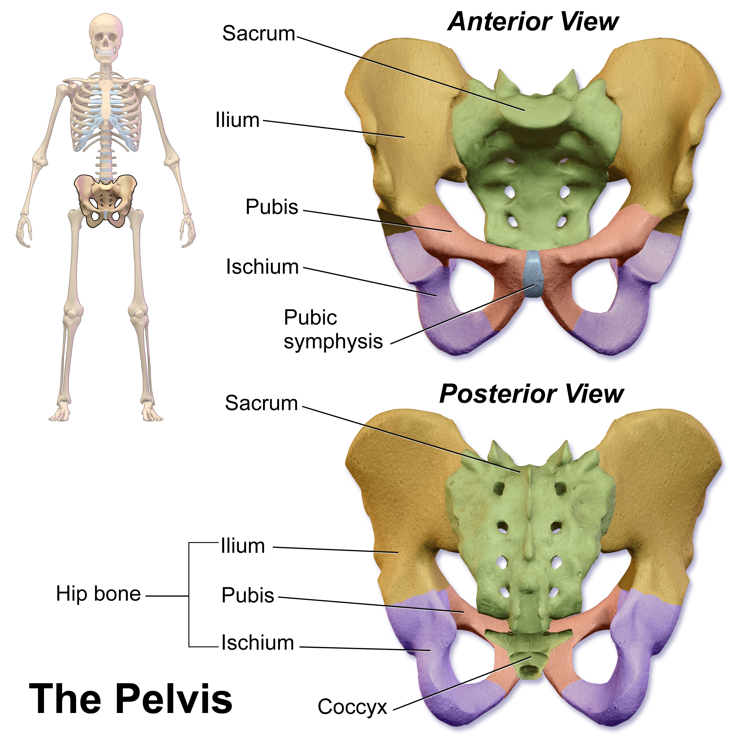
The pubic symphysis (: symphyses) is a secondary cartilaginous joint between the left and right superior rami of the pubis of the hip bones. It is in front of and below the urinary bladder. In males, the suspensory ligament of the penis attaches to the pubic symphysis. In females, the pubic symphysis is attached to the suspensory ligament of the clitoris. In most adults, it can be moved roughly 2 mm and with 1 degree rotation. This increases for women at the time of childbirth. The name comes from the Greek word ''symphysis'', meaning 'growing together'. Structure The pubic symphysis is a nonsynovial amphiarthrodial joint. The width of the pubic symphysis at the front is 3–5 mm greater than its width at the back. This joint is connected by fibrocartilage and may contain a fluid-filled cavity; the center is avascular, possibly due to the nature of the compressive forces passing through this joint, which may lead to harmful vascular disease. The ends of both pubi ... [...More Info...] [...Related Items...] OR: [Wikipedia] [Google] [Baidu] |
Iliac Tubercle
The iliac tubercle is located approximately posterior to the anterior superior iliac spine on the iliac crest in humans. The transverse plane that includes each of the tubercles (one from the left iliac tubercle and one from the right iliac tubercle) is called the transtubercular plane. The origin of the iliotibial tract is the iliac tubercle. The iliac tubercle is also the widest point of the iliac crest, and lies at the level of the L5 spinous process Each vertebra (: vertebrae) is an irregular bone with a complex structure composed of bone and some hyaline cartilage, that make up the vertebral column or spine, of vertebrates. The proportions of the vertebrae differ according to their spina .... References Bones of the pelvis Ilium (bone) {{musculoskeletal-stub ... [...More Info...] [...Related Items...] OR: [Wikipedia] [Google] [Baidu] |
Lumbar Vertebrae
The lumbar vertebrae are located between the thoracic vertebrae and pelvis. They form the lower part of the back in humans, and the tail end of the back in quadrupeds. In humans, there are five lumbar vertebrae. The term is used to describe the anatomy of humans and quadrupeds, such as horses, pigs, or cattle. These bones are found in particular cuts of meat, including tenderloin or sirloin steak. Human anatomy In human anatomy, the five vertebrae are between the rib cage and the pelvis. They are the largest segments of the vertebral column and are characterized by the absence of the foramen transversarium within the transverse process (since it is only found in the cervical region) and by the absence of facets on the sides of the body (as found only in the thoracic region). They are designated L1 to L5, starting at the top. The lumbar vertebrae help support the weight of the body, and permit movement. General characteristics The adjacent figure depicts the general cha ... [...More Info...] [...Related Items...] OR: [Wikipedia] [Google] [Baidu] |
Quadrants And Regions Of Abdomen
The human abdomen is divided into quadrants and regions by anatomy, anatomists and physicians for the purposes of study, medical diagnosis, diagnosis, and therapy, treatment. The division into four quadrants allows the localisation of pain and tenderness (medicine), tenderness, scars, lumps, and other items of interest, narrowing in on which organ (anatomy), organs and tissue (biology), tissues may be involved. The quadrants are referred to as the left lower quadrant, left upper quadrant, right upper quadrant and right lower quadrant. These terms are not used in comparative anatomy, since most other animals do not stand erect. The left lower quadrant includes the left iliac fossa and half of the Flank (anatomy), flank. The equivalent in other animals is ''left posterior quadrant''. The left upper quadrant extends from the umbilical plane to the left ribcage. This is the ''left anterior quadrant'' in other animals. The right upper quadrant extends from umbilical plane to the right ... [...More Info...] [...Related Items...] OR: [Wikipedia] [Google] [Baidu] |
Subcostal Plane
The subcostal plane is a transverse plane which bisects the body at the level of the 10th costal margin and the upper border of the third lumbar vertebra. See also *Inferior mesenteric artery *Quadrants and regions of abdomen The human abdomen is divided into quadrants and regions by anatomy, anatomists and physicians for the purposes of study, medical diagnosis, diagnosis, and therapy, treatment. The division into four quadrants allows the localisation of pain and te ... * Supracristal plane * Transpyloric plane * Transtubercular plane References External links * http://www.liv.ac.uk/HumanAnatomy/phd/mbchb/travel/surface1.html * http://www.qub.ac.uk/cskills/Abd%20exam.htm Anatomical planes {{Anatomy-stub ... [...More Info...] [...Related Items...] OR: [Wikipedia] [Google] [Baidu] |
Supracristal Plane
Supracristal plane (''Planum supracristale'') (or supracrestal plane) is an anatomical transverse plane lying at the upper most part of the pelvis, the iliac crest. According to Gray's Anatomy, this anatomical plane crosses the upper border of the spinous process of L4 (fourth lumbar vertebra). It passes through the umbilical region and the left and right lumbar regions. Clinical significance The supracristal plane can be used as a landmark for several nerve branches, as well as an approximate marker for the umbilicus (belly button). It is also used as the divider between the lower (left and right) and upper (left and right) quadrants of the abdomen (where the vertical midline divides left from right). It is also the level where the abdominal aorta bifurcates into the left and right common iliac artery and just superior to the union of the common iliac veins. It can help in the identification of the level of L3/L4 where a lumbar puncture can be done safely. See also * Intertu ... [...More Info...] [...Related Items...] OR: [Wikipedia] [Google] [Baidu] |
Transpyloric Plane
The transpyloric plane, also known as Addison's plane, is an imaginary transverse plane, horizontal plane, located halfway between the suprasternal notch of the manubrium and the upper border of the symphysis pubis at the level of the first lumbar vertebrae, L1. It lies roughly a hand's breadth beneath the xiphisternum or midway between the xiphisternum and the navel, umbilicus. The plane in most cases cuts through the pylorus of the stomach, the tips of the ninth costal cartilages and the lower border of the first lumbar vertebra. Structures crossed The transpyloric plane is clinically notable because it passes through several important abdominal structures. It also divides the Supracolic compartment, supracolic and infracolic compartments, with the liver, spleen and gastric fundus above it and the small intestine and colon below it. Lumbar vertebra and spinal cord The lower border of first Lumbar vertebrae, lumbar vertebra lies at the level of the transpyloric plane, according ... [...More Info...] [...Related Items...] OR: [Wikipedia] [Google] [Baidu] |


