|
Interleukin-20
Interleukin 20 (IL20) is a protein that is in humans encoded by the ''IL20'' gene which is located in close proximity to the IL-10 gene on the 1q32 chromosome. IL-20 is a part of an IL-20 subfamily which is a part of a larger IL-10 family. IL-20 subfamily also includes other cytokines, including IL-19, IL-20, IL-22, IL-24, and IL-26. Members of the cytokine IL-20 subfamily form an important link between the immune system and epithelial tissues due to the fact that receptors for these cytokines are highly expressed on epithelial cells and are almost exclusively produced by cells of the immune system. IL-20 requires an IL-β-subunit receptor ( IL-20RB) for signaling, which can form a functional heterodimeric receptor with either the α-subunit of the IL-20 receptor (IL-20RA) or the α1-subunit of the IL-22 receptor (IL-22RA1). Both of these receptor variants allow efficient IL-20 signaling. Receptors for IL-20 are expressed in the skin, lungs, ovary, testes, and placent ... [...More Info...] [...Related Items...] OR: [Wikipedia] [Google] [Baidu] |
Interleukin 19
Interleukin 19 (IL-19) is an immunosuppressive protein that belongs to the IL-10 cytokine subfamily. Human IL-19 is encoded by the IL-19 gene which codes for 9 exons and is located on chromosome 1. The IL-19 protein is composed of 159 amino acids and has a quaternary structure with alpha helix motifs and loops. IL-19 is preferentially expressed in monocytes, macrophages, and T and B lymphocytes, but interacts with immune cells ( macrophages, T cells, B cells) and non-immune cells ( endothelial cells and brain resident glial cells, etc). IL-19 initiates JAK-STAT signaling which activates genes and creates mRNA sequences ( transcription) that are translated into proteins (translation) which have downstream effector functions. IL-19 signaling uses IL-20 dimer receptor complexes that bind the IL-19 ligand, Janus kinases (JAKs), and the signal transducer and activator of transcription 3 ( STAT3) to initiate the molecular signaling cascade shown on the diagram on the right. Funct ... [...More Info...] [...Related Items...] OR: [Wikipedia] [Google] [Baidu] |
Interleukin 20 Receptor, Beta Subunit
Interleukin 20 receptor, beta subunit (IL20R2 or IL20RB) is a subunit of the interleukin-20 receptor and interleukin-22 receptor. It is believed to be involved in both pro-inflammatory and anti-inflammatory responses. IL20RB is found in many organ resident effector cells such as keratinocytes at the skin epidermis, osteoclasts, found in bones, and epithelial cells of the intestine and trachea. IL20RB is also found in some immune cells. The subunit has been linked with multiple diseases, including gastrointestinal diseases and glaucoma. Structure and function IL20RB is a β-chain with a short intracellular domain. IL20RB, along with the IL-20 receptor, alpha subunit, form the heterodimeric type I interleukin-20 receptor, which binds the cytokines IL-20 and IL-24. IL20RB also associates with IL-22 receptor, to form the heterodimeric type II interleukin-20 receptor, which also binds IL-20 and IL-24. Signaling After a cytokine binds both the IL20RB and the alpha subunit of ... [...More Info...] [...Related Items...] OR: [Wikipedia] [Google] [Baidu] |
Protein
Proteins are large biomolecules and macromolecules that comprise one or more long chains of amino acid residues. Proteins perform a vast array of functions within organisms, including catalysing metabolic reactions, DNA replication, responding to stimuli, providing structure to cells and organisms, and transporting molecules from one location to another. Proteins differ from one another primarily in their sequence of amino acids, which is dictated by the nucleotide sequence of their genes, and which usually results in protein folding into a specific 3D structure that determines its activity. A linear chain of amino acid residues is called a polypeptide. A protein contains at least one long polypeptide. Short polypeptides, containing less than 20–30 residues, are rarely considered to be proteins and are commonly called peptides. The individual amino acid residues are bonded together by peptide bonds and adjacent amino acid residues. The sequence of amino acid resid ... [...More Info...] [...Related Items...] OR: [Wikipedia] [Google] [Baidu] |
IL22RA1
Interleukin 22 receptor, alpha 1 is a protein that in humans is encoded by the IL22RA1 gene. Function The protein encoded by this gene belongs to the class II cytokine receptor family, and has been shown to be a receptor for interleukin 22 ( IL22). IL22 receptor is a protein complex that consists of this protein and interleukin 10 receptor, beta (IL10RB/CRFB4), a subunit also shared by the receptor complex for interleukin 10 (IL10). This gene and interleukin 28 receptor, alpha (IL28RA Interleukin 28 receptor, alpha subunit is a subunit for the interleukin-28 receptor. IL28RA is its human gene In biology, the word gene (from , ; "...Wilhelm Johannsen coined the word gene to describe the Mendelian units of heredity..." me ...) form a cytokine receptor gene cluster in the chromosomal region 1p36. References Further reading * * * * * * * * * Type II cytokine receptors {{gene-1-stub ... [...More Info...] [...Related Items...] OR: [Wikipedia] [Google] [Baidu] |
Interleukin 1 Beta
Interleukin-1 beta (IL-1β) also known as leukocytic pyrogen, leukocytic endogenous mediator, mononuclear cell factor, lymphocyte activating factor and other names, is a cytokine protein that in humans is encoded by the ''IL1B'' gene."Catabolin" is the name given by Jeremy Saklatvala for IL-1 alpha. There are two genes for interleukin-1 (IL-1): IL-1 alpha and IL-1 beta (this gene). IL-1β precursor is cleaved by cytosolic caspase 1 (interleukin 1 beta convertase) to form mature IL-1β. Function The fever-producing property of human leukocytic pyrogen (interleukin 1) was purified by Dinarello in 1977 with a specific activity of 10–20 nanograms/kg. In 1979, Dinarello reported that purified human leukocytic pyrogen was the same molecule that was described by Igal Gery in 1972. He named it lymphocyte-activating factor (LAF) because it was a lymphocyte mitogen. It was not until 1984 that interleukin 1 was discovered to consist of two distinct proteins, now called interleukin- ... [...More Info...] [...Related Items...] OR: [Wikipedia] [Google] [Baidu] |
Fibroblast
A fibroblast is a type of biological cell that synthesizes the extracellular matrix and collagen, produces the structural framework ( stroma) for animal tissues, and plays a critical role in wound healing. Fibroblasts are the most common cells of connective tissue in animals. Structure Fibroblasts have a branched cytoplasm surrounding an elliptical, speckled nucleus having two or more nucleoli. Active fibroblasts can be recognized by their abundant rough endoplasmic reticulum. Inactive fibroblasts (called fibrocytes) are smaller, spindle-shaped, and have a reduced amount of rough endoplasmic reticulum. Although disjointed and scattered when they have to cover a large space, fibroblasts, when crowded, often locally align in parallel clusters. Unlike the epithelial cells lining the body structures, fibroblasts do not form flat monolayers and are not restricted by a polarizing attachment to a basal lamina on one side, although they may contribute to basal lamina components in so ... [...More Info...] [...Related Items...] OR: [Wikipedia] [Google] [Baidu] |
Keratinocyte
Keratinocytes are the primary type of cell found in the epidermis, the outermost layer of the skin. In humans, they constitute 90% of epidermal skin cells. Basal cells in the basal layer (''stratum basale'') of the skin are sometimes referred to as basal keratinocytes. Keratinocytes form a barrier against environmental damage by heat, UV radiation, water loss, pathogenic bacteria, fungi, parasites, and viruses. A number of structural proteins, enzymes, lipids, and antimicrobial peptides contribute to maintain the important barrier function of the skin. Keratinocytes differentiate from epidermal stem cells in the lower part of the epidermis and migrate towards the surface, finally becoming corneocytes and eventually be shed off, which happens every 40 to 56 days in humans. Function The primary function of keratinocytes is the formation of a barrier against environmental damage by heat, UV radiation, water loss, pathogenic bacteria, fungi, parasites, and viruses. Pathogens i ... [...More Info...] [...Related Items...] OR: [Wikipedia] [Google] [Baidu] |
Dendritic Cell
Dendritic cells (DCs) are antigen-presenting cells (also known as ''accessory cells'') of the mammalian immune system. Their main function is to process antigen material and present it on the cell surface to the T cells of the immune system. They act as messengers between the innate and the adaptive immune systems. Dendritic cells are present in those tissues that are in contact with the external environment, such as the skin (where there is a specialized dendritic cell type called the Langerhans cell) and the inner lining of the nose, lungs, stomach and intestines. They can also be found in an immature state in the blood. Once activated, they migrate to the lymph nodes where they interact with T cells and B cells to initiate and shape the adaptive immune response. At certain development stages they grow branched projections, the '' dendrites'' that give the cell its name (δένδρον or déndron being Greek for 'tree'). While similar in appearance, these are stru ... [...More Info...] [...Related Items...] OR: [Wikipedia] [Google] [Baidu] |
Granulocyte
Granulocytes are cells in the innate immune system characterized by the presence of specific granules in their cytoplasm. Such granules distinguish them from the various agranulocytes. All myeloblastic granulocytes are polymorphonuclear. They have varying shapes (morphology) of the nucleus (segmented, irregular; often lobed into three segments); and are referred to as polymorphonuclear leukocytes (PMN, PML, or PMNL). In common terms, ''polymorphonuclear granulocyte'' refers specifically to " neutrophil granulocytes", the most abundant of the granulocytes; the other types ( eosinophils, basophils, and mast cells) have varying morphology. Granulocytes are produced via granulopoiesis in the bone marrow. Types There are four types of granulocytes (full name polymorphonuclear granulocytes): * Basophils * Eosinophils * Neutrophils * Mast cells Except for the mast cells, their names are derived from their staining characteristics; for example, the most abundant granulocyte ... [...More Info...] [...Related Items...] OR: [Wikipedia] [Google] [Baidu] |
Monocyte
Monocytes are a type of leukocyte or white blood cell. They are the largest type of leukocyte in blood and can differentiate into macrophages and conventional dendritic cells. As a part of the vertebrate innate immune system monocytes also influence adaptive immune responses and exert tissue repair functions. There are at least three subclasses of monocytes in human blood based on their phenotypic receptors. Structure Monocytes are amoeboid in appearance, and have nongranulated cytoplasm. Thus they are classified as agranulocytes, although they might occasionally display some azurophil granules and/or vacuoles. With a diameter of 15–22 μm, monocytes are the largest cell type in peripheral blood. Monocytes are mononuclear cells and the ellipsoidal nucleus is often lobulated/indented, causing a bean-shaped or kidney-shaped appearance. Monocytes compose 2% to 10% of all leukocytes in the human body. Development Monocytes are produced by the bone marrow from precursors ... [...More Info...] [...Related Items...] OR: [Wikipedia] [Google] [Baidu] |
Myeloid Cells
A myelocyte is a young cell of the granulocytic series, occurring normally in bone marrow (can be found in circulating blood when caused by certain diseases). Structure When stained with the usual dyes, the cytoplasm is distinctly basophilic and relatively more abundant than in myeloblasts or promyelocytes, even though myelocytes are smaller cells. Numerous cytoplasmic granules are present in the more mature forms of myelocytes. Neutrophilic and eosinophilic granules are peroxidase-positive, while basophilic granules are not. The nuclear chromatin is coarser than that observed in a promyelocyte, but it is relatively faintly stained and lacks a well-defined membrane. The nucleus is fairly regular in contour (not indented), and seems to be 'buried' beneath the numerous cytoplasmic granules. (If the nucleus were indented, it would likely be a metamyelocyte.) Measurement There is an internationally agreed method of counting blasts, with results from M1 upwards. Development Pr ... [...More Info...] [...Related Items...] OR: [Wikipedia] [Google] [Baidu] |
Placenta
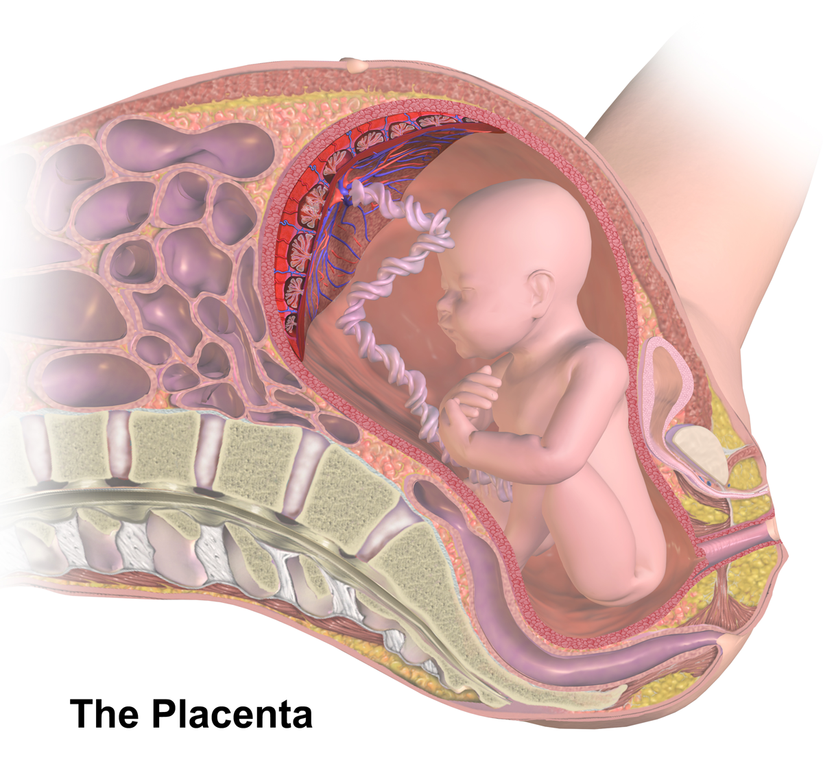
The placenta is a temporary embryonic and later fetal organ (anatomy), organ that begins embryonic development, developing from the blastocyst shortly after implantation (embryology), implantation. It plays critical roles in facilitating nutrient, gas and waste exchange between the physically separate maternal and fetal circulations, and is an important Endocrine system, endocrine organ, producing hormones that regulate both Maternal physiological changes in pregnancy, maternal and fetal physiology during pregnancy. The placenta connects to the fetus via the umbilical cord, and on the opposite aspect to the maternal uterus in a species-dependent manner. In humans, a thin layer of maternal decidual (Endometrium, endometrial) tissue comes away with the placenta when it is expelled from the uterus following birth (sometimes incorrectly referred to as the 'maternal part' of the placenta). Placentas are a defining characteristic of placental mammals, but are also found in marsupials an ... [...More Info...] [...Related Items...] OR: [Wikipedia] [Google] [Baidu] |


.jpg)


.png)

