|
Ingestive Behaviors
Ingestive behaviors encompass all eating and drinking behaviors. These actions are influenced by physiological regulatory mechanisms; these mechanisms exist to control and establish homeostasis within the human body. Disruptions in these ingestive regulatory mechanisms can result in eating disorders such as obesity, anorexia, and bulimia. Research has confirmed that physiological mechanisms play an important role in homeostasis; however, human food intake must also be evaluated within the context of non-physiological determinants present in human life. Within laboratory environments, hunger and satiety are factors that can be controlled and tested. Outside of experiments though, social constraints may influence the size and number of daily meals. Initiating ingestion Body weight regulation requires a balance between food intake and energy expenditure. Two mechanisms are required to maintain a relatively constant body weight: one must increase motivation to eat if long-term reservo ... [...More Info...] [...Related Items...] OR: [Wikipedia] [Google] [Baidu] |
Homeostasis
In biology, homeostasis (British English, British also homoeostasis; ) is the state of steady internal physics, physical and chemistry, chemical conditions maintained by organism, living systems. This is the condition of optimal functioning for the organism and includes many variables, such as body temperature and fluid balance, being kept within certain pre-set limits (homeostatic range). Other variables include the pH of extracellular fluid, the concentrations of sodium, potassium, and calcium ions, as well as the blood sugar level, and these need to be regulated despite changes in the environment, diet, or level of activity. Each of these variables is controlled by one or more regulators or homeostatic mechanisms, which together maintain life. Homeostasis is brought about by a natural resistance to change when already in optimal conditions, and equilibrium is maintained by many regulatory mechanisms; it is thought to be the central motivation for all organic action. All home ... [...More Info...] [...Related Items...] OR: [Wikipedia] [Google] [Baidu] |
Adipose Tissue
Adipose tissue (also known as body fat or simply fat) is a loose connective tissue composed mostly of adipocytes. It also contains the stromal vascular fraction (SVF) of cells including preadipocytes, fibroblasts, Blood vessel, vascular endothelial cells and a variety of White blood cell, immune cells such as adipose tissue macrophages. Its main role is to store energy in the form of lipids, although it also cushions and Thermal insulation, insulates the body. Previously treated as being hormonally inert, in recent years adipose tissue has been recognized as a major endocrine organ, as it produces hormones such as leptin, estrogen, resistin, and cytokines (especially TNF-alpha, TNFα). In obesity, adipose tissue is implicated in the chronic release of pro-inflammatory markers known as adipokines, which are responsible for the development of metabolic syndromea constellation of diseases including type 2 diabetes, cardiovascular disease and atherosclerosis. Adipose tissue is d ... [...More Info...] [...Related Items...] OR: [Wikipedia] [Google] [Baidu] |
Melanin Concentrating Hormone
Melanin-concentrating hormone (MCH), also known as pro-melanin stimulating hormone (PMCH), is a cyclic 19-amino acid orexigenic hypothalamic peptide originally isolated from the pituitary gland of teleost fish, where it controls skin pigmentation. In mammals it is involved in the regulation of feeding behavior, mood, sleep-wake cycle and energy balance. Structure MCH is a cyclic 19-amino acid neuropeptide, as it is a polypeptide chain that is able to act as a neurotransmitter. MCH neurons are mainly concentrated in the lateral hypothalamic area, zona incerta, and the incerto-hypothalamic area, but they are also located, in much smaller amounts, in the paramedian pontine reticular formation (PPRF), medial preoptic area, laterodorsal tegmental nucleus, and the olfactory tubercle. MCH is activated by binding to two G-coupled protein receptors ( GCPRs), MCHR1 and MCHR2. MCHR2 has only been identified in certain species such as humans, dogs, ferrets, and rhesus monkeys, while ... [...More Info...] [...Related Items...] OR: [Wikipedia] [Google] [Baidu] |
Orexin
Orexin (), also known as hypocretin, is a neuropeptide that regulates arousal, wakefulness, and appetite. It exists in the forms of orexin-A and orexin-B. The most common form of narcolepsy, type 1, in which the individual experiences brief losses of muscle tone ("drop attacks" or cataplexy), is caused by a lack of orexin in the brain due to destruction of the cells that produce it.Stanford Center for NarcolepsFAQ(retrieved 27-Mar-2012) There are 50,000–80,000 orexin-producing neurons in the human brain, located predominantly in the perifornical area and lateral hypothalamus. They project widely throughout the central nervous system, regulating wakefulness, feeding, and other behaviours. There are two types of orexin peptide and two types of orexin receptor. Orexin was discovered in 1998 almost simultaneously by two independent groups of researchers working on the rat brain. One group named it orexin, from ''orexis,'' meaning "appetite" in Greek; the other group named it hy ... [...More Info...] [...Related Items...] OR: [Wikipedia] [Google] [Baidu] |
Lateral Hypothalamus
The lateral hypothalamus (LH), also called the lateral hypothalamic area (LHA), contains the primary orexinergic nucleus within the hypothalamus that widely projects throughout the nervous system; this system of neurons mediates an array of cognitive and physical processes, such as promoting feeding behavior and arousal, reducing pain perception, and regulating body temperature, digestive functions, and blood pressure, among many others. Clinically significant disorders that involve dysfunctions of the orexinergic projection system include narcolepsy, motility disorders or functional gastrointestinal disorders involving visceral hypersensitivity (e.g., irritable bowel syndrome), and eating disorders. The neurotransmitter glutamate and the endocannabinoids (e.g., anandamide) and the orexin neuropeptides orexin-A and orexin-B are the primary signaling neurochemicals in orexin neurons; pathway-specific neurochemicals include GABA, melanin-concentrating hormone, nociceptin, gluc ... [...More Info...] [...Related Items...] OR: [Wikipedia] [Google] [Baidu] |
Forebrain
In the anatomy of the brain of vertebrates, the forebrain or prosencephalon is the rostral (forward-most) portion of the brain. The forebrain controls body temperature, reproductive functions, eating, sleeping, and the display of emotions. Vesicles of the forebrain (prosencephalon), the midbrain (mesencephalon), and hindbrain (rhombencephalon) are the three primary brain vesicles during the early development of the nervous system. At the five-vesicle stage, the forebrain separates into the diencephalon (thalamus, hypothalamus, subthalamus, and epithalamus) and the telencephalon which develops into the cerebrum. The cerebrum consists of the cerebral cortex, underlying white matter, and the basal ganglia. In humans, by 5 weeks in utero it is visible as a single portion toward the front of the fetus. At 8 weeks in utero, the forebrain splits into the left and right cerebral hemispheres. When the embryonic forebrain fails to divide the brain into two lobes, it results i ... [...More Info...] [...Related Items...] OR: [Wikipedia] [Google] [Baidu] |
Nucleus Of The Solitary Tract
The solitary nucleus (SN) (nucleus of the solitary tract, nucleus solitarius, or nucleus tractus solitarii) is a series of neurons whose cell bodies form a roughly vertical column of grey matter in the medulla oblongata of the brainstem. Their axons form the bulk of the enclosed solitary tract. The solitary nucleus can be divided into different parts including dorsomedial, dorsolateral, and ventrolateral subnuclei. The solitary nucleus receives general visceral and special visceral inputs from the facial nerve (CN VII), glossopharyngeal nerve (CN IX) and vagus nerve (CN X); it receives and relays stimuli related to taste and visceral sensation. It sends outputs to various parts of the brain, such as the hypothalamus, thalamus, and reticular formation, forming circuits that contribute to autonomic regulation. Cells along the length of the SN are arranged roughly in accordance with function; for instance, cells involved in taste are located in the rostral part, while those rece ... [...More Info...] [...Related Items...] OR: [Wikipedia] [Google] [Baidu] |
Area Postrema
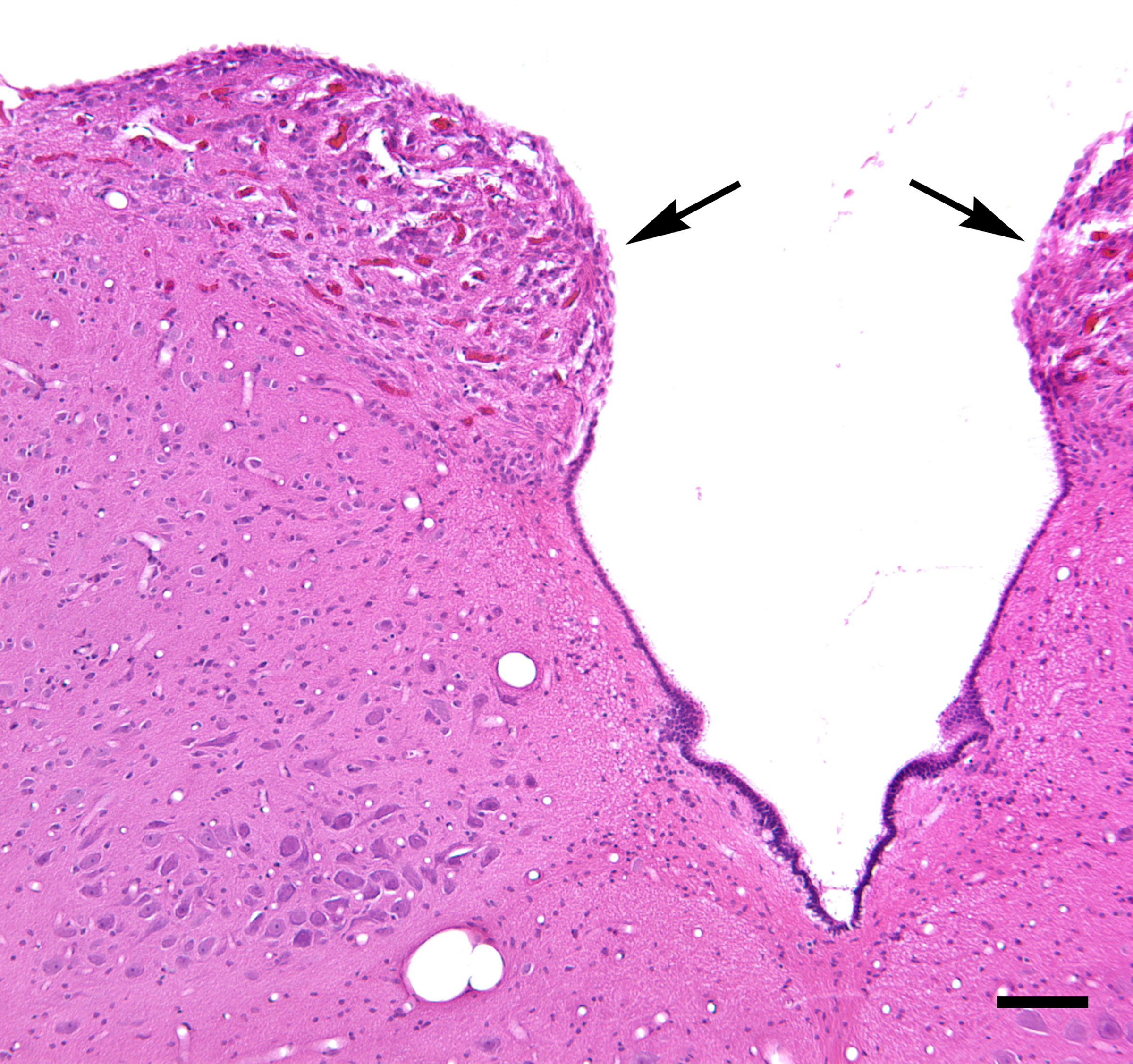
The area postrema, a paired structure in the medulla oblongata of the brainstem, is a circumventricular organ having permeable capillaries and sensory neurons that enable its role to detect circulating chemical messengers in the blood and transduce them into neural signals and networks. Its position adjacent to the bilateral nuclei of the solitary tract and role as a sensory transducer allow it to integrate blood-to-brain autonomic functions. Such roles of the area postrema include its detection of circulating hormones involved in vomiting, thirst, hunger, and blood pressure control. Structure The area postrema is a paired protuberance found at the inferoposterior limit of the fourth ventricle. Specialized ependymal cells are found within the area postrema. These cells differ slightly from the majority of ependymal cells (ependymocytes), forming a unicellular epithelial lining of the ventricles and central canal. The area postrema is separated from the vagal trigone by ... [...More Info...] [...Related Items...] OR: [Wikipedia] [Google] [Baidu] |
Brain Stem
The brainstem (or brain stem) is the posterior stalk-like part of the brain that connects the cerebrum with the spinal cord. In the human brain the brainstem is composed of the midbrain, the pons, and the medulla oblongata. The midbrain is continuous with the thalamus of the diencephalon through the tentorial notch, and sometimes the diencephalon is included in the brainstem. The brainstem is very small, making up around only 2.6 percent of the brain's total weight. It has the critical roles of regulating heart and respiratory function, helping to control heart rate and breathing rate. It also provides the main motor and sensory nerve supply to the face and neck via the cranial nerves. Ten pairs of cranial nerves come from the brainstem. Other roles include the regulation of the central nervous system and the body's sleep cycle. It is also of prime importance in the conveyance of motor and sensory pathways from the rest of the brain to the body, and from the body back ... [...More Info...] [...Related Items...] OR: [Wikipedia] [Google] [Baidu] |
Leptin
Leptin (from Ancient Greek, Greek λεπτός ''leptos'', "thin" or "light" or "small"), also known as obese protein, is a protein hormone predominantly made by adipocytes (cells of adipose tissue). Its primary role is likely to regulate long-term Energy homeostasis, energy balance. As one of the major signals of energy status, leptin levels influence appetite, Hunger (physiology), satiety, and motivated behaviors oriented toward the maintenance of energy reserves (e.g., feeding, foraging behaviors). The amount of circulating leptin correlates with the amount of energy reserves, mainly Triglyceride, triglycerides stored in adipose tissue. High leptin levels are interpreted by the brain that energy reserves are high, whereas low leptin levels indicate that energy reserves are low, in the process adapting the organism to Starvation response, starvation through a variety of metabolic, endocrine, neurobiochemical, and behavioral changes. Leptin is coded for by the ''LEP'' gene ... [...More Info...] [...Related Items...] OR: [Wikipedia] [Google] [Baidu] |
Cholecystokinin
Cholecystokinin (CCK or CCK-PZ; from Greek ''chole'', "bile"; ''cysto'', "sac"; ''kinin'', "move"; hence, ''move the bile-sac (gallbladder)'') is a peptide hormone of the gastrointestinal system responsible for stimulating the digestion of fat and protein. Cholecystokinin, formerly called pancreozymin, is synthesized and secreted by enteroendocrine cells in the duodenum, the first segment of the small intestine. Its presence causes the release of pancreatic juice from the pancreas and bile from the gallbladder. History Evidence that the small intestine controls the release of bile was uncovered as early as 1856, when French physiologist Claude Bernard showed that when dilute acetic acid was applied to the orifice of the bile duct, the duct released bile into the duodenum. In 1903, the French physiologist showed that this reflex was not mediated by the nervous system. In 1904, the French physiologist Charles Fleig showed that the discharge of bile was mediated by a substance t ... [...More Info...] [...Related Items...] OR: [Wikipedia] [Google] [Baidu] |


