|
Inflammatory Myofibroblastic Tumor
Inflammatory myofibroblastic tumor (IMT) is a rare neoplasm of the mesodermal cells that form the connective tissues which support virtually all of the organs and tissues of the body. IMT was formerly termed inflammatory pseudotumor. Currently, however, inflammatory pseudotumor designates a large and heterogeneous group of soft tissue tumors that includes inflammatory myofibroblastic tumor, plasma cell granuloma, xanthomatous pseudotumor, solitary mast cell granuloma, inflammatory fibrosarcoma, pseudosarcomatous myofibroblastic proliferation, myofibroblastoma, inflammatory myofibrohistiocytic proliferation, and other tumors that develop from connective tissue cells. Inflammatory pseudotumour is a generic term applied to various neoplastic and non-neoplastic tissue lesions which share a common microscopic appearance consisting of spindle cells and a prominent presence of the white blood cells that populate chronic or, less commonly, acute inflamed tissues. Inflammatory myofibrobla ... [...More Info...] [...Related Items...] OR: [Wikipedia] [Google] [Baidu] |
Micrograph
A micrograph is an image, captured photographically or digitally, taken through a microscope or similar device to show a magnify, magnified image of an object. This is opposed to a macrograph or photomacrograph, an image which is also taken on a microscope but is only slightly magnified, usually less than 10 times. Micrography is the practice or art of using microscopes to make photographs. A photographic micrograph is a photomicrograph, and one taken with an electron microscope is an electron micrograph. A micrograph contains extensive details of microstructure. A wealth of information can be obtained from a simple micrograph like behavior of the material under different conditions, the phases found in the system, failure analysis, grain size estimation, elemental analysis and so on. Micrographs are widely used in all fields of microscopy. Types Photomicrograph A light micrograph or photomicrograph is a micrograph prepared using an optical microscope, a process referred to ... [...More Info...] [...Related Items...] OR: [Wikipedia] [Google] [Baidu] |
Epithelioid Cells
Epithelioid cells (also called epithelioid histiocytes) are derivatives of activated macrophages resembling epithelial cells. Structure and function Structurally, epithelioid cells (when examined by light microscopy after stained with hematoxylin and eosin), are elongated, with finely granular, pale eosinophilic (pink) cytoplasm, and central, ovoid nuclei (oval or elongate), which are less dense than that of a lymphocyte. They have indistinct shape and often appear to merge into one another, forming aggregates known as giant cells. When examined by transmission electron microscopy in epithelioid cells in the field of Golgi lamellar complex are taped not only zonated, but also sleek vesicles with dense center, and also great many (more than 100) large granulas with diameters up to 340 nm and with finegranular matrix more light than in macrophage granulas, sometimes with perigranular halo. “The most prominent feature of these cells is the enormous Golgi area; up to 6 indi ... [...More Info...] [...Related Items...] OR: [Wikipedia] [Google] [Baidu] |
Surveillance, Epidemiology, And End Results
The Surveillance, Epidemiology, and End Results (SEER) program of the National Cancer Institute (NCI) is a source of epidemiologic information on the incidence and survival rates of cancer in the United States. The Program SEER collects and publishes cancer incidence and survival data from population-based cancer registries covering approximately 45.9% of the population of the United States. SEER coverage includes 39.6% of Whites, 43.5% of African Americans, 64.9% of Hispanics, 59.3% of American Indians and Alaska Natives, 68.2% of Asians, and 69.9% of Hawaiian/ Pacific Islanders. The SEER Program registries routinely collect data on patient demographics, primary tumor site, tumor morphology and stage at diagnosis, first course of treatment, and follow-up for vital status. The SEER Program is the only comprehensive source of population-based information in the United States that includes stage of cancer at the time of diagnosis and patient survival data. History SEER began col ... [...More Info...] [...Related Items...] OR: [Wikipedia] [Google] [Baidu] |
Spinal Cord
The spinal cord is a long, thin, tubular structure made up of nervous tissue that extends from the medulla oblongata in the lower brainstem to the lumbar region of the vertebral column (backbone) of vertebrate animals. The center of the spinal cord is hollow and contains a structure called the central canal, which contains cerebrospinal fluid. The spinal cord is also covered by meninges and enclosed by the neural arches. Together, the brain and spinal cord make up the central nervous system. In humans, the spinal cord is a continuation of the brainstem and anatomically begins at the occipital bone, passing out of the foramen magnum and then enters the spinal canal at the beginning of the cervical vertebrae. The spinal cord extends down to between the first and second lumbar vertebrae, where it tapers to become the cauda equina. The enclosing bony vertebral column protects the relatively shorter spinal cord. It is around long in adult men and around long in adult women. The diam ... [...More Info...] [...Related Items...] OR: [Wikipedia] [Google] [Baidu] |
Meninges
In anatomy, the meninges (; meninx ; ) are the three membranes that envelop the brain and spinal cord. In mammals, the meninges are the dura mater, the arachnoid mater, and the pia mater. Cerebrospinal fluid is located in the subarachnoid space between the arachnoid mater and the pia mater. The primary function of the meninges is to protect the central nervous system. Structure Dura mater The dura mater (), is a thick, durable membrane, closest to the Human skull, skull and vertebrae. The dura mater, the outermost part, is a loosely arranged, fibroelastic layer of cells, characterized by multiple interdigitating cell processes, no extracellular collagen, and significant extracellular spaces. The middle region is a mostly fibrous portion. It consists of two layers: the endosteal layer, which lies closest to the skull, and the inner meningeal layer, which lies closer to the brain. It contains larger blood vessels that split into the capillaries in the pia mater. It is composed ... [...More Info...] [...Related Items...] OR: [Wikipedia] [Google] [Baidu] |
Central Nervous System
The central nervous system (CNS) is the part of the nervous system consisting primarily of the brain, spinal cord and retina. The CNS is so named because the brain integrates the received information and coordinates and influences the activity of all parts of the bodies of bilateria, bilaterally symmetric and triploblastic animals—that is, all multicellular animals except sponges and Coelenterata, diploblasts. It is a structure composed of nervous tissue positioned along the Anatomical_terms_of_location#Rostral,_cranial,_and_caudal, rostral (nose end) to caudal (tail end) axis of the body and may have an enlarged section at the rostral end which is a brain. Only arthropods, cephalopods and vertebrates have a true brain, though precursor structures exist in onychophorans, gastropods and lancelets. The rest of this article exclusively discusses the vertebrate central nervous system, which is radically distinct from all other animals. Overview In vertebrates, the brain and spinal ... [...More Info...] [...Related Items...] OR: [Wikipedia] [Google] [Baidu] |
Peripheral Nervous System
The peripheral nervous system (PNS) is one of two components that make up the nervous system of Bilateria, bilateral animals, with the other part being the central nervous system (CNS). The PNS consists of nerves and ganglia, which lie outside the brain and the spinal cord. The main function of the PNS is to connect the CNS to the Limb (anatomy), limbs and Organ (anatomy), organs, essentially serving as a relay between the brain and spinal cord and the rest of the body. Unlike the CNS, the PNS is not protected by the vertebral column and skull, or by the blood–brain barrier, which leaves it exposed to toxins. The peripheral nervous system can be divided into a somatic nervous system, somatic division and an autonomic nervous system, autonomic division. Each of these can further be differentiated into a sensory and a motor sector. In the somatic nervous system, the cranial nerves are part of the PNS with the exceptions of the olfactory nerve and epithelia and the optic nerve (c ... [...More Info...] [...Related Items...] OR: [Wikipedia] [Google] [Baidu] |
Orbit (anatomy)
In anatomy Anatomy () is the branch of morphology concerned with the study of the internal structure of organisms and their parts. Anatomy is a branch of natural science that deals with the structural organization of living things. It is an old scien ..., the orbit is the Body cavity, cavity or socket/hole of the skull in which the eye and Accessory visual structures, its appendages are situated. "Orbit" can refer to the bony socket, or it can also be used to imply the contents. In the adult human, the volume of the orbit is about , of which the eye occupies . The orbital contents comprise the eye, the Orbital fascia, orbital and retrobulbar fascia, extraocular muscles, cranial nerves optic nerve, II, oculomotor nerve, III, trochlear nerve, IV, trigeminal nerve, V, and abducens nerve, VI, blood vessels, fat, the lacrimal gland with its Lacrimal sac, sac and nasolacrimal duct, duct, the eyelids, Medial palpebral ligament, medial and Lateral palpebral raphe, lateral palpebr ... [...More Info...] [...Related Items...] OR: [Wikipedia] [Google] [Baidu] |
Spermatic Cord
The spermatic cord is the cord-like structure in males formed by the vas deferens (''ductus deferens'') and surrounding tissue that runs from the deep inguinal ring down to each testicle. Its serosal covering, the tunica vaginalis, is an extension of the peritoneum that passes through the transversalis fascia. Each testicle develops in the lower thoracic and upper lumbar region and migrates into the scrotum. During its descent it carries along with it the vas deferens, its vessels, nerves etc. There is one on each side. Structure The spermatic cord is ensheathed in three layers of tissue: * '' external spermatic fascia'', an extension of the innominate fascia that overlies the aponeurosis of the external oblique muscle. * '' cremasteric muscle and fascia'', formed from a continuation of the internal oblique muscle and its fascia. * '' internal spermatic fascia'', continuous with the transversalis fascia. The normal diameter of the spermatic cord is about 16 mm (range 11 ... [...More Info...] [...Related Items...] OR: [Wikipedia] [Google] [Baidu] |
Greater Omentum
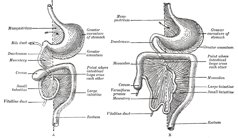
Greater may refer to: *Greatness, the state of being great *Greater than, in inequality * ''Greater'' (film), a 2016 American film *Greater (flamingo), the oldest flamingo on record * "Greater" (song), by MercyMe, 2014 * Greater Bank, an Australian bank * Greater Media, an American media company See also * Irredentism usually named as Greater ''Nation A nation is a type of social organization where a collective Identity (social science), identity, a national identity, has emerged from a combination of shared features across a given population, such as language, history, ethnicity, culture, t ...''. Examples include Greater Hungary, Greater Romania * * {{Disambiguation ... [...More Info...] [...Related Items...] OR: [Wikipedia] [Google] [Baidu] |
Mesentery
In human anatomy, the mesentery is an Organ (anatomy), organ that attaches the intestines to the posterior abdominal wall, consisting of a double fold of the peritoneum. It helps (among other functions) in storing Adipose tissue, fat and allowing blood vessels, lymphatics, and nerves to supply the intestines. The (the part of the mesentery that attaches the colon to the abdominal wall) was formerly thought to be a fragmented structure, with all named parts—the ascending, transverse, descending, and sigmoid mesocolons, the mesoappendix, and the mesorectum—separately terminating their insertion into the posterior abdominal wall. However, in 2012, new microscopy, microscopic and electron microscope, electron microscopic histology, examinations showed the mesocolon to be a single structure derived from the duodenojejunal flexure and extending to the distal mesorectal layer. Thus the mesentery is an internal organ. Structure The mesentery of the small intestine arises from th ... [...More Info...] [...Related Items...] OR: [Wikipedia] [Google] [Baidu] |
Anaplastic Lymphoma Kinase
Anaplastic lymphoma kinase (ALK) also known as ALK tyrosine kinase receptor or CD246 (cluster of differentiation 246) is an enzyme that in humans is encoded by the ''ALK'' gene. Identification Anaplastic lymphoma kinase (ALK) was originally discovered in 1994 in anaplastic large-cell lymphoma (ALCL) cells. ALCL is caused by a (2;5)(p23:q35) chromosomal translocation that generates the fusion protein NPM-ALK, in which the kinase domain of ALK is fused to the amino-terminal part of the nucleophosmin (NPM) protein. Dimerization of NPM constitutively activates the ALK kinase domain. The full-length protein ALK was identified in 1997 by two groups. The deduced amino acid sequences revealed that ALK was a novel receptor tyrosine kinase (RTK), having an extracellular ligand-binding domain, a transmembrane domain, and an intracellular tyrosine kinase domain. While the tyrosine kinase domain of human ALK shares a high degree of similarity with that of the insulin receptor, its ... [...More Info...] [...Related Items...] OR: [Wikipedia] [Google] [Baidu] |


