|
Iliohypogastric Nerve
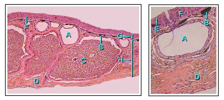
The iliohypogastric nerve is a nerve that originates from the lumbar plexus that supplies sensation to skin over the lateral gluteal and hypogastric regions and motor to the internal oblique muscles and transverse abdominal muscles. Structure Origin The iliohypogastric nerve originates from the superior branch of the anterior ramus of spinal nerve L1. It also receives fibers from T12 via the subcostal nerve. The branch below it is the ilioinguinal nerve. Course It emerges from the upper lateral border of the psoas major. It then crosses in front of the quadratus lumborum muscle to an area superior to the iliac crest. It runs behind the kidneys. Just superior to the iliac crest, it pierces the posterior part of the transversus abdominis muscle and continues anteriorly in the abdominal wall between the transversus abdominis and internal oblique muscles. It divides into a lateral cutaneous branch and an anterior cutaneous branch between the transversus abdominis muscle a ... [...More Info...] [...Related Items...] OR: [Wikipedia] [Google] [Baidu] |
Lumbar Plexus
The lumbar plexus is a web of nerves (a nerve plexus) in the lumbar region of the body which forms part of the larger lumbosacral plexus. It is formed by the divisions of the first four lumbar nerves (L1–L4) and from contributions of the subcostal nerve (T12), which is the last thoracic nerve. Additionally, the ventral rami of the fourth lumbar nerve pass communicating branches, the lumbosacral trunk, to the sacral plexus. The nerves of the lumbar plexus pass in front of the hip joint and mainly support the anterior part of the thigh.''Thieme Atlas of anatomy'' (2006), pp 470-471 The plexus is formed lateral to the intervertebral foramina and passes through psoas major. Its smaller motor branches are distributed directly to psoas major, while the larger branches leave the muscle at various sites to run obliquely down through the pelvis to leave under the inguinal ligament with the exception of the obturator nerve which exits the pelvis through the obturator foramen. ... [...More Info...] [...Related Items...] OR: [Wikipedia] [Google] [Baidu] |
Ilioinguinal Nerve
The ilioinguinal nerve is a branch of the first lumbar nerve (L1). It separates from the first lumbar nerve along with the larger iliohypogastric nerve. It emerges from the lateral border of the psoas major just inferior to the iliohypogastric, and passes obliquely across the quadratus lumborum and iliacus. The ilioinguinal nerve then perforates the transversus abdominis near the anterior part of the iliac crest, and communicates with the iliohypogastric nerve between the transversus and the internal oblique muscle. It then pierces the internal oblique muscle, distributing filaments to it, and then accompanies the spermatic cord (in males) or the round ligament of uterus (in females) through the superficial inguinal ring. Its fibres are then distributed to the skin of the upper and medial part of the thigh, and to the following locations in the male and female: * In the male (" anterior scrotal nerve"): to the skin over the root of the penis and upper part of the scrotum. ... [...More Info...] [...Related Items...] OR: [Wikipedia] [Google] [Baidu] |
Medical Error
A medical error is a preventable adverse effect of care (" iatrogenesis"), whether or not it is evident or harmful to the patient. This might include an inaccurate or incomplete diagnosis or treatment of a disease, injury, syndrome, behavior, infection, or other ailments. The incidence of medical errors varies depending on the setting. The World Health Organization has named adverse outcomes due to patient care that is unsafe as the 14th causes of disability and death in the world, with an estimated 1/300 people may be harmed by healthcare practices around the world. Definitions A medical error occurs when a health-care provider chooses an inappropriate method of care or improperly executes an appropriate method of care. Medical errors are often described as human errors in healthcare. There are many types of medical error, from minor to major, and causality understanding and assessing if the likelihood that the specific event or factor was responsible for the negative outc ... [...More Info...] [...Related Items...] OR: [Wikipedia] [Google] [Baidu] |
Hypogastrium
The hypogastrium (also called the hypogastric region or suprapubic region) is a region of the abdomen located below the umbilical region. Etymology The roots of the word ''hypogastrium'' mean "below the stomach"; the roots of ''suprapubic'' mean "above the pubic bone". Boundaries The upper limit is the umbilicus while the pubis bone constitutes its lower limit. The lateral boundaries are formed are drawing straight lines through the midway between the anterior superior iliac spine The anterior superior iliac spine (ASIS) is a bony projection of the iliac bone, and an important landmark of surface anatomy. It refers to the anterior extremity of the iliac crest of the pelvis. It provides attachment for the inguinal ligament, ... and symphisis pubis. References External links * Abdomen {{Anatomy-stub ... [...More Info...] [...Related Items...] OR: [Wikipedia] [Google] [Baidu] |
Subcutaneous Inguinal Ring
The inguinal canal is a passage in the anterior abdominal wall on each side of the body (one on each side of the midline), which in males, convey the spermatic cords and in females, the round ligament of the uterus. The inguinal canals are larger and more prominent in males. Structure The inguinal canals are situated just above the medial half of the inguinal ligament. The canals are approximately 4 to 6 cm long, angled anteroinferiorly and medially. In males, its diameter is normally 2 cm (±1 cm in standard deviation) at the deep inguinal ring.The diameter has been estimated to be ±2.2cm ±1.08cm in Africans, and 2.1 cm ±0.41cm in Europeans. A first-order approximation is to visualize each canal as a cylinder. Walls To help define the boundaries, these canals are often further approximated as boxes with six sides. Not including the two rings, the remaining four sides are usually called the "anterior wall", "inferior wall ("floor")", "superior wall ("roof")", and "pos ... [...More Info...] [...Related Items...] OR: [Wikipedia] [Google] [Baidu] |
Aponeurosis
An aponeurosis (; : aponeuroses) is a flattened tendon by which muscle attaches to bone or fascia. Aponeuroses exhibit an ordered arrangement of collagen fibres, thus attaining high tensile strength in a particular direction while being vulnerable to tensional or shear forces in other directions. They have a shiny, whitish-silvery color, are histologically similar to tendons, and are very sparingly supplied with blood vessels and nerves. When dissected, aponeuroses are papery and peel off by sections. The primary regions with thick aponeuroses are in the ventral abdominal region, the dorsal lumbar region, the ventriculus in birds, and the palmar (palms) and plantar (soles) regions. Anatomy Anterior abdominal aponeuroses The anterior abdominal aponeuroses are located just superficial to the rectus abdominis muscle. It has for its borders the external oblique, pectoralis muscles, and the latissimus dorsi. Posterior lumbar aponeuroses The posterior lumbar aponeuroses are sit ... [...More Info...] [...Related Items...] OR: [Wikipedia] [Google] [Baidu] |
Cutaneous
Skin is the layer of usually soft, flexible outer tissue covering the body of a vertebrate animal, with three main functions: protection, regulation, and sensation. Other animal coverings, such as the arthropod exoskeleton, have different developmental origin, structure and chemical composition. The adjective cutaneous means "of the skin" (from Latin ''cutis'' 'skin'). In mammals, the skin is an organ of the integumentary system made up of multiple layers of ectodermal tissue and guards the underlying muscles, bones, ligaments, and internal organs. Skin of a different nature exists in amphibians, reptiles, and birds. Skin (including cutaneous and subcutaneous tissues) plays crucial roles in formation, structure, and function of extraskeletal apparatus such as horns of bovids (e.g., cattle) and rhinos, cervids' antlers, giraffids' ossicones, armadillos' osteoderm, and os penis/os clitoris. All mammals have some hair on their skin, even marine mammals like whales, dolphins ... [...More Info...] [...Related Items...] OR: [Wikipedia] [Google] [Baidu] |
Abdominal External Oblique Muscle
The abdominal external oblique muscle (also external oblique muscle or exterior oblique) is the largest and outermost of the three flat Abdomen#Muscles, abdominal muscles of the lateral anterior abdomen. Structure The external oblique is situated on the lateral and anterior parts of the abdomen. It is broad, thin, and irregularly quadrilateral, its muscular portion occupying the side, its aponeurosis the anterior wall of the abdomen. In most humans, the oblique is not visible, due to subcutaneous adipose, fat deposits and the small size of the muscle. It arises from eight fleshy digitations, each from the external surfaces and inferior borders of the fifth to twelfth ribs (lower eight ribs). These digitations are arranged in an oblique line which runs inferiorly and anteriorly, with the upper digitations being attached close to the cartilages of the corresponding ribs, the lowest to the apex of the cartilage of the last rib, the intermediate ones to the ribs at some distance fr ... [...More Info...] [...Related Items...] OR: [Wikipedia] [Google] [Baidu] |
Transverse Abdominal Muscle
The transverse abdominal muscle (TVA), also known as the transverse abdominis, transversalis muscle and transversus abdominis muscle, is a muscle layer of the anterior and lateral (front and side) abdominal wall, deep to (layered below) the internal oblique muscle. It serves to compress and retain the contents of the abdomen as well as assist in exhalation. Structure The transverse abdominal, so called for the direction of its fibers, is the innermost of the flat muscles of the abdomen. It is positioned immediately deep to the internal oblique muscle. The transverse abdominal arises as fleshy fibers, from the lateral third of the inguinal ligament, from the anterior three-fourths of the inner lip of the iliac crest, from the inner surfaces of the cartilages of the lower six ribs, interdigitating with the diaphragm, and from the thoracolumbar fascia. It ends anteriorly in a broad aponeurosis (the Spigelian fascia), the lower fibers of which curve inferomedially (medially and do ... [...More Info...] [...Related Items...] OR: [Wikipedia] [Google] [Baidu] |
Kidney
In humans, the kidneys are two reddish-brown bean-shaped blood-filtering organ (anatomy), organs that are a multilobar, multipapillary form of mammalian kidneys, usually without signs of external lobulation. They are located on the left and right in the retroperitoneal space, and in adult humans are about in length. They receive blood from the paired renal artery, renal arteries; blood exits into the paired renal veins. Each kidney is attached to a ureter, a tube that carries excreted urine to the urinary bladder, bladder. The kidney participates in the control of the volume of various body fluids, fluid osmolality, Acid-base homeostasis, acid-base balance, various electrolyte concentrations, and removal of toxins. Filtration occurs in the glomerulus (kidney), glomerulus: one-fifth of the blood volume that enters the kidneys is filtered. Examples of substances reabsorbed are solute-free water, sodium, bicarbonate, glucose, and amino acids. Examples of substances secreted are hy ... [...More Info...] [...Related Items...] OR: [Wikipedia] [Google] [Baidu] |
Iliac Crest
The crest of the ilium (or iliac crest) is the superior border of the wing of ilium and the superolateral margin of the greater pelvis. Structure The iliac crest stretches posteriorly from the anterior superior iliac spine (ASIS) to the posterior superior iliac spine (PSIS). Behind the ASIS, it divides into an outer and inner lip separated by the intermediate zone. The outer lip bulges laterally into the iliac tubercle.Platzer (2004), p 186 Palpation, Palpable in its entire length, the crest is convex superiorly but is sinuously curved, being concave inward in front, concave outward behind.Palastanga (2006), p 243 It is thinner at the center than at the extremities. Development The iliac crest is derived from endochondral bone. Function To the external lip are attached the ''Tensor fasciae latae'', ''abdominal external oblique muscle, Obliquus externus abdominis'', and ''Latissimus dorsi muscle, Latissimus dorsi'', and along its whole length the ''fascia lata''; to the int ... [...More Info...] [...Related Items...] OR: [Wikipedia] [Google] [Baidu] |
Quadratus Lumborum Muscle
The quadratus lumborum muscle, informally called the ''QL'', is a paired muscle of the left and right posterior abdominal wall. It is the deepest abdominal muscle, and commonly referred to as a back muscle. Each muscle of the pair is an irregular quadrilateral in shape, hence the name. The quadratus lumborum muscles originate from the wings of the ilium; their insertions are on the transverse processes of the upper four lumbar vertebrae Each vertebra (: vertebrae) is an irregular bone with a complex structure composed of bone and some hyaline cartilage, that make up the vertebral column or spine, of vertebrates. The proportions of the vertebrae differ according to their spinal ... plus the lower posterior border of the twelfth rib. Contraction of one of the pair of muscles causes lateral flexion of the Lumbar vertebrae, lumbar spine, ''elevation'' of the pelvis, or both. Contraction of both causes ''extension'' of the lumbar spine. A disorder of the quadratus lumborum mu ... [...More Info...] [...Related Items...] OR: [Wikipedia] [Google] [Baidu] |


