|
Horseshoe Kidney
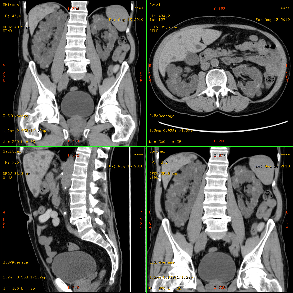
Horseshoe kidney, also known as ''ren arcuatus'' (in Latin), renal fusion or super kidney, is a congenital disorder affecting about 1 in 500 people that is more common in men, often asymptomatic, and usually diagnosed incidentally. In this disorder, the patient's kidneys fuse to form a horseshoe-shape during development in the womb. The fused part is the isthmus of the horseshoe kidney. The abnormal anatomy can affect kidney drainage resulting in increased frequency of kidney stones and urinary tract infections as well as increase risk of certain renal cancers. Fusion abnormalities of the kidney can be categorized into two groups: horseshoe kidney and Crossed renal ectopia, crossed fused ectopia. The 'horseshoe kidney' is the most common renal fusion anomaly. Signs and symptoms Although often asymptomatic, the most common presenting symptom of patients with a horseshoe kidney is abdominal or flank pain. However, presentation is often non-specific. Approximately a third of patient ... [...More Info...] [...Related Items...] OR: [Wikipedia] [Google] [Baidu] |
Congenital Disorder
A birth defect is an abnormal condition that is present at childbirth, birth, regardless of its cause. Birth defects may result in disability, disabilities that may be physical disability, physical, intellectual disability, intellectual, or developmental disability, developmental. The disabilities can range from mild to severe. Birth defects are divided into two main types: structural disorders in which problems are seen with the shape of a body part and functional disorders in which problems exist with how a body part works. Functional disorders include metabolic disorder, metabolic and degenerative disease, degenerative disorders. Some birth defects include both structural and functional disorders. Birth defects may result from genetic disorder, genetic or chromosome abnormality, chromosomal disorders, exposure to certain medications or chemicals, or certain vertically transmitted infection, infections during pregnancy. Risk factors include folate deficiency, alcohol drink, d ... [...More Info...] [...Related Items...] OR: [Wikipedia] [Google] [Baidu] |
Myelomeningocele
Spina bifida (SB; ; Latin for 'split spine') is a birth defect in which there is incomplete closing of the spine and the membranes around the spinal cord during early development in pregnancy. There are three main types: spina bifida occulta, meningocele and myelomeningocele. Meningocele and myelomeningocele may be grouped as spina bifida cystica. The most common location is the lower back, but in rare cases it may be in the middle back or neck. Occulta has no or only mild signs, which may include a hairy patch, dimple, dark spot or swelling on the back at the site of the gap in the spine. Meningocele typically causes mild problems, with a sac of fluid present at the gap in the spine. Myelomeningocele, also known as open spina bifida, is the most severe form. Problems associated with this form include poor ability to walk, impaired bladder or bowel control, accumulation of fluid in the brain, a tethered spinal cord and latex allergy. Some experts believe such an allergy ca ... [...More Info...] [...Related Items...] OR: [Wikipedia] [Google] [Baidu] |
Edward Syndrome
Trisomy 18, also known as Edwards syndrome, is a genetic disorder caused by the presence of a third copy of all or part of chromosome 18. Many parts of the body are affected. Babies are often born small and have heart defects. Other features include a small head, small jaw, clenched fists with overlapping fingers, and severe intellectual disability. Most cases of trisomy 18 occur due to problems during the formation of the reproductive cells or during early development. The chance of this condition occurring increases with the mother's age. Rarely, cases may be inherited. Occasionally, not all cells have the extra chromosome, known as mosaic trisomy, and symptoms in these cases may be less severe. An ultrasound during pregnancy can increase suspicion for the condition, which can be confirmed by amniocentesis. Treatment is supportive. After having one child with the condition, the risk of having a second is typically around one percent. It is the second-most common conditio ... [...More Info...] [...Related Items...] OR: [Wikipedia] [Google] [Baidu] |
Patau Syndrome
Patau syndrome is a syndrome caused by a chromosomal abnormality, in which some or all of the cells of the body contain extra genetic material from chromosome 13. The extra genetic material disrupts normal development, causing multiple and complex organ defects. This can occur either because each cell contains a full extra copy of chromosome 13 (a disorder known as trisomy 13 or trisomy D or T13), or because each cell contains an extra partial copy of the chromosome, or because there are two different lines of cells—one healthy with the correct number of chromosomes 13 and one that contains an extra copy of the chromosome—mosaic Patau syndrome. Full trisomy 13 is caused by nondisjunction of chromosomes during meiosis; the mosaic form is caused by nondisjunction during mitosis. Like all nondisjunction conditions (such as Down syndrome and Edwards syndrome), the risk of this syndrome in the offspring increases with maternal age at pregnancy, with about 31 years being th ... [...More Info...] [...Related Items...] OR: [Wikipedia] [Google] [Baidu] |
Turner Syndrome
Turner syndrome (TS), commonly known as 45,X, or 45,X0,Also written as 45,XO. is a chromosomal disorder in which cells of females have only one X chromosome instead of two, or are partially missing an X chromosome (sex chromosome monosomy) leading to the complete or partial deletion of the pseudoautosomal regions (PAR1, PAR2) in the affected X chromosome. Typically, people have two sex chromosomes (XX for females or XY for males). The chromosomal abnormality is often present in just some cells, in which case it is known as Turner syndrome with mosaicism. 45,X0 with mosaicism can occur in males or females, but Turner syndrome without mosaicism only occurs in females. Signs and symptoms vary among those affected but often include additional skin folds on the neck, arched palate, low-set ears, low hairline at the nape of the neck, short stature, and lymphedema of the hands and feet. Those affected do not normally develop menstrual periods or mammary glands without hormone trea ... [...More Info...] [...Related Items...] OR: [Wikipedia] [Google] [Baidu] |
Polycystic Kidney Disease
Polycystic kidney disease (PKD or PCKD, also known as polycystic kidney syndrome) is a genetic disorder in which the renal tubules become structurally abnormal, resulting in the development and growth of multiple cysts within the kidney. These cysts may begin to develop in utero, in infancy, in childhood, or in adulthood. Cysts are non-functioning tubules filled with fluid pumped into them, which range in size from microscopic to enormous, crushing adjacent normal tubules and eventually rendering them non-functional as well. PKD is caused by abnormal genes that produce a specific abnormal protein; this protein has an adverse effect on tubule development. PKD is a general term for two types, each having their own pathology and genetic cause: autosomal dominant polycystic kidney disease (ADPKD) and autosomal recessive polycystic kidney disease (ARPKD). The abnormal gene exists in all cells in the body; as a result, cysts may occur in the liver, seminal vesicles, and pancreas. T ... [...More Info...] [...Related Items...] OR: [Wikipedia] [Google] [Baidu] |
Undescended Testis
Cryptorchidism, also known as undescended testis, is the failure of one or both testes to descend into the scrotum. The word is . It is the most common birth defect of the male genital tract. About 3% of full-term and 30% of premature infant boys are born with at least one undescended testis. However, about 80% of cryptorchid testes descend by the first year of life (the majority within three months), making the true incidence of cryptorchidism around 1% overall. Cryptorchidism may develop after infancy, sometimes as late as young adulthood, but that is exceptional. Cryptorchidism is distinct from monorchism, the condition of having only one testicle. Though the condition may occur on one or both sides, it more commonly affects the right testis. A testis absent from the normal scrotal position may be: # Anywhere along the "path of descent" from high in the posterior (retroperitoneal) abdomen, just below the kidney, to the inguinal ring # In the inguinal canal # Ectopic, having ... [...More Info...] [...Related Items...] OR: [Wikipedia] [Google] [Baidu] |
Hypospadias
Hypospadias is a common malformation in fetal development of the penis in which the urethra does not open from its usual location on the head of the penis. It is the second-most common birth defect of the male reproductive system, affecting about one of every 250 males at birth, although when including milder cases, is found in up to 4% of newborn males. Roughly 90% of cases are the less serious distal hypospadias, in which the urethral opening (the Urinary meatus, meatus) is on or near the head of the penis (Glans penis, glans). The remainder have proximal hypospadias, in which the meatus is all the way back on the shaft of the penis, near or within the scrotum. Shiny tissue or anything that typically forms the urethra instead extends from the meatus to the tip of the glans; this tissue is called the urethral plate. In most cases, the foreskin is less developed and does not wrap completely around the penis, leaving the underside of the glans uncovered. Also, a downward bending of ... [...More Info...] [...Related Items...] OR: [Wikipedia] [Google] [Baidu] |
Bicornuate Uterus
A bicornuate uterus or bicornate uterus (from the Latin ''cornū'', meaning "horn"), is a type of müllerian anomalies, Müllerian anomaly in the human uterus, where there is a deep indentation at the Uterus#Structure, fundus (top) of the uterus. Pathophysiology A bicornuate uterus develops during embryogenesis. It occurs when the proximal (upper) portions of the paramesonephric ducts do not fuse, but the distal portions that develops into the lower uterine segment, cervix, and upper vagina fuse normally. Diagnosis Diagnosis of bicornuate uterus typically involves imaging of the uterus with 2D or 3D Gynecologic ultrasonography, ultrasound, hysterosalpingography, or magnetic resonance imaging (MRI). On imaging, a bicornuate uterus can be distinguished from a septate uterus by the angle between the cornua (intercornual angle): less than 75 degrees in a septate uterus, and greater than 105 degrees in a bicornuate uterus. Measuring the depth of the cleft between the cornua (fundal ... [...More Info...] [...Related Items...] OR: [Wikipedia] [Google] [Baidu] |
Septate Vagina
A vaginal septum is a vaginal anomaly that is partition within the vagina; such a septum could be either longitudinal or transverse. In some affected women, the septum is partial or does not extend the length or width of the vagina. Pain during intercourse can be a symptom. A longitudinal vaginal septum develops during embryogenesis when there is an incomplete fusion of the lower parts of the two Müllerian ducts. As a result, there may appear to be two openings to the vagina. There may be associated duplications of the more cranial parts of the Müllerian derivatives, a double cervix, and either a uterine septum or uterus didelphys (double uterus). A transverse septum forms during embryogenesis when the Müllerian ducts do not fuse to the urogenital sinus. A complete transverse septum can occur across the vagina at different levels. Menstrual flow can be blocked, and is a cause of primary amenorrhea. The accumulation of menstrual debris behind the septum is termed cryptomenorrh ... [...More Info...] [...Related Items...] OR: [Wikipedia] [Google] [Baidu] |
Micrognathia
Micrognathism is a condition where the jaw is undersized. It is also sometimes called mandibular hypoplasia. It is common in infants, but is usually self-corrected during growth, due to the jaws' increasing in size. It may be a cause of abnormal tooth alignment and in severe cases can hamper feeding. It can also, both in adults and children, make intubation difficult, either during anesthesia or in emergency situations. Causes According to the NCBI, the following conditions feature micrognathism: * 11q partial monosomy syndrome * 3-methylglutaconic aciduria, type VIIB * 46,XY sex reversal 4 * 4p partial monosomy syndrome * Achard syndrome * Acrofacial dysostosis Cincinnati type * Acrofacial dysostosis Rodriguez type * Acrofacial dysostosis, Catania type * Acromegaloid facial appearance syndrome * Adams-Oliver syndrome 2 * Agnathia- otocephaly complex * ALG1-congenital disorder of glycosylation * Alveolar capillary dysplasia with pulmonary venous misalignment * Amish ... [...More Info...] [...Related Items...] OR: [Wikipedia] [Google] [Baidu] |


