|
Harada–Ito Procedure
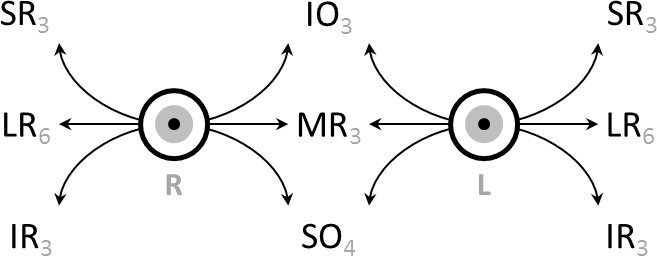
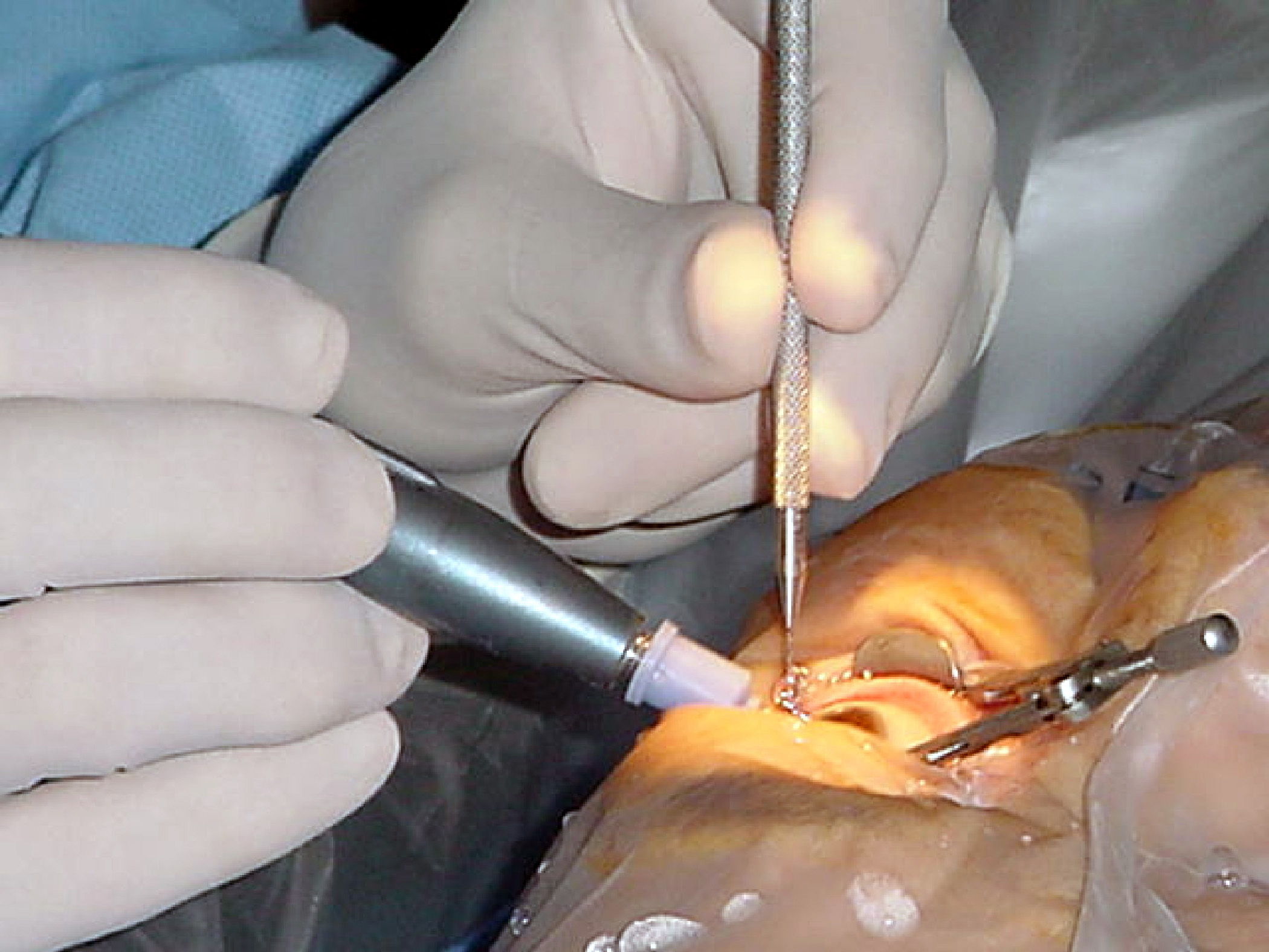
The Harada–Ito procedure is an eye muscle operation designed to improve the excyclotorsion experienced by some patients with cranial nerve IV palsy. In this procedure, the superior oblique tendon is split, and the anterior fibers – the fibers most responsible for incyclotorsion – are moved anteriorly and laterally. This selectively stretches and tightens these fibers, enhancing the incyclotorsion power of the superior oblique. The most common indication for the Harada–Ito procedure is bilateral acquired cranial nerve IV palsy following closed head trauma (particularly automobile accidents). In this clinical situation the vertical imbalance is often less symptomatically bothersome to the patient than the induced excyclotorsion. Affected patients have a particularly annoying type of double vision (diplopia), wherein the images are twisted (excyclotorted). Patients with cranial nerve IV palsy whose complaints are not specifically limited to to ... [...More Info...] [...Related Items...] OR: [Wikipedia] [Google] [Baidu] |
Eye Muscle
The extraocular muscles (extrinsic ocular muscles), are the seven extrinsic muscles of the human eye. Six of the extraocular muscles, the four recti muscles, and the superior and inferior oblique muscles, control movement of the eye and the other muscle, the levator palpebrae superioris, controls eyelid elevation. The actions of the six muscles responsible for eye movement depend on the position of the eye at the time of muscle contraction. Structure Since only a small part of the eye called the fovea provides sharp vision, the eye must move to follow a target. Eye movements must be precise and fast. This is seen in scenarios like reading, where the reader must shift gaze constantly. Although under voluntary control, most eye movement is accomplished without conscious effort. Precisely how the integration between voluntary and involuntary control of the eye occurs is a subject of continuing research."eye, human."Encyclopædia Britannica from Encyclopædia Britannica 2006 U ... [...More Info...] [...Related Items...] OR: [Wikipedia] [Google] [Baidu] |
Excyclotorsion
Eye movement includes the voluntary or involuntary movement of the eyes. Eye movements are used by a number of organisms (e.g. primates, rodents, flies, birds, fish, cats, crabs, octopus) to fixate, inspect and track visual objects of interests. A special type of eye movement, rapid eye movement, occurs during REM sleep. The eyes are the visual organs of the human body, and move using a system of six muscles. The retina, a specialised type of tissue containing photoreceptors, senses light. These specialised cells convert light into electrochemical signals. These signals travel along the optic nerve fibers to the brain, where they are interpreted as vision in the visual cortex. Primates and many other vertebrates use three types of voluntary eye movement to track objects of interest: smooth pursuit, vergence shifts and saccades. These types of movements appear to be initiated by a small cortical region in the brain's frontal lobe. This is corroborated by removal of the fronta ... [...More Info...] [...Related Items...] OR: [Wikipedia] [Google] [Baidu] |
Cranial Nerve IV Palsy
Fourth cranial nerve palsy or trochlear nerve palsy, is a condition affecting cranial nerve 4 (IV), the trochlear nerve, which is one of the cranial nerves. It causes weakness or paralysis of the superior oblique muscle that it innervates. This condition often causes vertical or near vertical double vision as the weakened muscle prevents the eyes from moving in the same direction together. Because the trochlear nerve is the thinnest and has the longest intracranial course of the cranial nerves, it is particularly vulnerable to traumatic injury. To compensate for the double-vision resulting from the weakness of the superior oblique, patients characteristically tilt their head down and to the side opposite the affected muscle. When present at birth, it is known as congenital fourth nerve palsy. See also * Harada–Ito procedure The Harada–Ito procedure is an eye muscle operation designed to improve the excyclotorsion experienced by some patients with cranial nerve IV p ... [...More Info...] [...Related Items...] OR: [Wikipedia] [Google] [Baidu] |
Superior Oblique
The superior oblique muscle, or obliquus oculi superior, is a fusiform muscle originating in the upper, medial side of the orbit (i.e. from beside the nose) which abducts, depresses and internally rotates the eye. It is the only extraocular muscle innervated by the trochlear nerve (the fourth cranial nerve). Structure The superior oblique muscle loops through a pulley-like structure (the trochlea of superior oblique) and inserts into the sclera on the posterotemporal surface of the eyeball. It is the pulley system that gives superior oblique its actions, causing depression of the eyeball despite being inserted on the superior surface. The superior oblique arises immediately above the margin of the optic foramen, superior and medial to the origin of the superior rectus, and, passing forward, ends in a rounded tendon, which plays in a fibrocartilaginous ring or pulley attached to the trochlear fossa of the frontal bone. The contiguous surfaces of the tendon and ring are lined by ... [...More Info...] [...Related Items...] OR: [Wikipedia] [Google] [Baidu] |
Incyclotorsion
Eye movement includes the voluntary or involuntary movement of the eyes. Eye movements are used by a number of organisms (e.g. primates, rodents, flies, birds, fish, cats, crabs, octopus) to fixate, inspect and track visual objects of interests. A special type of eye movement, rapid eye movement, occurs during REM sleep. The eyes are the visual organs of the human body, and move using a system of six muscles. The retina, a specialised type of tissue containing photoreceptors, senses light. These specialised cells convert light into electrochemical signals. These signals travel along the optic nerve fibers to the brain, where they are interpreted as vision in the visual cortex. Primates and many other vertebrates use three types of voluntary eye movement to track objects of interest: smooth pursuit, vergence shifts and saccades. These types of movements appear to be initiated by a small cortical region in the brain's frontal lobe. This is corroborated by removal o ... [...More Info...] [...Related Items...] OR: [Wikipedia] [Google] [Baidu] |
Head Trauma
A head injury is any injury that results in trauma to the skull or brain. The terms ''traumatic brain injury'' and ''head injury'' are often used interchangeably in the medical literature. Because head injuries cover such a broad scope of injuries, there are many causes—including accidents, falls, physical assault, or traffic accidents—that can cause head injuries. The number of new cases is 1.7 million in the United States each year, with about 3% of these incidents leading to death. Adults have head injuries more frequently than any age group resulting from falls, motor vehicle crashes, colliding or being struck by an object, or assaults. Children, however, may experience head injuries from accidental falls or intentional causes (such as being struck or shaken) leading to hospitalization. Acquired brain injury (ABI) is a term used to differentiate brain injuries occurring after birth from injury, from a genetic disorder, or from a congenital disorder. Unlike a broke ... [...More Info...] [...Related Items...] OR: [Wikipedia] [Google] [Baidu] |
Diplopia
Diplopia is the simultaneous perception of two images of a single object that may be displaced horizontally or vertically in relation to each other. Also called double vision, it is a loss of visual focus under regular conditions, and is often voluntary. However, when occurring involuntarily, it results in impaired function of the extraocular muscles, where both eyes are still functional, but they cannot turn to target the desired object. Problems with these muscles may be due to mechanical problems, disorders of the neuromuscular junction, disorders of the cranial nerves (III, IV, and VI) that innervate the muscles, and occasionally disorders involving the supranuclear oculomotor pathways or ingestion of toxins. Diplopia can be one of the first signs of a systemic disease, particularly to a muscular or neurological process, and it may disrupt a person's balance, movement, or reading abilities. Causes Diplopia has a diverse range of ophthalmologic, infectious, autoimmune ... [...More Info...] [...Related Items...] OR: [Wikipedia] [Google] [Baidu] |
Inferior Oblique
The inferior oblique muscle or obliquus oculi inferior is a thin, narrow muscle placed near the anterior margin of the floor of the orbit. The inferior oblique is one of the extraocular muscles, and is attached to the maxillary bone (origin) and the posterior, inferior, lateral surface of the eye (insertion). The inferior oblique is innervated by the inferior branch of the oculomotor nerve. Structure The inferior oblique arises from the orbital surface of the maxilla, lateral to the lacrimal groove. Unlike the other extraocular muscles (recti and superior oblique), the inferior oblique muscle does ''not'' originate from the common tendinous ring (annulus of Zinn). Passing lateralward, backward, and upward, between the inferior rectus and the floor of the orbit, and just underneath the lateral rectus muscle, the inferior oblique inserts onto the scleral surface between the inferior rectus and lateral rectus. In humans, the muscle is about 35 mm long. Innervation The inf ... [...More Info...] [...Related Items...] OR: [Wikipedia] [Google] [Baidu] |
Strabismus
Strabismus is a vision disorder in which the eyes do not properly align with each other when looking at an object. The eye that is focused on an object can alternate. The condition may be present occasionally or constantly. If present during a large part of childhood, it may result in amblyopia, or lazy eyes, and loss of depth perception. If onset is during adulthood, it is more likely to result in diplopia, double vision. Strabismus can occur due to muscle dysfunction, farsightedness, problems in the brain, trauma or infections. Risk factors include premature birth, cerebral palsy and a family history of the condition. Types include esotropia, where the eyes are crossed ("cross eyed"); exotropia, where the eyes diverge ("lazy eyed" or "wall eyed"); and hypertropia or hypotropia where they are vertically misaligned. They can also be classified by whether the problem is present in all directions a person looks (comitant) or varies by direction (incomitant). Diagnosis may be mad ... [...More Info...] [...Related Items...] OR: [Wikipedia] [Google] [Baidu] |
Eye Surgery
Eye surgery, also known as ophthalmic or ocular surgery, is surgery performed on the eye or its adnexa, by an ophthalmologist or sometimes, an optometrist. Eye surgery is synonymous with ophthalmology. The eye is a very fragile organ, and requires extreme care before, during, and after a surgical procedure to minimize or prevent further damage. An expert eye surgeon is responsible for selecting the appropriate surgical procedure for the patient, and for taking the necessary safety precautions. Mentions of eye surgery can be found in several ancient texts dating back as early as 1800 BC, with cataract treatment starting in the fifth century BC. Today it continues to be a widely practiced type of surgery, with various techniques having been developed for treating eye problems. Preparation and precautions Since the eye is heavily supplied by nerves, anesthesia is essential. Local anesthesia is most commonly used. Topical anesthesia using lidocaine topical gel is often used f ... [...More Info...] [...Related Items...] OR: [Wikipedia] [Google] [Baidu] |


