|
Hampton's Hump
Hampton's hump, also called Hampton hump, is a radiologic sign which consists of a shallow wedge-shaped opacity in the periphery of the lung with its base against the pleural surface. It is named after Aubrey Otis Hampton, who first described it in 1940. Hampton's hump along with Westermark sign may aid in the diagnosis of pulmonary embolism Pulmonary embolism (PE) is a blockage of an pulmonary artery, artery in the lungs by a substance that has moved from elsewhere in the body through the bloodstream (embolism). Symptoms of a PE may include dyspnea, shortness of breath, chest pain ..., although they are rare and their sensitivities and interoperator reliabilities are low. If the sign is present in an image, there is a high chance that the person has a pulmonary embolism, but when the sign is absent a pulmonary embolism is not ruled out. References {{Radiologic signs Radiologic signs ... [...More Info...] [...Related Items...] OR: [Wikipedia] [Google] [Baidu] |
Radiologic Sign
A radiologic sign is an objective indication of some medical fact (that is, a medical sign) that is detected by a physician during radiologic examination with medical imaging (for example, via an X-ray, CT scan, MRI scan, or sonographic scan). Examples * Double decidual sac sign * Face of the giant panda sign * Football sign * Golden S sign * Hampton's hump * Hilum overlay sign * Kerley lines * Mickey Mouse sign * Omental cake * Peribronchial cuffing * Pneumatosis intestinalis * Rigler's sign * Westermark sign In chest radiograph A chest radiograph, chest X-ray (CXR), or chest film is a projection radiograph of the chest used to diagnose conditions affecting the chest, its contents, and nearby structures. Chest radiographs are the most common film ... See also * List of radiologic signs References {{Radiologic signs * ... [...More Info...] [...Related Items...] OR: [Wikipedia] [Google] [Baidu] |
Lung
The lungs are the primary Organ (biology), organs of the respiratory system in many animals, including humans. In mammals and most other tetrapods, two lungs are located near the Vertebral column, backbone on either side of the heart. Their function in the respiratory system is to extract oxygen from the atmosphere and transfer it into the bloodstream, and to release carbon dioxide from the bloodstream into the atmosphere, in a process of gas exchange. Respiration is driven by different muscular systems in different species. Mammals, reptiles and birds use their musculoskeletal systems to support and foster breathing. In early tetrapods, air was driven into the lungs by the pharyngeal muscles via buccal pumping, a mechanism still seen in amphibians. In humans, the primary muscle that drives breathing is the Thoracic diaphragm, diaphragm. The lungs also provide airflow that makes Animal communication#Auditory, vocalisation including speech possible. Humans have two lungs, a ri ... [...More Info...] [...Related Items...] OR: [Wikipedia] [Google] [Baidu] |
Aubrey Otis Hampton
Aubrey Otis Hampton (September 10, 1900 in Copeville, Texas – July 17, 1955 in Weare, New Hampshire) was an American radiologist remembered for describing Hampton's hump and Hampton's line. He graduated from Baylor College of Medicine in 1925, undertook his internship in Dallas and worked at the Massachusetts General Hospital from 1926. He became chief of radiology at Massachusetts General in 1941, serving as chief of radiology at the Walter Reed Army Hospital in Washington, D.C. Washington, D.C., formally the District of Columbia and commonly known as Washington or D.C., is the capital city and federal district of the United States. The city is on the Potomac River, across from Virginia, and shares land borders with ... from 1942 to 1945. Hampton was said to be one of the most accurate radiologists in diagnosing during his era. Hampton died in 1955, aged 54, and is buried in the Hillside Cemetery, in Weare, New Hampshire. Sources''Wonders of Radiology'' p. 62-65 Ex ... [...More Info...] [...Related Items...] OR: [Wikipedia] [Google] [Baidu] |
Westermark Sign
In chest radiograph A chest radiograph, chest X-ray (CXR), or chest film is a projection radiograph of the chest used to diagnose conditions affecting the chest, its contents, and nearby structures. Chest radiographs are the most common film taken in medicine. L ...y, the Westermark sign is a sign that represents a focus of oligemia (hypovolemia) (leading to collapse of vessel) seen distal to a pulmonary embolism (PE). While the chest x-ray is normal in the majority of PE cases, the Westermark sign is seen in 2% of patients. Essentially, this is a plain X-ray version of a filling defect as seen on computed tomography pulmonary arteriogram. The sign results from a combination of: # the dilation of the pulmonary arteries proximal to the embolus and # the collapse of the distal vasculature creating the appearance of a sharp cut off on chest radiography. Sensitivity and specificity The Westermark sign, like Hampton's hump (a wedge shaped, pleural based consolidation asso ... [...More Info...] [...Related Items...] OR: [Wikipedia] [Google] [Baidu] |
Pulmonary Embolism
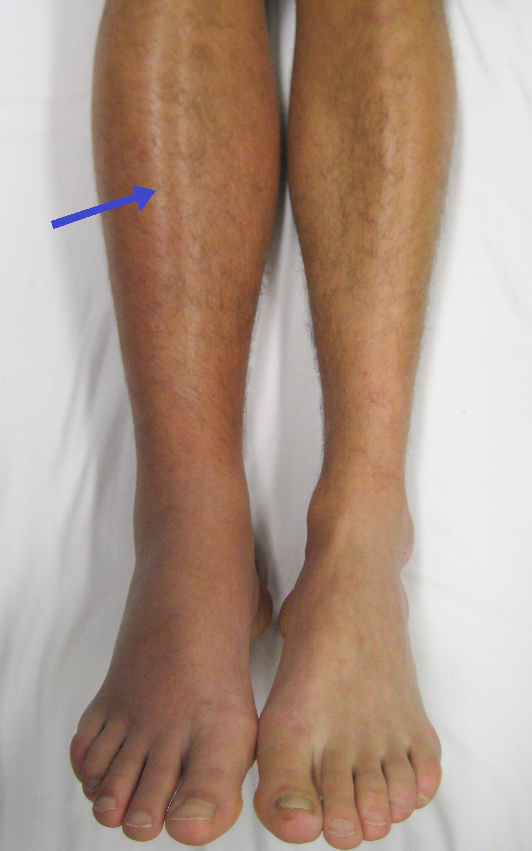
Pulmonary embolism (PE) is a blockage of an pulmonary artery, artery in the lungs by a substance that has moved from elsewhere in the body through the bloodstream (embolism). Symptoms of a PE may include dyspnea, shortness of breath, chest pain particularly upon breathing in, and coughing up blood. Symptoms of a deep vein thrombosis, blood clot in the leg may also be present, such as a erythema, red, warm, swollen, and painful leg. Signs of a PE include low blood oxygen saturation, oxygen levels, tachypnea, rapid breathing, tachycardia, rapid heart rate, and sometimes a mild fever. Severe cases can lead to Syncope (medicine), passing out, shock (circulatory), abnormally low blood pressure, obstructive shock, and cardiac arrest, sudden death. PE usually results from a blood clot in the leg that travels to the lung. The risk of blood clots is increased by advanced age, cancer, prolonged bed rest and immobilization, smoking, stroke, long-haul travel over 4 hours, certain genetics, ... [...More Info...] [...Related Items...] OR: [Wikipedia] [Google] [Baidu] |


