|
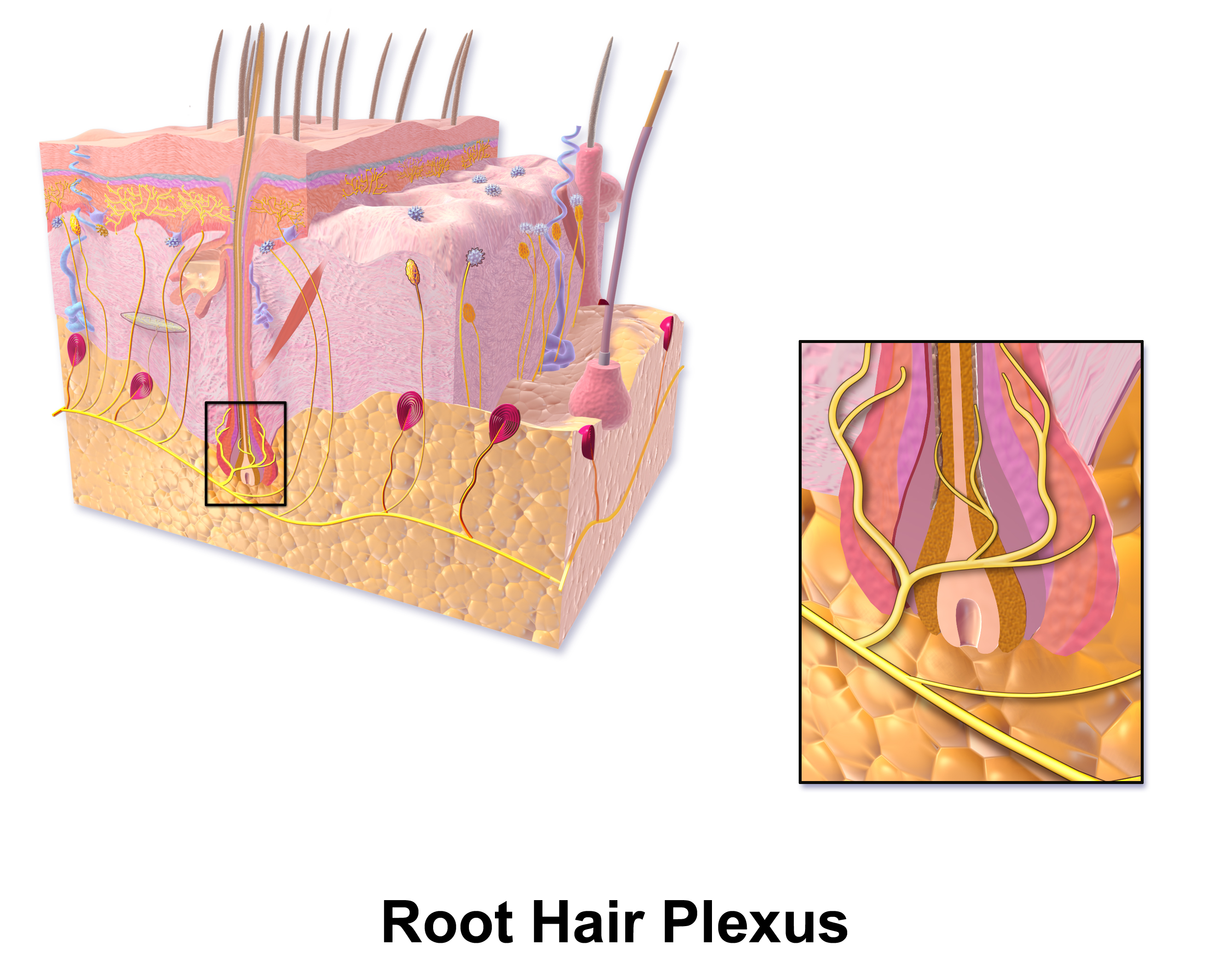
Hair Plexus
A hair plexus or root hair plexus is a special group of nerve fiber endings and serves as a very sensitive mechanoreceptor for touch sensation. Each hair plexus forms a network around a hair follicle and is a receptor, which means it sends and receives nerve impulses to and from the brain when the hair moves. Endings of sensory nerve fibers which form a plexus around a hair follicle in hairy skin. They are mechanoreceptors conveying touch sensation. Specifically, crude touch and pressure sensation conveyed through the spinocervical tract, which is located in the posterior part of the lateral funiculus and terminates in the lateral cervical nucleus The lateral cervical Nucleus (neuroanatomy), nucleus is a scattered nucleus located dorsally in the lateral funiculus in the first three cervical segments of the Spine (anatomy), spine. The Spinocervical pathway, spinocervical and Spinothalamic tra ....Nolte, John. Nolte’s The Human Brain E-Book: An Introduction to its Functional Ana ... [...More Info...] [...Related Items...] OR: [Wikipedia] [Google] [Baidu] |
Blausen 0806 Skin RootHairPlexus
Blausen Medical Communications, Inc. is the creator and owner of a library of two- and three-dimensional medical and scientific images and animations, a developer of information technology allowing access to that content, and a business focused on licensing and distributing the content. It was founded by Bruce Blausen in Houston, Texas, in 1991, and is privately held. Background Blausen Medical Communications, Inc. (BMC) is a privately held company founded by Bruce Blausen in Houston, Texas in 1991. BMC created and owns a library of medical and scientific images and animations, and has developed information technology tools allowing access to the library; as well, it licenses and otherwise works to distribute the content. As of this date, BMC's animation library comprised approximately 1,500 animations and over 27,000 two- and three-dimensional images designed for point-of-care patient education, which could be accessed by consumers or professional caregivers (primarily via ... [...More Info...] [...Related Items...] OR: [Wikipedia] [Google] [Baidu] |
Nerve Fiber
An axon (from Greek ἄξων ''áxōn'', axis), or nerve fiber (or nerve fibre: see spelling differences), is a long, slender projection of a nerve cell, or neuron, in vertebrates, that typically conducts electrical impulses known as action potentials away from the nerve cell body. The function of the axon is to transmit information to different neurons, muscles, and glands. In certain sensory neurons ( pseudounipolar neurons), such as those for touch and warmth, the axons are called afferent nerve fibers and the electrical impulse travels along these from the periphery to the cell body and from the cell body to the spinal cord along another branch of the same axon. Axon dysfunction can be the cause of many inherited and acquired neurological disorders that affect both the peripheral and central neurons. Nerve fibers are classed into three types group A nerve fibers, group B nerve fibers, and group C nerve fibers. Groups A and B are myelinated, and group C are unmyelinate ... [...More Info...] [...Related Items...] OR: [Wikipedia] [Google] [Baidu] |
Mechanoreceptor
A mechanoreceptor, also called mechanoceptor, is a sensory receptor that responds to mechanical pressure or distortion. Mechanoreceptors are innervated by sensory neurons that convert mechanical pressure into electrical signals that, in animals, are sent to the central nervous system. Vertebrate mechanoreceptors Cutaneous mechanoreceptors Cutaneous mechanoreceptors respond to mechanical stimuli that result from physical interaction, including pressure and vibration. They are located in the skin, like other cutaneous receptors. They are all innervated by Aβ fibers, except the mechanorecepting free nerve endings, which are innervated by Aδ fibers. Cutaneous mechanoreceptors can be categorized by what kind of sensation they perceive, by the rate of adaptation, and by morphology. Furthermore, each has a different receptive field. By sensation *The Slowly Adapting type 1 (SA1) mechanoreceptor, with the Merkel corpuscle end-organ (also known as Merkel discs) detect sustaine ... [...More Info...] [...Related Items...] OR: [Wikipedia] [Google] [Baidu] |
Hair Follicle
The hair follicle is an organ found in mammalian skin. It resides in the dermal layer of the skin and is made up of 20 different cell types, each with distinct functions. The hair follicle regulates hair growth via a complex interaction between hormones, neuropeptides, and immune cells. This complex interaction induces the hair follicle to produce different types of hair as seen on different parts of the body. For example, terminal hairs grow on the scalp and lanugo hairs are seen covering the bodies of fetuses in the uterus and in some newborn babies. The process of hair growth occurs in distinct sequential stages. The first stage is called ''anagen'' and is the active growth phase, ''telogen'' is the resting stage, ''catagen'' is the regression of the hair follicle phase, ''exogen'' is the active shedding of hair phase and lastly ''kenogen'' is the phase between the empty hair follicle and the growth of new hair. The function of hair in humans has long been a subject ... [...More Info...] [...Related Items...] OR: [Wikipedia] [Google] [Baidu] |
Receptor (biochemistry)
In biochemistry and pharmacology, receptors are chemical structures, composed of protein, that receive and transduce signals that may be integrated into biological systems. These signals are typically chemical messengers which bind to a receptor and cause some form of cellular/tissue response, e.g. a change in the electrical activity of a cell. There are three main ways the action of the receptor can be classified: relay of signal, amplification, or integration. Relaying sends the signal onward, amplification increases the effect of a single ligand, and integration allows the signal to be incorporated into another biochemical pathway. Receptor proteins can be classified by their location. Transmembrane receptors include ligand-gated ion channels, G protein-coupled receptors, and enzyme-linked hormone receptors. Intracellular receptors are those found inside the cell, and include cytoplasmic receptors and nuclear receptors. A molecule that binds to a receptor is called a li ... [...More Info...] [...Related Items...] OR: [Wikipedia] [Google] [Baidu] |
Brain
The brain is an organ that serves as the center of the nervous system in all vertebrate and most invertebrate animals. It consists of nervous tissue and is typically located in the head ( cephalization), usually near organs for special senses such as vision, hearing and olfaction. Being the most specialized organ, it is responsible for receiving information from the sensory nervous system, processing those information (thought, cognition, and intelligence) and the coordination of motor control (muscle activity and endocrine system). While invertebrate brains arise from paired segmental ganglia (each of which is only responsible for the respective body segment) of the ventral nerve cord, vertebrate brains develop axially from the midline dorsal nerve cord as a vesicular enlargement at the rostral end of the neural tube, with centralized control over all body segments. All vertebrate brains can be embryonically divided into three parts: the forebrain (prosencep ... [...More Info...] [...Related Items...] OR: [Wikipedia] [Google] [Baidu] |
Mechanoreceptor
A mechanoreceptor, also called mechanoceptor, is a sensory receptor that responds to mechanical pressure or distortion. Mechanoreceptors are innervated by sensory neurons that convert mechanical pressure into electrical signals that, in animals, are sent to the central nervous system. Vertebrate mechanoreceptors Cutaneous mechanoreceptors Cutaneous mechanoreceptors respond to mechanical stimuli that result from physical interaction, including pressure and vibration. They are located in the skin, like other cutaneous receptors. They are all innervated by Aβ fibers, except the mechanorecepting free nerve endings, which are innervated by Aδ fibers. Cutaneous mechanoreceptors can be categorized by what kind of sensation they perceive, by the rate of adaptation, and by morphology. Furthermore, each has a different receptive field. By sensation *The Slowly Adapting type 1 (SA1) mechanoreceptor, with the Merkel corpuscle end-organ (also known as Merkel discs) detect sustaine ... [...More Info...] [...Related Items...] OR: [Wikipedia] [Google] [Baidu] |
Spinocervical Tract
The spinocervical pathway is a four-neuron, fast-conducting, tactile/pressure pathway from the spinal cord to sensory cortex The sensory cortex can refer informally to the primary somatosensory cortex, or it can be used as a term for the primary and secondary cortices of the different senses (two cortices each, on left and right hemisphere): the visual cortex on the .... It involves the primary sensory neuron in the dorsal root ganglion, second order cell in the posterior horn of spinal cord, dorsal horn, third order cell in the lateral cervical nucleus near C1 and C2, and a fourth order cell in Isothalamus#Tactile_lemniscal_territory_VPC.3DVPL.2BVPM, VPL thalamus. It is well developed in the cat, but vestigial in humans. References [...More Info...] [...Related Items...] OR: [Wikipedia] [Google] [Baidu] |
Lateral Funiculus
The most lateral of the bundles of the anterior nerve roots In anatomy and neurology, the ventral root of spinal nerve, anterior root, or motor root is the efferent motor root of a spinal nerve. At its distal end, the ventral root joins with the dorsal root The dorsal root of spinal nerve (or posterior r ... is generally taken as a dividing line that separates the anterolateral system into two parts. These are the anterior funiculus, between the anterior median fissure and the most lateral of the anterior nerve roots, and the lateral funiculus (or lateral column) between the exit of these roots and the posterolateral sulcus. The lateral funiculus transmits the contralateral corticospinal and spinothalamic tracts. A lateral cutting of the spinal cord results in the transection of both ipsilateral posterior column and lateral funiculus and this produces Brown-Séquard syndrome.Kaplan Qbook - USMLE Step 1 - 5th edition - page References Central nervous system { ... [...More Info...] [...Related Items...] OR: [Wikipedia] [Google] [Baidu] |
Lateral Cervical Nucleus
The lateral cervical Nucleus (neuroanatomy), nucleus is a scattered nucleus located dorsally in the lateral funiculus in the first three cervical segments of the Spine (anatomy), spine. The Spinocervical pathway, spinocervical and Spinothalamic tract, spinothalamic tracts synapse in the lateral cervical nucleus; the spinocervical tract projects ipsilaterally while the spinothalamic tract projects contralaterally. The axons of neurons from the lateral cervical nucleus cross the midline, join the medial lemniscus as it forms in the caudal Medulla oblongata, medulla and ascend to the ventral posterolateral nucleus.Nolte, John. Nolte’s The Human Brain E-Book: An Introduction to its Functional Anatomy. Elsevier Health Sciences, 2015. References Spinal cord {{Neuroanatomy-stub ... [...More Info...] [...Related Items...] OR: [Wikipedia] [Google] [Baidu] |


